
Renin angiotensin system in BP regulation flow chart
Reading File
The RAS is a key hormonal cascade where renin cleaves angiotensinogen to Angiotensin I, which is then converted by ACE to Angiotensin II - the main effector that raises BP via vasoconstriction and aldosterone release.
Searching sources...
Finding Sources
Finding Sources
I have great sources. Let me read the key sections from Guyton (physiology), Robbins (pathology - has a diagram), and Katzung (pharmacology) simultaneously.
Reading File
Reading File
Reading File
Excellent - I found the key diagram from Robbins. Let me fetch both textbook images simultaneously.
Loading Image
Loading Image
Reading File
I now have everything I need - two excellent diagrams and comprehensive textbook content. Here is the full response:
Renin-Angiotensin System in Blood Pressure Regulation
Overview Flow Chart (Robbins & Kumar Basic Pathology, Fig. 8.3)
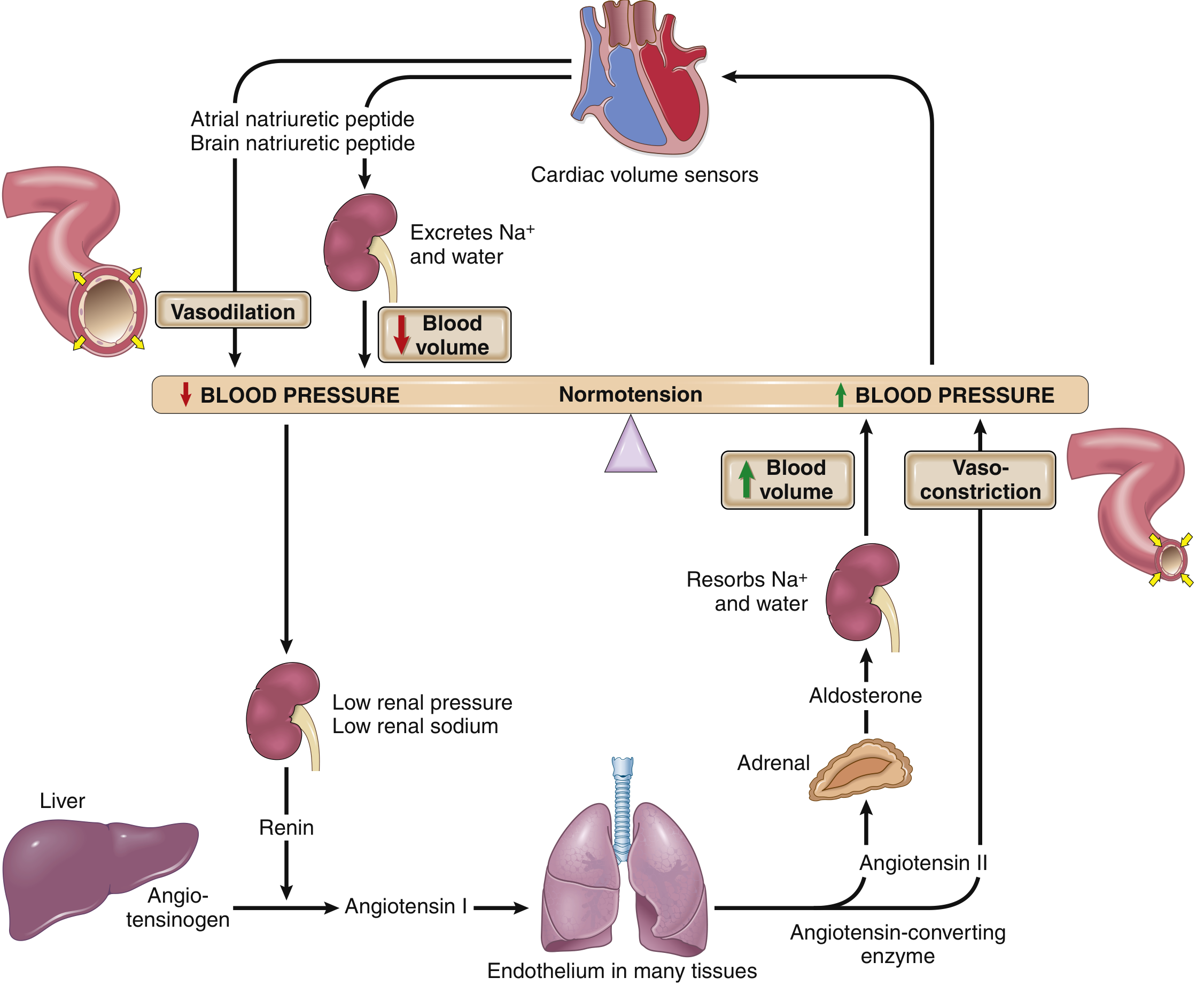
This classic diagram shows how low BP triggers the RAAS cascade (right side raises BP) and how high BP triggers natriuretic peptides to lower it (left side lowers BP).
Full Molecular Pathway (Katzung Basic & Clinical Pharmacology, Fig. 17-3)
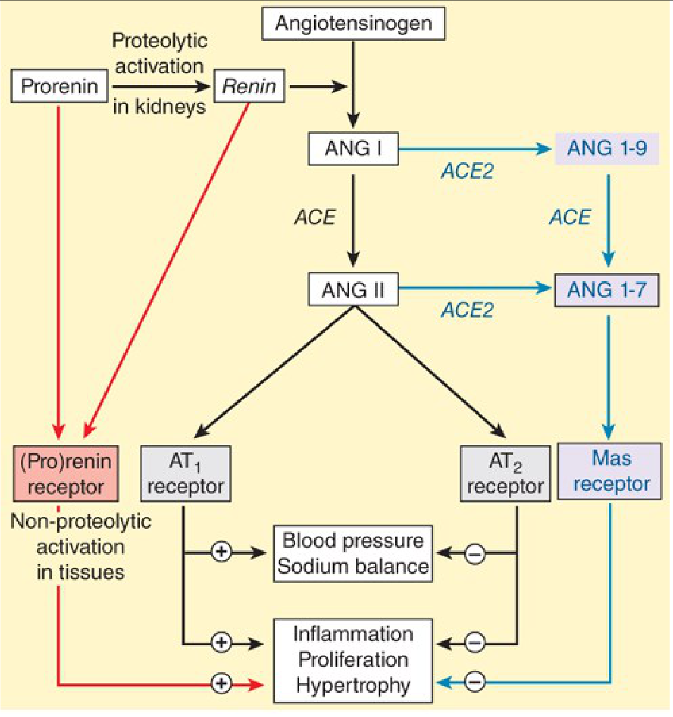
This diagram shows both the classical pathway (black) and the newer counter-regulatory pathways (blue = ANG 1-7/Mas receptor; red = prorenin receptor).
Step-by-Step Flow of the Classical RAAS
↓ Blood Pressure / ↓ Renal Perfusion / ↓ Renal Na⁺
            ↓
  Juxtaglomerular (JG) cells of kidney
  → Release RENIN
  (also triggered by: β1-adrenergic stimulation, PGE2;
   inhibited by: ANG II feedback, high Ca²⁺, ANP)
            ↓
  ANGIOTENSINOGEN (α-globulin, made in liver)
  ──[Renin cleaves]──→ ANGIOTENSIN I (10 aa, inactive)
            ↓
  ACE (Angiotensin-Converting Enzyme)
  — expressed on vascular endothelium, especially lungs —
  ──[ACE cleaves 2 aa]──→ ANGIOTENSIN II (8 aa, highly active)
            ↓
       AT₁ Receptors
      ↙              ↘
VASOCONSTRICTION    ADRENAL CORTEX
(arteriolar SMC)     → Aldosterone secretion
↑ Peripheral         → ↑ Na⁺ & H₂O reabsorption
  resistance           in distal tubule/collecting duct
                     → ↑ Blood volume
            ↓                  ↓
     ↑ BLOOD PRESSURE ←────────┘
Three Mechanisms by Which Ang II Raises BP
| Mechanism | Target | Effect | Timing |
|---|---|---|---|
| Direct vasoconstriction | Arteriolar smooth muscle (AT₁) | ↑ Peripheral resistance | Rapid (10-15 sec) |
| Aldosterone release | Adrenal zona glomerulosa | ↑ Na⁺/H₂O retention → ↑ blood volume | Slow (hours-days) |
| Direct renal effect | Tubular cells + efferent arteriole constriction | ↑ Na⁺/H₂O reabsorption, ↓ peritubular capillary pressure | Intermediate |
Ang II is ~40× more potent than norepinephrine on a molar basis. The vasoconstrictor response takes ~20 minutes to become fully active - slower than baroreceptor reflexes but more sustained. - Katzung, Basic & Clinical Pharmacology 16e
Counter-Regulatory (Protective) Arm
The system has a built-in counter-balance:
-
ACE2 converts ANG I → ANG 1-9, and ANG II → ANG 1-7
-
ANG 1-7 acts on the Mas receptor → vasodilation, anti-inflammatory, anti-proliferative
-
This opposes AT₁-mediated effects and limits excessive vasoconstriction
-
ANP/BNP (released from cardiac volume sensors when BP is high):
- Cause natriuresis/diuresis → ↓ blood volume
- Cause vasodilation
- Directly inhibit renin release
Triggers for Renin Release (Stimuli)
- Low renal artery pressure - JG cells sense stretch; hypotension = less stretch = more renin
- Low tubular Na⁺ at macula densa - detected when GFR falls (e.g., heart failure)
- Sympathetic activation - norepinephrine via β₁-adrenoceptors on JG cells (links stress/volume loss to RAAS)
- Prostaglandins (PGE₂) - paracrine stimulation
Inhibitors of Renin Release
- ANG II (short-loop negative feedback via AT₁)
- High renal perfusion pressure (stretch)
- ANP/BNP
- High Na⁺ at macula densa
Drug Targets in the RAAS
| Drug Class | Site of Action | Example |
|---|---|---|
| Direct renin inhibitors | Block renin | Aliskiren |
| ACE inhibitors | Block ACE | Enalapril, Lisinopril |
| ARBs | Block AT₁ receptor | Losartan, Valsartan |
| Aldosterone antagonists | Block aldosterone effect | Spironolactone |
| β-blockers | Reduce renin release (β₁) | Metoprolol |
Sources:
- Robbins & Kumar Basic Pathology, Fig. 8.3 - BP regulation interplay
- Guyton and Hall Textbook of Medical Physiology - Chapter 19, RAS in arterial pressure control
- Katzung Basic & Clinical Pharmacology 16e - Chapter 17, Renin-Angiotensin System
This is a shared conversation. Sign in to Orris to start your own chat.