
ryle' s tube
A Ryle's tube is a nasogastric (NG) tube used for feeding or gastric drainage. It's a long, flexible, single-lumen tube passed through the nose, down the esophagus, into the stomach. Key points: made of PVC or rubber, typically 90-105 cm long, with multiple side holes at the tip, and radio-opaque for X-ray confirmation of placement.
Searching sources...
Reading File
Searching the Web
Ryle's tube nasogastric tube uses insertion technique
Finding Sources
Finding Sources
Reading Web Page
https://www.medikabazaar.com/blogs/ryles-tube-uses
Now let me read the relevant textbook sections for detailed content:
Reading File
Reading File
Reading File
Reading File
Loading Image
Here is a thorough overview of the Ryle's tube, compiled from surgical textbooks and clinical sources:
Ryle's Tube (Nasogastric Tube)
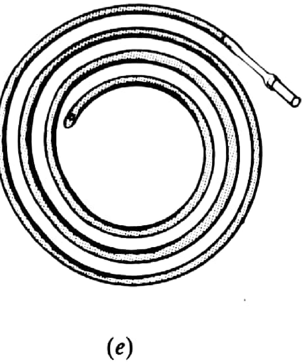
What is It?
A Ryle's tube is a nasogastric (NG) tube - a flexible, single-lumen tube passed through a nostril, down the pharynx and oesophagus, and into the stomach. It is named after John Alfred Ryle, a British physician who described it in the early 20th century.
Modern versions are made of radio-opaque PVC (polyvinylchloride), approximately 125 cm long, with multiple holes near the rounded distal tip. Depth-of-insertion markings are printed along the length. It is a non-vented tube (unlike the vented Salem sump tube).
Key Features
| Feature | Detail |
|---|---|
| Material | High-grade PVC (radio-opaque) |
| Length | ~125 cm |
| Lumen | Single (non-vented) |
| Tip | Rounded, soft, with side ports/holes |
| Markings | Graduated cm markings for insertion depth |
| Radio-opacity | Visible on X-ray for placement confirmation |
Indications (Uses)
1. Gastric Decompression
The most common surgical use. In intestinal obstruction or ileus, the Ryle's tube is placed on free drainage with 4-hourly aspiration, or on continuous/intermittent suction, to decompress the stomach and reduce aspiration risk - particularly before anaesthesia. (Bailey and Love's Surgery, 28th Ed.)
2. Enteral (Tube) Feeding
Used for short-term enteral nutrition in patients who cannot maintain adequate oral intake. The wide calibre allows both feeding and aspiration of gastric contents. The PVC material becomes brittle over time, so the tube should be changed every 2 weeks. - Bailey and Love's Surgery, 28th Ed.
3. Gastric Function Tests (Acid Secretion Studies)
Historically the primary use - used to aspirate gastric juice to measure basal acid output (BAO) and maximum acid output (MAO) in response to stimulants (histamine, pentagastrin, insulin). Normal basal secretion: < 70 mL in the resting stomach. - S Das, Manual on Clinical Surgery, 13th Ed.
4. Medication Administration
Drugs can be given directly into the stomach via the tube.
5. Gastric Lavage
Used in toxicological emergencies for gastric washout.
Insertion Procedure
- Preparation: Patient is positioned semirecumbent. Nostril and pharynx are sprayed with 3% lignocaine in isotonic saline for local anaesthesia. The tube is well lubricated (liquid paraffin or water-soluble gel).
- Insertion: Tube is passed through one nostril, directed posteriorly (not superiorly) toward the nasopharynx.
- Advancement: As the tube reaches the back of the throat, the patient is asked to swallow repeatedly - this pulls the tube down through the oesophagus. A sip of water via straw can assist.
- Depth estimation: A common method is to loop the tube over the patient's ear, place the tip at the xiphoid process, and use that distance (~55 cm in adults) as the target insertion length.
- Securing: Tape the tube to the nose once at the correct depth.
- Confirmation of placement:
- Aspiration of gastric contents (pH < 5.5 on pH indicator strips)
- Chest/upper abdominal X-ray (gold standard - the radio-opaque line confirms tip position)
- Auscultation while injecting air is unreliable and should NOT be used alone.
Complications
Minor (resolve on removal):
- Epistaxis
- Rhinitis / sinusitis
- Pharyngitis / sore throat
- Gagging / discomfort
Serious:
- Pulmonary misplacement - tube passed into trachea/bronchus; feeding into lungs is potentially fatal
- Aspiration pneumonia
- Oesophageal injury / perforation
- Intracranial placement (in patients with basilar skull fractures - an absolute contraindication to nasal insertion)
- Tube knotting (forceful removal)
Ryle's vs Other NG Tubes
| Tube | Type | Key Difference |
|---|---|---|
| Ryle's | Single-lumen, non-vented | Classic; wide bore; good for drainage + feeding |
| Salem Sump | Double-lumen, vented | Vented - prevents stomach wall suction injury; better for continuous drainage |
| Levin | Single-lumen | Similar to Ryle's; slightly different design |
| Fine-bore (8-12 Fr) | Single-lumen | Preferred for long-term feeding (>2 weeks); better tolerated; less risk of erosions |
Contraindications
- Suspected basilar skull fracture (risk of intracranial passage)
- Severe mid-face / nasal trauma
- Oesophageal varices / stricture (relative)
- Recent oesophagogastric surgery (relative)
Sources:
- Bailey and Love's Short Practice of Surgery, 28th Ed. - Tube feeding, Supportive management
- S Das, A Manual on Clinical Surgery, 13th Ed. - Special investigations
- Pye's Surgical Handicraft, 22nd Ed. - NG tube types
- StatPearls - Nasogastric Tube Insertion (NIH)
feeding tube
Feeding tubes are devices used to deliver nutrition, fluids, and medications directly into the GI tract when a patient cannot eat adequately by mouth. Types include nasogastric (NG), nasojejunal (NJ), gastrostomy (PEG), and jejunostomy tubes, chosen based on duration of need and patient anatomy.
Searching sources...
Finding Sources
Searching the Web
feeding tube types indications enteral nutrition clinical overview
Finding Sources
Reading Web Page
https://www.ncbi.nlm.nih.gov/books/NBK532876
Reading File
Reading File
Reading File
Reading File
Loading Image
Here is a comprehensive guide to feeding tubes, drawing from multiple surgical and medical textbooks:
Feeding Tubes (Enteral Nutrition Access)
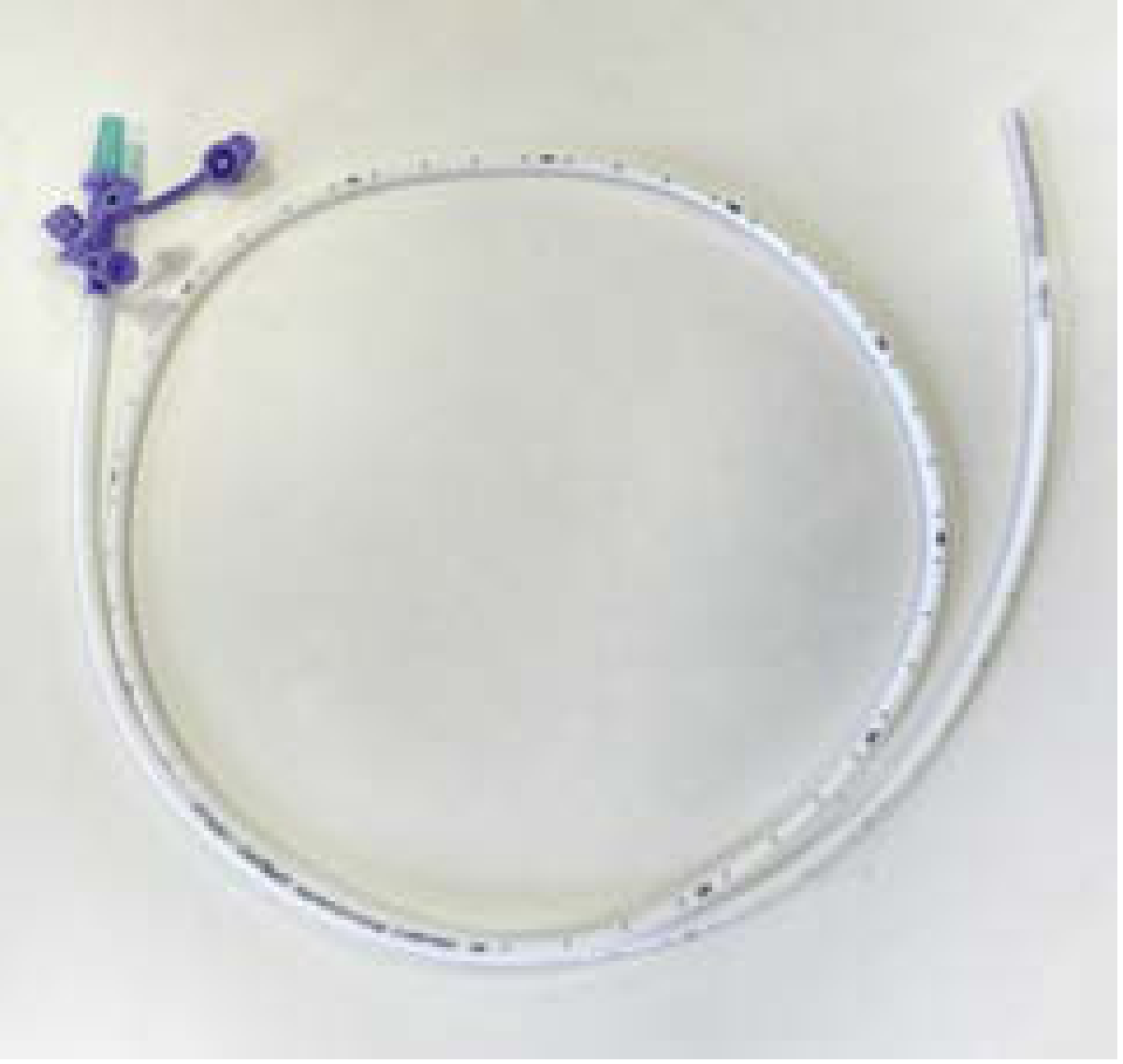
Overview
A feeding tube delivers liquid nutrition, fluids, and medications directly into the GI tract when a patient cannot eat orally. The route and device chosen depend primarily on how long feeding will be needed and where in the GI tract delivery is required.
Enteral nutrition is always preferred over parenteral (IV) nutrition when the GI tract is functional - it maintains gut integrity, stimulates biliary/pancreatic secretions, and reduces infectious complications.
Indications for Tube Feeding
- Dysphagia (stroke, neurological disease, head and neck cancer)
- Severe neurologic impairment (reduced consciousness, dementia)
- Oropharyngeal or proximal GI tract pathology (tumors, trauma)
- Facial trauma
- Inadequate oral intake despite supplements
- Critical illness (start within 24-48 hours once hemodynamically stable)
- Prolonged post-operative period with anticipated poor oral intake
Types of Feeding Tubes
1. Nasogastric (NG) / Ryle's Tube
- Passed through the nose into the stomach
- Used for short-term feeding (< 4-6 weeks)
- Wide bore - can both feed and aspirate gastric contents
- PVC; change every 2 weeks
- Risk of rhinitis, pharyngitis, oesophageal erosions with prolonged use
2. Nasoenteric Tube (NET) - Nasoduodenal / Nasojejunal (NJ)
- Passed through the nose into the duodenum or jejunum (post-pyloric)
- Made of silicone or polyurethane; commonly 12 or 14F diameter
- Used when gastric feeding is not tolerated (gastroparesis, high aspiration risk)
- Bypasses the stomach - feeding must be delivered by continuous pump (not bolus), as the normal regulation of gastric emptying is bypassed
- Rate: typically begin at 15-40 mL/hr, advance to max ~125 mL/hr directly into small bowel
- Placement confirmed by abdominal X-ray
- Duration: also short-term (< 4-6 weeks) - Current Surgical Therapy, 14th Ed.
3. Percutaneous Endoscopic Gastrostomy (PEG)
- Tube placed directly through the abdominal wall into the stomach under endoscopic guidance
- Used for long-term feeding (> 4-6 weeks) or when nasal placement is impossible
- No need for laparotomy or general anaesthesia - done at bedside with monitored anesthesia care
- Technique ("pull" method):
- Gastroscope advanced into stomach; tip transilluminates anterior gastric wall
- Small abdominal incision over area of maximal transillumination
- Angiocatheter inserted; wire passed into gastric lumen, snared, pulled out through mouth
- PEG tube looped onto wire and pulled through the abdominal wall
- Inner bumper confirmed in correct position; tube secured externally
- May be difficult in obese patients (difficult transillumination); fluoroscopic approaches are an alternative
- Goldman-Cecil Medicine; Mulholland and Greenfield's Surgery, 7th Ed.
4. PEG-J (Gastrojejunal) Tube
- A jejunal extension is passed through a gastrostomy into the jejunum
- Allows simultaneous jejunal feeding + gastric venting/decompression
- Used in gastroparesis, after oesophagectomy/gastrectomy
5. Feeding Jejunostomy (Surgical)
- A loop of proximal jejunum (typically 20-40 cm distal to the ligament of Treitz) is brought up and secured to the abdominal wall, with the tube passed transabdominally
- Witzel jejunostomy: tube is covered for a short distance with bowel serosa for stability
- Placed via laparoscopic approach during major abdominal procedures when prolonged feeding is anticipated
- Used when the stomach is absent or non-functional - Mulholland and Greenfield's Surgery, 7th Ed.
6. Surgical Gastrostomy (Stamm)
- Open or laparoscopic technique; small incision into gastric body with concentric purse-string sutures securing a large-bore tube
- Less common now that PEG is available
Choosing the Right Tube
| Duration | Route | Preferred Tube |
|---|---|---|
| < 4-6 weeks | Gastric | Nasogastric (NG/Ryle's) |
| < 4-6 weeks | Post-pyloric | Nasoenteric (NJ/ND) tube |
| > 4-6 weeks | Gastric | PEG |
| > 4-6 weeks | Post-pyloric | PEG-J or feeding jejunostomy |
| Post-upper GI surgery | Small bowel | Surgical feeding jejunostomy |
Feeding Regimens
| Mode | How | When |
|---|---|---|
| Bolus | 300-500 mL intermittently | Intragastric only; mimics normal meals; good for mobile patients |
| Continuous | Pump, 20-24 hrs/day | Preferred for post-pyloric and critically ill patients |
| Cyclic | Pump overnight | Long-term home patients; allows daytime mobility |
Starting rates: 15-20 mL/hr (critically ill / prolonged starvation) up to 40-50 mL/hr (routine), advancing to 75-125 mL/hr as tolerated. - Goldman-Cecil Medicine
Enteral Formulas
| Type | Description | Use |
|---|---|---|
| Polymeric (standard) | Whole proteins, complex carbs, fats; 1 kcal/mL | Most patients with intact GI function |
| High-protein/energy-rich | ~1.5 kcal/mL | Fluid-restricted patients |
| Partially/fully hydrolysed | Pre-digested proteins, simple sugars | Malabsorption, short gut, pancreatitis |
| Disease-specific | Tailored formulations | Renal, hepatic, diabetic patients |
Advantages of Intragastric vs Post-pyloric Feeding
Intragastric (NG/PEG):
- Stomach acts as a reservoir - tolerable as bolus feeds
- Stimulates biliary and pancreatic secretions - trophic for the small bowel
- Gastric secretions dilute osmolarity, reducing diarrhoea risk
- Risk: aspiration in obtunded or neurologically impaired patients
Post-pyloric (NJ/jejunostomy):
- Lower aspiration risk (bypasses stomach)
- Must be continuous pump feeding only (no reservoir function)
- Maximum ~125 mL/hr into the small bowel
Complications
Tube-Related (Mechanical)
- Misplacement into the tracheobronchial tree - potentially fatal if feeds are instilled into lungs
- Tube blockage (prevent by regular water flushing; chymotrypsin may unblock; guidewires are avoided due to perforation risk)
- Tube dislodgement (especially jejunostomy tubes)
- Epistaxis, sinusitis, pharyngitis (nasal tubes)
- Peristomal leakage, skin excoriation (PEG/jejunostomy)
- Peritonitis (if dislodged tube is replaced into peritoneal cavity)
Infectious
- Aspiration pneumonia (most serious)
- Peristomal infection
Formula-Related
- Diarrhoea (up to 50% in critically ill; more common with bolus feeding)
- Nausea, vomiting, abdominal distension - usually from gastroparesis
- Osmotic diarrhoea (high osmolarity formulas)
- Refeeding syndrome - hypophosphataemia on restarting feeds after prolonged starvation; can cause cardiac arrhythmias, respiratory failure, lactic acidosis. Start at low rates (15-20 mL/hr) in at-risk patients.
Important Clinical Points
- Placement must always be confirmed radiographically before starting feeds - auscultation ("air bubble" test) is unreliable
- Position patients at 30-45° head elevation during feeding and for 1-2 hours afterward to reduce aspiration risk
- PVC Ryle's tubes should be changed every 2 weeks (becomes brittle)
- For fine-bore tubes (8-12Fr), never use a guidewire to unblock - perforation risk
- Enteral nutrition can be deferred 24-48 hours in critically ill patients without adverse effects
- Do not start enteral feeding in haemodynamic shock (on high-dose vasopressors) - risk of non-occlusive mesenteric ischaemia
Sources:
- Current Surgical Therapy, 14th Ed. - PEG/NET access
- Goldman-Cecil Medicine, International Ed. - Enteral nutrition
- Mulholland and Greenfield's Surgery, 7th Ed. - Enteral access, jejunostomy
- StatPearls - Enteral Feeding (NIH)
- Merck Manual - Enteral Nutrition
suction catheter
A suction catheter is a flexible tube connected to a suction source, used to clear secretions from the airway (oral, nasal, tracheal) or body cavities. Common types include the Yankauer (rigid, oropharyngeal) and flexible/soft catheters (for nasopharyngeal and endotracheal suctioning).
Searching sources...
Finding Sources
Searching the Web
suction catheter types uses technique airway suctioning clinical
Reading File
Reading File
Reading File
Finding Sources
Reading File
Finding Sources
Loading Image
Finding Sources
Here is a full clinical overview of suction catheters, compiled from emergency medicine and surgical textbooks:
Suction Catheter
What is a Suction Catheter?
A suction catheter is a hollow, flexible tube connected to a vacuum/suction source, used to remove secretions, blood, vomitus, or other material from the airway or body cavities. It is an essential piece of equipment for airway management, particularly when a patient cannot clear their own secretions.
Types of Suction Catheters
1. Yankauer (Tonsil-Tip) Suction
- Rigid or semi-rigid, angled plastic catheter
- Used for oropharyngeal suctioning - clears blood, secretions from the mouth and pharynx
- Rounded tip minimises soft tissue trauma
- Limitation: not large-bore enough to clear vomitus effectively
- Most commonly used in emergency airway management
2. Large-Bore Dental / Wide-Bore Suction Tip (e.g., HI-D Big Stick)
- Large-bore rigid tip (5/8 to 3/4 inch inner diameter)
- Best for clearing vomitus and particulate matter from the upper airway - standard catheters (1/4 inch) become obstructed by thick/particulate material
- Should be available at the bedside during all emergency airway management
3. Flexible / Soft Suction Catheter
- Soft, pliable catheter for nasopharyngeal, nasotracheal, and endotracheal suctioning
- Available in multiple sizes (French gauge)
- Tips may be:
- Single end-hole
- Multiple side ports (reduces mucosal invagination and trauma)
- Coude (directional) tip - angled tip for selective suctioning of the left or right main stem bronchus
4. Closed-System (In-Line) Suction Catheter
- Catheter enclosed in a sterile plastic sheath, permanently attached to the ventilator circuit
- Allows tracheal suctioning without disconnecting the ventilator - continuous oxygen delivery and positive pressure ventilation are maintained
- Used for ventilator-dependent patients (ICU, intubated patients)
- Reduces infection risk (avoids exposure to environment)
5. Bulb Syringe
- Simple hand-operated suction device
- Used in neonates and infants for clearing nose and oropharynx (e.g., at delivery, after amniotic fluid)
Catheter Sizing
Formula for tracheal suction catheter:
Catheter size (French) = 2 × (ETT size - 2)
For example: ETT size 8.0 → catheter = 2 × (8-2) = 12 Fr
Key rule: The suction catheter should occupy less than 50% of the internal diameter of the endotracheal/tracheostomy tube in adults and children (< 70% in neonates). Using an oversized catheter causes excessive evacuation of airway gases and atelectasis.
| Patient | Max Suction Pressure |
|---|---|
| Neonates / Paediatric | < 80-120 mmHg |
| Adults | < 150-200 mmHg |
Indications
- Oropharyngeal: Blood, vomitus, secretions during emergency airway management
- Nasopharyngeal: Secretions in infants, unconscious patients who cannot expectorate
- Endotracheal / Tracheostomy: Retained secretions in intubated or tracheostomised patients; signs include:
- Audible secretions / rattling breath sounds
- Sawtooth pattern on ventilator waveform
- Increased airway resistance / peak pressures
- Visible secretions in the airway tube
- Selective bronchial: Coude-tip catheter to suction a specific lung zone
Procedure & Technique (Tracheal/Tracheostomy Suctioning)
- Monitoring: Attach continuous pulse oximetry, cardiac monitor, and capnography if available
- Position: Awake patients - sitting upright, head neutral. Ventilated patients - head of bed elevated 30°
- Preoxygenate: At least 30-60 seconds of 100% FiO₂ (ventilated) or 10-15 L high-flow O₂ (non-ventilated). Routine instillation of normal saline is no longer recommended - no proven benefit
- Aseptic technique: Sterile gloves, sterile catheter throughout
- Insert catheter through the inner cannula without applying suction
- Advance to desired depth. If carina is reached, a vigorous cough reflex is triggered - withdraw 1-2 cm
- Apply suction while slowly withdrawing the catheter with a gentle rotating motion
- Duration: Maximum 10-15 seconds per pass; no more than 3 passes in succession
- Monitor throughout for desaturation, dysrhythmia, or rise in end-tidal CO₂ - stop immediately if these occur
- Re-oxygenate between passes
Complications
Hypoxaemia
- Most common complication - from interruption of ventilation + aspiration of airway gases + atelectasis
- Can trigger increased ICP, dysrhythmias, or death
- Prevented by preoxygenation and limiting duration to ≤15 seconds
- Use in-line (closed) catheters in ventilated patients to avoid desaturation
Dysrhythmias
- Caused by hypoxia, vagal stimulation (bradycardia, hypotension), catecholamine release
- Bradycardia in hypoxia → risk of ventricular fibrillation
- Pre-treat at-risk patients (especially infants) with nebulised or IV atropine
- Stop suctioning immediately if dysrhythmia develops
Raised Intracranial Pressure (ICP)
- ET suctioning triggers cough → raised intrathoracic pressure → reduced cerebral perfusion pressure
- Prevention: adequate sedation, preoxygenation, hyperventilation (increase RR to ~30/min for 1 min pre-suction), intratracheal lidocaine to blunt the cough reflex
Mucosal Injury
- Invagination of mucosa into catheter side ports
- Risk reduced by multi-port tip catheters, avoiding excessive suction pressure, and rotating the catheter on withdrawal
Atelectasis
- Caused by negative pressure evacuating airway gases
- Resolved by limiting catheter size (<50% ETT lumen) and limiting vacuum pressure
Others
- Bleeding (epistaxis with nasal suction, tracheal bleeding)
- Infection / tracheitis (use strict aseptic technique)
- Bronchospasm
Key Safety Rules (AARC Guidelines)
| Rule | Detail |
|---|---|
| Indication | Suction only when needed (not on a fixed schedule) |
| Preoxygenate | Always before suctioning |
| Catheter size | < 50% of ETT lumen (adults/paeds); < 70% neonates |
| Suction pressure | ≤ 120 mmHg neonates/paeds; ≤ 200 mmHg adults |
| Duration | Maximum 15 seconds per pass |
| Deep suctioning | Only when shallow suctioning is ineffective |
| Saline instillation | Not recommended routinely |
| Ventilated patients | Use closed (in-line) suction system |
| Confirmation | Auscultation while pushing air is unreliable - do not rely on it alone |
Sources:
- Roberts and Hedges' Clinical Procedures in Emergency Medicine, Chapters 3 & 7 - Suctioning technique, equipment, complications
- Tintinalli's Emergency Medicine - Emergency airway suctioning
- Scott-Brown's Otorhinolaryngology - Tracheostomy suctioning
- AARC Clinical Practice Guidelines - Artificial Airway Suctioning
- StatPearls - Surgical Airway Suctioning
k-90 tube
The "K-90 tube" isn't immediately familiar to me as a standard medical term - it could refer to a specific catheter, drainage tube, or feeding tube used in a regional/institutional context. Let me search for it.
Searching sources...
Finding Sources
Searching the Web
K-90 tube medical catheter uses
Reading Web Page
https://meddeygo.com/collections/k90-catheter
Finding Sources
Reading File
Searching the Web
K90 urethral catheter Robinson catheter straight tip India specification
Reading Web Page
https://meddeygo.com/collections/k90-catheter
Finding Sources
Reading File
Reading File
Here is a complete overview of the K-90 tube / catheter:
K-90 Tube (Urethral Catheter)
What is a K-90 Tube?
The K-90 (also written K90, product code 90-22) is a non-retaining, straight-tip urethral catheter used primarily for short-term or intermittent bladder drainage. It is the Indian/South Asian trade designation for what is internationally known as a Robinson catheter - one of the oldest and most widely used urethral catheter designs.
The defining feature is the absence of a balloon - unlike a Foley catheter, it cannot be left in place. It is designed for the "in-and-out" (intermittent) technique: inserted to drain the bladder, then immediately removed.
Key Features
| Feature | Detail |
|---|---|
| Type | Non-retaining / intermittent / straight catheter |
| Tip | Rounded, atraumatic straight tip |
| Balloon | None (not self-retaining) |
| Lumen | Single lumen (one drainage port) |
| Material | PVC (medical grade) or silicone; some rubber versions |
| Length | ~40 cm (standard adult) |
| Radio-opaque | Some brands have an X-ray opaque line |
| Use | Single-use, sterile, disposable |
| Packaging | Individually packed |
Sizes (French Gauge)
| Color Code | Size (FG) | Outer Diameter | Typical Use |
|---|---|---|---|
| White | 12 FG | 4.0 mm | Standard adult female |
| Green | 14 FG | 4.7 mm | Standard adult male |
| Orange | 16 FG | 5.3 mm | Adult male with BPH / enlarged prostate |
| Range | 6 FG - 24 FG | - | Full range available |
French (Fr) to mm conversion: Diameter (mm) = Fr ÷ 3. So 12 Fr = 4 mm, 14 Fr = 4.7 mm, 16 Fr = 5.3 mm.
Indications / Uses
1. Acute Urinary Retention (AUR)
Most common emergency use. Provides immediate bladder decompression when a patient cannot void, without committing to an indwelling catheter. A 14-16 FG K-90 is typically used in adults.
2. Intermittent Self-Catheterisation (ISC)
Used in patients with neurogenic bladder (spinal cord injury, multiple sclerosis, spina bifida) who need to drain the bladder at regular intervals - typically 4-6 times per day. Prevents bladder over-distension, detrusor damage, and back-pressure on the kidneys.
3. Sterile Urine Specimen Collection
Single in-and-out catheterisation provides an uncontaminated mid-stream equivalent for microbiological culture, especially when a clean-catch specimen is unreliable.
4. Post-Void Residual (PVR) Measurement
Inserted after the patient voids to measure exactly how much urine remains in the bladder - important in assessing bladder outlet obstruction, detrusor underactivity, and neurogenic bladder.
5. Clot Evacuation / Haematuria
A wide-bore K-90 can be used to irrigate and clear blood clots from the bladder in gross haematuria, before placing a 3-way Foley for continuous irrigation.
6. Peri-operative Bladder Drainage
Empties the bladder immediately before surgery or a procedure without leaving an indwelling catheter.
K-90 vs Foley Catheter - Key Differences
| Feature | K-90 (Robinson) | Foley Catheter |
|---|---|---|
| Balloon | No | Yes (inflated with sterile water) |
| Retained in bladder | No (must be removed) | Yes (indwelling) |
| Use duration | Intermittent / single-use | Short or long-term indwelling |
| Lumens | 1 (drainage only) | 2 (drainage + balloon) or 3 (+irrigation) |
| Infection risk | Lower (no dwell time) | Higher (catheter-associated UTI - CAUTI) |
| Main use | Intermittent drainage, specimens | Continuous drainage, post-op, ICU |
Insertion Technique (Same as Urethral Catheterisation)
- Position: Supine (frog-legged for female); supine with penis held straight and taut for male
- Preparation: Sterile field, sterile gloves, antiseptic cleansing of meatus
- Lubrication: 10-15 mL sterile lidocaine/lubricant jelly injected into the meatus (male); lubricant jelly on catheter tip (female)
- Insert gently with slow steady pressure; slight resistance at the external sphincter is normal - maintain steady pressure until it yields (asking patient to take a deep breath helps)
- Drain urine completely
- Remove immediately - the catheter is not left in place
- Important: Never force the catheter; blood at the meatus, a perineal haematoma, or a high-riding prostate suggest urethral injury - do not attempt catheterisation
Contraindications
- Suspected urethral disruption (pelvic trauma, blood at meatus, perineal haematoma)
- Recent urethral/bladder neck reconstructive surgery
- Known urethral stricture (relative - may need dilatation with filiform bougies first)
- Acute prostatitis / urethritis (relative)
Complications
- Urethral trauma / false passage (from forceful insertion)
- Urinary tract infection (risk is low with intermittent technique but increases with repeated use)
- Haematuria
- Urethral stricture formation (with repeated traumatic catheterisation)
- Paraphimosis in uncircumcised males (if foreskin not reduced after insertion)
Popular Indian Brands
| Brand | Material | Size Range | Feature |
|---|---|---|---|
| Romsons Nel-Cath | PVC | 6-24 FG | Atraumatic side eyes |
| Angel K90 | PVC | 8-18 FG | Radio-opaque line |
| BARD Robinson | Red rubber | 10-22 FG | Flexible |
| HMD Uro-Cath | PVC | Standard | High-purity connectors |
| Poly Medicure | PVC | Full range | Cost-effective |
Sources:
- Pfenninger and Fowler's Procedures for Primary Care, 3rd Ed. - Robinson catheter, equipment
- Campbell-Walsh-Wein Urology - Types of urethral catheters, sizing, technique
- Meddeygo - K90 Catheter Overview
difference between feeding tube and ryle's tube
A Ryle's tube IS a type of feeding tube (nasogastric), but in clinical practice the term "feeding tube" often refers specifically to the narrow-bore fine feeding tube, while Ryle's tube refers to the wider-bore nasogastric tube. The key difference is calibre, material, and primary purpose.
Searching sources...
Finding Sources
Searching the Web
difference between Ryle's tube and fine bore feeding tube nasogastric comparison
Finding Sources
Reading Web Page
https://patient.info/doctor/gastroenterology/nasogastric-ryl…
Finding Sources
Finding Sources
Finding Sources
Reading File
Excellent - I now have authoritative textbook content. Here is the definitive comparison:
Ryle's Tube vs Fine-Bore Feeding Tube
First, the important clarification: a Ryle's tube is a type of nasogastric (NG) tube, and it can be used for feeding. But in clinical practice, when clinicians say "feeding tube" as a distinct category, they usually mean the fine-bore feeding tube - a narrower, softer, longer-lasting device. Here is how the two differ:
Side-by-Side Comparison
| Feature | Ryle's Tube | Fine-Bore Feeding Tube |
|---|---|---|
| Also known as | Wide-bore NG tube | Enteral feeding tube, silk/soft NG tube |
| Bore (calibre) | Wide bore (~14-18 Fr) | Fine bore (8-12 Fr) |
| Material | High-grade PVC (polyvinylchloride) | Polyurethane or silicone |
| Flexibility | Stiffer / less flexible | Soft and pliable |
| Radio-opacity | Yes (visible on X-ray) | Yes (most have radio-opaque tip or line) |
| Guidewire needed for insertion? | No | Often yes - internal guidewire/stylet to stiffen it during insertion (must be removed after) |
| Duration of use | Up to 2 weeks (PVC becomes brittle) | Up to 6 weeks (polyurethane/silicone unaffected by gastric acid) |
| Can aspirate gastric contents? | Yes - wide calibre allows aspiration | No (or difficult) - too narrow to aspirate reliably |
| Gastric decompression? | Yes - suitable for drainage + suction | No - not suitable for drainage |
| Feeding? | Yes (short-term) | Yes (preferred for longer-term feeding) |
| Tube blockage risk | Lower (wider lumen) | Higher - requires daily water flushing; chymotrypsin to unblock if needed; never use a guidewire to unblock (perforation risk) |
| Patient comfort | Less comfortable - causes more rhinitis, pharyngitis, oesophageal erosions | Much better tolerated - less interference with eating, drinking, and sleeping |
| Interferes with eating/drinking? | More likely | Less likely |
| When preferred | Post-op drainage, intestinal obstruction, short-term feeding (< 2 weeks), gastric function tests | Longer-term feeding (2-6 weeks), patients who need to eat/drink orally as well |
The Core Clinical Rule
"Use a Ryle's tube when you also need to drain or decompress the stomach. Use a fine-bore feeding tube when feeding is the only goal and comfort/duration matters."
- Bailey and Love's Surgery, 28th Ed.
When to Use Which
Use a Ryle's Tube (Wide-Bore NG) when:
- Gastric decompression is needed (intestinal obstruction, ileus, post-op)
- Short-term feeding (< 2 weeks)
- You need to both feed and aspirate gastric residuals
- Gastric function tests / acid secretion studies
- Gastric lavage
- Emergency use (easier to insert without a stylet)
Use a Fine-Bore Feeding Tube when:
- Feeding is the only purpose (no drainage needed)
- Longer-term enteral nutrition (2-6 weeks)
- Patient needs to remain comfortable and continue eating/drinking orally
- Patients with sensitive airways (less rhinitis, pharyngitis, oesophageal erosion risk)
- Patients who are awake and ambulant
Use Neither (switch to PEG/gastrostomy) when:
- Feeding will be needed for > 4-6 weeks
Insertion Differences
| Step | Ryle's Tube | Fine-Bore Feeding Tube |
|---|---|---|
| Stylet / guidewire | Not needed | Required (internal guidewire stiffens the soft tube) - must be removed after placement confirmation |
| Insertion technique | Same NG technique | Same, but more care needed as tube is floppy |
| Placement confirmation | pH < 5 on aspirate OR CXR | CXR preferred (aspirate difficult due to narrow bore); pH strips if aspirate obtained |
Material Matters
| Material | Tube Type | Effect of Gastric Acid | Max Duration |
|---|---|---|---|
| PVC | Ryle's tube | Becomes brittle over time | 2 weeks - must change |
| Polyurethane | Fine-bore feeding tube | Unaffected by gastric acid | Up to 6 weeks |
| Silicone | Fine-bore feeding tube | Unaffected by gastric acid | Up to 6 weeks |
Summary in One Line
A Ryle's tube is wide, stiff, PVC, dual-purpose (feed + drain), short-term (≤2 weeks). A fine-bore feeding tube is narrow, soft, polyurethane/silicone, feeding-only, longer-term (up to 6 weeks). Both are nasogastric tubes - the difference is calibre, material, purpose, and duration.
Source: Bailey and Love's Short Practice of Surgery, 28th Ed. - Tube feeding; Patient.info - Nasogastric (Ryle's) Tubes
This is a shared conversation. Sign in to Orris to start your own chat.