
Gray and white matter in brain and spinal cord difference
Reading File
Gray matter contains neuronal cell bodies, dendrites, and unmyelinated axons - it's where synaptic processing happens. White matter is composed mainly of myelinated axons that connect different regions. Their spatial arrangement is inverted between the brain and spinal cord: in the brain, gray matter is on the outside (cortex) with white matter inside, while in the spinal cord, gray matter is centrally located (butterfly/H-shape) with white matter surrounding it.
Searching sources...
Finding Sources
Finding Sources
Reading File
Reading File
Reading File
Loading Image
Loading Image
Finding Sources
Reading File
Here is a complete, textbook-sourced breakdown of gray and white matter differences in the brain vs. spinal cord.
Gray Matter vs. White Matter - Brain and Spinal Cord
What Are They?
| Feature | Gray Matter | White Matter |
|---|---|---|
| Main contents | Neuronal cell bodies, dendrites, unmyelinated axons, glia | Myelinated axons, glial cells, blood vessels |
| Color (fresh tissue) | Grayish-tan (no myelin) | White (due to myelin lipid content) |
| Synapses | Present - all synaptic activity occurs here | Absent |
| Function | Local processing, integration, computation | Long-distance signal transmission between regions |
| Organized as | Cortex (surface layer) or nuclei (deep clusters) | Tracts / fascicles / commissures |
"Synapses occur only in the gray matter." - Histology: A Text and Atlas, p. 1000
Key Difference: Spatial Arrangement is INVERTED
This is the most tested and clinically important difference:
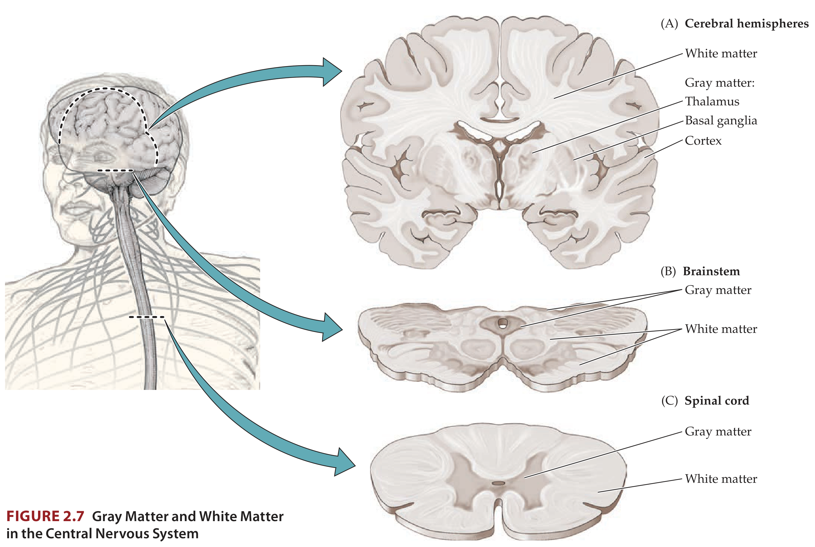
Brain (Cerebral Hemispheres):
- Gray matter = OUTSIDE (forms the cerebral cortex, the outermost layer)
- White matter = INSIDE (inner core/medulla connecting different cortical and subcortical regions)
- Additional gray matter islands deep inside = nuclei (basal ganglia, thalamus)
Spinal Cord:
- Gray matter = INSIDE (centrally located, butterfly/H-shaped surrounding the central canal)
- White matter = OUTSIDE (peripheral shell of ascending and descending tracts)
"In the cerebral hemispheres the gray matter cortex is outside, while the white matter is inside. In the spinal cord the opposite is true: White matter pathways lie on the outside, while the gray matter is in the center." - Neuroanatomy through Clinical Cases, 3rd Ed.
Histological Cross-Section of the Spinal Cord
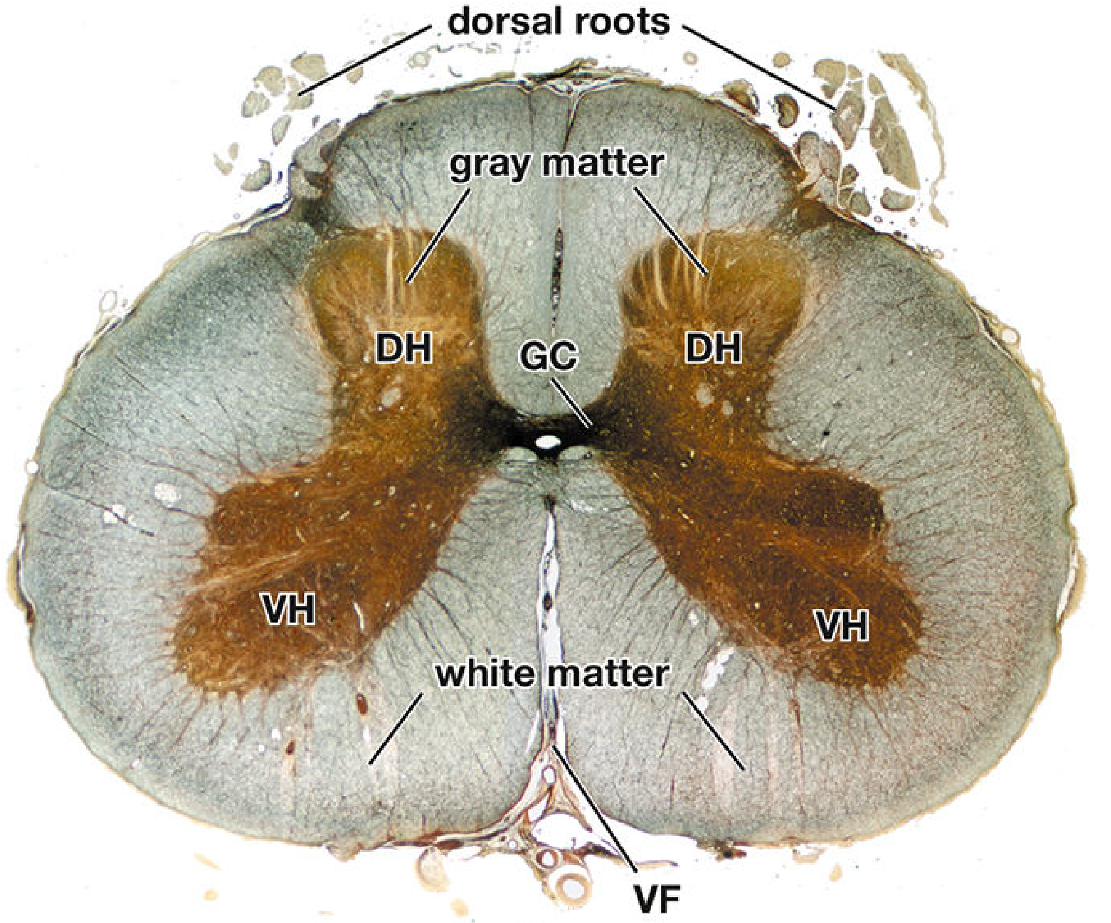
The spinal cord gray matter is divided into three horns:
- Dorsal horn - receives sensory (afferent) input
- Ventral horn - contains large motor neurons (anterior horn cells) whose axons form ventral roots - efferent output
- Lateral horn - present in thoracic and upper lumbar segments; contains autonomic preganglionic neurons
The surrounding white matter is divided into three columns (funiculi):
- Dorsal columns - ascending fine touch/proprioception
- Lateral columns - corticospinal (motor) + spinothalamic (pain/temp)
- Ventral columns - vestibulospinal and other descending pathways
Brainstem - The Exception
The brainstem is intermediate: gray matter and white matter are interdigitated on both the inside and outside, though most of the outer surface is white matter. Cranial nerve nuclei (gray matter) are scattered throughout the brainstem interior alongside white matter tracts.
Summary Table
| Location | Gray Matter Position | White Matter Position |
|---|---|---|
| Cerebral hemispheres | Outer cortex + deep nuclei | Inner core |
| Cerebellum | Outer cortex + deep nuclei | Inner core |
| Brainstem | Both inner and outer (mixed) | Predominantly outer surface |
| Spinal cord | Inner core (butterfly shape) | Outer shell (3 funiculi) |
Sources: Neuroanatomy through Clinical Cases, 3rd Ed. | Histology: A Text and Atlas with Correlated Cell and Molecular Biology | Neuroscience: Exploring the Brain, 5th Ed. | Gray's Anatomy for Students
This is a shared conversation. Sign in to Orris to start your own chat.