
Arcuate line
Reading File
The arcuate line (linea arcuata) is a key anatomical landmark on the posterior surface of the rectus sheath, marking where the posterior lamina of the sheath ends, typically at the midpoint between the umbilicus and pubic symphysis.
Searching sources...
Finding Sources
Finding Sources
Reading File
Reading File
Reading File
Loading Image
Loading Image
Arcuate Line (Linea Semicircularis / Line of Douglas)
Definition and Location
The arcuate line is a crescent-shaped (bow-shaped) demarcation on the inner (posterior) surface of the rectus sheath, found roughly one-third to one-half of the distance from the pubic crest to the umbilicus (approximately at the level of the anterior superior iliac spine). Below this line, the posterior wall of the rectus sheath is absent. It is also called the linea semicircularis or the semicircular line of Douglas.
Composition of the Rectus Sheath: Above vs. Below
This is the central anatomical fact the arcuate line defines.
| Layer | Above Arcuate Line | Below Arcuate Line |
|---|---|---|
| Anterior rectus sheath | External oblique aponeurosis + anterior lamella of internal oblique | External oblique + internal oblique + transversus abdominis (all three) |
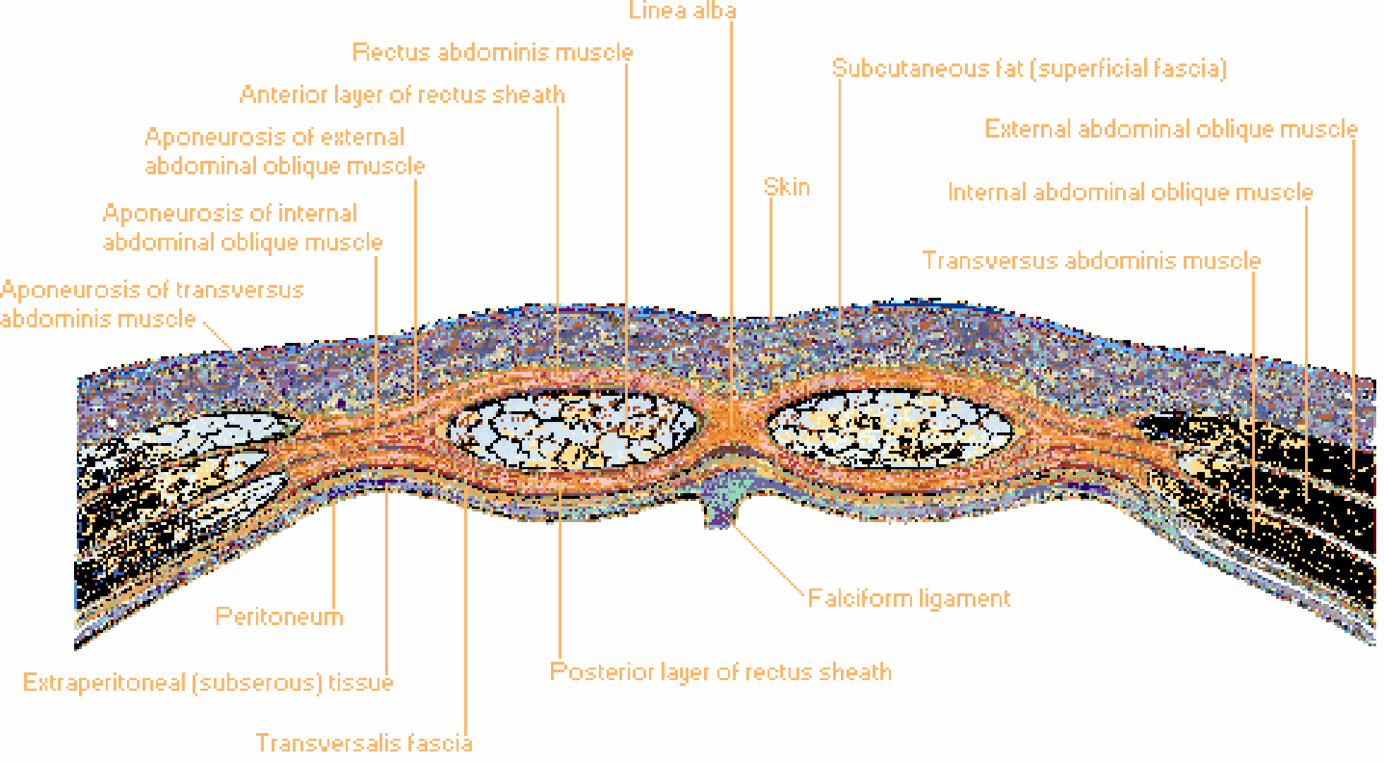
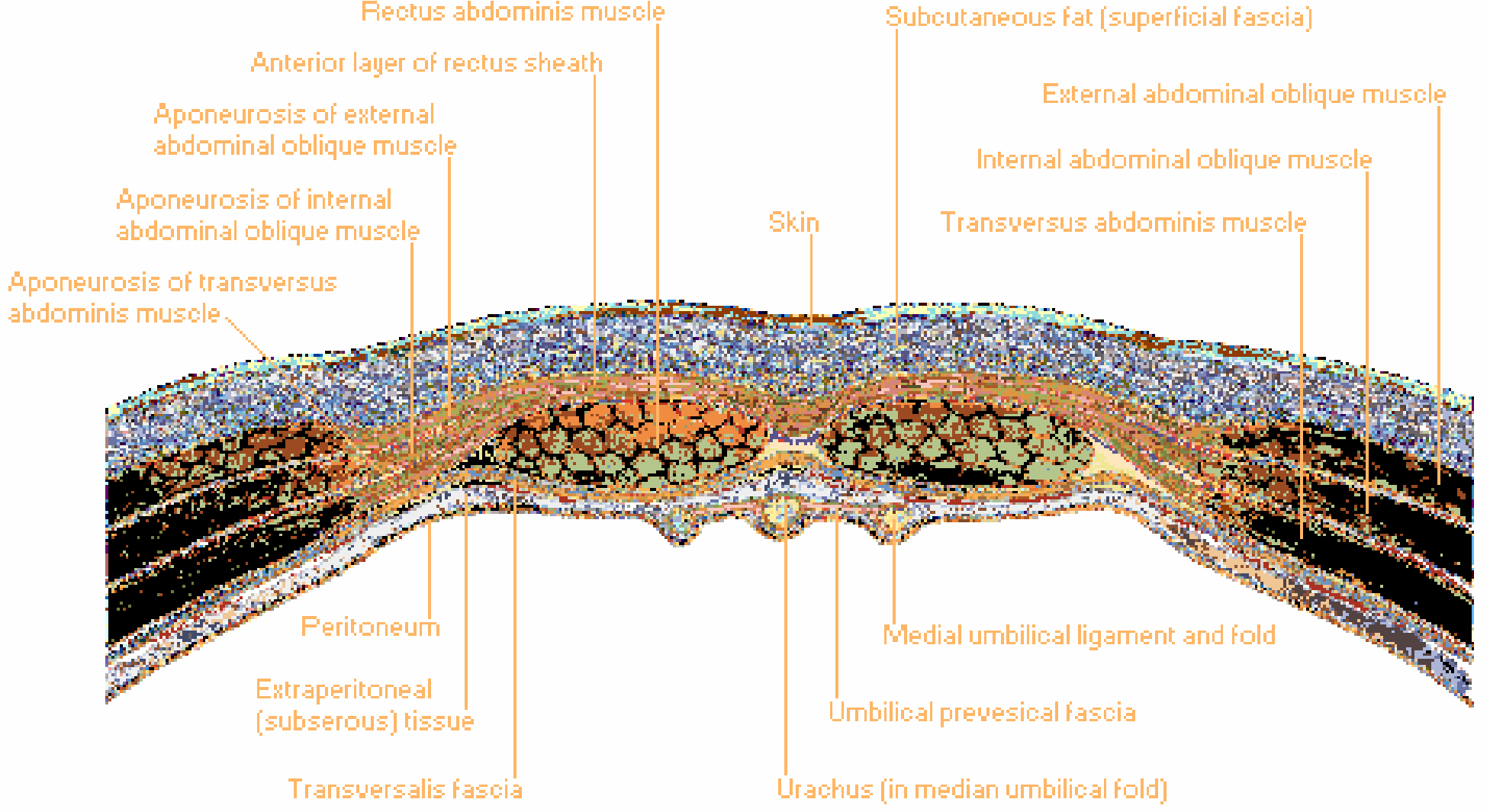
| Posterior rectus sheath | Posterior lamella of internal oblique + transversus abdominis aponeurosis | Absent - only transversalis fascia remains |
| Rectus abdominis lies on... | Posterior layer of rectus sheath | Transversalis fascia only |
Key rule: Below the arcuate line, the internal oblique aponeurosis does NOT split - it passes entirely anterior to the rectus abdominis, joining the external oblique and transversus abdominis aponeuroses to form a thickened anterior sheath, leaving no posterior fascial layer.
Cross-Sectional Anatomy (Netter's)
Above the arcuate line - the internal oblique aponeurosis splits to form anterior and posterior layers of the rectus sheath; external oblique joins the anterior layer, transversus abdominis joins the posterior layer:
Below the arcuate line - the internal oblique aponeurosis does not split; all aponeurotic layers pass anterior; no posterior sheath; rectus abdominis lies directly on transversalis fascia:
Clinical Significance
1. Spigelian Hernia
The arcuate line is the most clinically important landmark in Spigelian hernia. These hernias occur at the junction of the arcuate line and the linea semilunaris - precisely because the posterior rectus sheath is absent below the arcuate line, creating an anatomical weak point in the Spigelian fascia (the aponeurotic portion of the transversus abdominis between the linea semilunaris and the lateral edge of the rectus). The deep inferior epigastric vessels also perforate the rectus abdominis muscle at this level, further weakening the fascia. - Current Surgical Therapy 14e
2. Inferior Epigastric Artery
The inferior epigastric artery (from the external iliac) enters the rectus sheath near the arcuate line from below and travels upward within/behind the rectus muscle to anastomose with the superior epigastric artery.
3. Surgical Incisions
- Surgeons making lower midline or Pfannenstiel incisions must be aware that the posterior sheath is absent below the arcuate line - the peritoneum/preperitoneal fat is encountered more directly.
- In laparoscopic port placement or rectus sheath blocks, the arcuate line marks where the posterior sheath can no longer be used as a guide.
4. Rectus Sheath Hematoma
Bleeding below the arcuate line (e.g., from inferior epigastric vessel injury) spreads more freely because there is no fascial compartment to contain it - it can dissect widely in the preperitoneal space, whereas above the arcuate line it is more contained within the sheath.
Second Use: Arcuate Line of the Ilium (Pelvic Context)
The term "arcuate line" is also used for the arcuate line of the ilium - the smooth, curved ridge on the medial surface of the ilium that, together with the pecten pubis, pubic crest, and sacral promontory, forms the linea terminalis (pelvic brim). This boundary separates the greater pelvis (above) from the lesser (true) pelvis (below). - General Anatomy and Musculoskeletal System (THIEME Atlas)
Summary
| Feature | Detail |
|---|---|
| Other names | Linea semicircularis, semicircular line of Douglas |
| Location | ~1/3 to 1/2 of the way from pubic crest to umbilicus |
| What changes here | Posterior rectus sheath disappears |
| Below: posterior covering | Transversalis fascia only |
| Key vessel at this level | Deep inferior epigastric vessels enter the rectus here |
| Clinical relevance | Spigelian hernia, rectus sheath hematoma, surgical anatomy |
Sources: Netter's Atlas of Human Anatomy, p. 404-405 | Schwartz's Principles of Surgery 11e | Current Surgical Therapy 14e
This is a shared conversation. Sign in to Orris to start your own chat.