
High grade fever, hypotension, tachycardia
high grade fever hypotension tachycardia differential diagnosis sepsis
septic shock diagnosis management
septic shock pathophysiology diagram sepsis
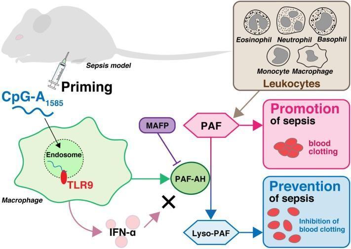
This medical schematic diagram illustrates the pathophysiology and therapeutic priming mechanism in a murine sepsis model. At the cellular level, the diagram depicts a macrophage endocytosing the molecule CpG-A1585, which subsequently binds to Toll-like receptor 9 (TLR9) within an endosome. This signaling pathway triggers the activation of platelet-activating factor acetylhydrolase (PAF-AH). While CpG-A1585 also induces interferon-alpha (IFN-α), a cross mark indicates that IFN-α does not contribute to PAF-AH activation. The diagram further details a leukocyte panel (eosinophils, neutrophils, basophils, monocytes, and macrophages) that produces platelet-activating factor (PAF), a mediator shown to promote sepsis and pathological blood clotting. PAF-AH serves as a critical regulatory enzyme by converting pro-septic PAF into Lyso-PAF, which prevents sepsis through the inhibition of blood clotting. Additionally, the specific inhibitor MAFP is shown antagonizing PAF-AH activity. This illustration highlights the clinical relevance of TLR9-mediated enzymatic pathways in mitigating endotoxin shock and disseminated intravascular coagulation (DIC).
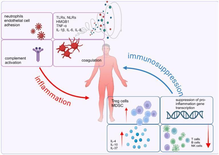
This pathophysiology diagram illustrates the dysregulated host immune response in sepsis, characterized by an imbalance between concurrent pro-inflammatory and anti-inflammatory pathways. Centered on a human figure, the left side (red 'inflammation' arrow) details the acute systemic inflammatory response syndrome (SIRS), featuring neutrophil-endothelial cell adhesion, complement activation, and coagulation cascades. A molecular cloud identifies key mediators including DAMPs/PAMPs (HMGB1, TLRs, NLRs) and pro-inflammatory cytokines (TNF-α, IL-1β, IL-6, IL-8). Conversely, the right side (blue 'immunosuppression' arrow) represents the compensatory anti-inflammatory response syndrome (CARS). This includes the expansion of suppressor cells (Treg cells, MDSCs) and anti-inflammatory cytokines (IL-4, IL-10, IL-37), the suppression of pro-inflammatory gene transcription, and the depletion or exhaustion of effector cells (T cells, B cells, and NK cells). The diagram serves as an educational tool for understanding the complex signaling pathways and cellular interactions that drive multi-organ dysfunction and immune homeostasis failure during septic shock.
Clinical Significance
1. Most likely diagnosis: Sepsis / Septic Shock
- Sepsis = life-threatening organ dysfunction caused by a dysregulated host response to infection.
- Septic shock = sepsis with persistent hypotension requiring vasopressors to keep MAP ≥65 mmHg despite adequate fluid resuscitation, plus lactate >2 mmol/L. Mortality is around 40% (The Washington Manual of Medical Therapeutics, p. 295).
- Older SIRS criteria (still useful at the bedside) included tachycardia >90 bpm, tachypnea >20/min or PaCO2 <32, temperature >38°C or <36°C, and WBC <4,000 or >12,000/µL - your patient already has 2 of these plus hemodynamic compromise, which should trigger a sepsis workup (qSOFA/SOFA scoring, blood cultures, lactate, source identification).
- Management priority: blood cultures before antibiotics, broad-spectrum antibiotics within the first hour, 30 mL/kg IV crystalloid (balanced solutions like lactated Ringer's preferred over normal saline) within the first hour, and norepinephrine as the first-line vasopressor if MAP remains <65 mmHg after fluids (The Washington Manual of Medical Therapeutics, p. 295).
2. Important mimics/differentials to rule out
| Condition | Distinguishing clue |
|---|---|
| Toxic shock syndrome (staph or strep) | Abrupt-onset high fever, hypotension, diffuse erythematous rash with later desquamation, multiorgan dysfunction; often linked to tampon use, wound packing, or skin/soft-tissue infection (K.J. Lee's Essential Otolaryngology, Goldman-Cecil Medicine, p. 266) |
| Gram-negative bacteremia / Vibrio vulnificus septicemia | High fever, tachycardia, hypotension, often after seafood exposure or wound in seawater, one-third progress to septic shock (Fitzpatrick's Dermatology, p. 3707) |
| Cutaneous anthrax with systemic spread | Fever, tachycardia, hypotension with a characteristic eschar (Fitzpatrick's Dermatology, p. 3014) |
| Ascending cholangitis (Charcot triad plus sepsis) | Fever/rigors, jaundice, RUQ pain, progressing to tachycardia, hypotension and altered mental status (Reynolds pentad) (Rosen's Emergency Medicine, p. 3484) |
| Cardiac tamponade with fever | Tachycardia, hypotension, narrow pulse pressure, pulsus paradoxus; fever if the cause is infectious/neoplastic pericarditis (Roberts and Hedges' Clinical Procedures in Emergency Medicine, p. 2803) |
| Adrenal crisis, acute pancreatitis, PE, MI, transfusion reaction, thyroid storm, severe drug/alcohol withdrawal | Non-infectious "sepsis mimics" that must be considered when cultures are negative (AAFP review on sepsis) |
| Drug toxicity (e.g., amphotericin B overdose) | Fever, hypotension, tachycardia, rigors, plus nephrotoxicity/electrolyte wasting on history of recent drug administration (Tintinalli's Emergency Medicine, p. 1229) |
3. Immediate bedside approach
- Confirm shock: repeat vitals, check capillary refill/skin perfusion, mental status.
- Draw blood cultures x2, lactate, CBC, CMP, coagulation panel, procalcitonin/CRP if available.
- Start empiric broad-spectrum antibiotics within 1 hour once cultures are drawn.
- Begin 30 mL/kg crystalloid bolus, reassess volume responsiveness.
- Search for a source: skin/soft tissue exam (rash, wounds), abdominal exam (biliary source), urinary source, pulmonary source (imaging), recent procedures/foreign bodies (tampon, packing) for toxic shock.
- If hypotension persists after fluids, start norepinephrine and consider ICU-level monitoring/echocardiography to assess cardiac function.