
Short notes on notochord
Reading File
The notochord is a transient embryonic structure derived from mesoderm that forms the axial skeleton of early embryos, induces neural tube formation, and later becomes the nucleus pulposus of intervertebral discs.
Searching sources...
Finding Sources
Finding Sources
Reading File
Reading File
Reading File
Finding Sources
Finding Sources
Finding Sources
Reading File
Here are short notes on the notochord, drawn from authoritative embryology textbooks:
Notochord - Short Notes
Definition
The notochord is a transient, midline, rod-like structure of mesodermal origin that forms the central axis of the early embryo. It is the defining feature of all chordates.
Development (Formation)
- Timing: Begins forming around day 16 of embryogenesis, during the 3rd week.
- Origin: Prenotochordal cells invaginate through the primitive node (cranial part of the primitive streak) and migrate cranially in the midline between ectoderm and endoderm.
- Process:
- Cells from the primitive node migrate cranially, forming the notochordal process (a hollow cellular tube).
- The notochordal process acquires a lumen - the notochordal canal.
- The floor of the notochordal process fuses with the underlying endoderm, then degenerates, forming the notochordal plate.
- The notochordal plate folds and proliferates to form the definitive notochord - a solid cellular cord.
- The neurenteric canal temporarily connects the amniotic cavity and umbilical vesicle; it is normally obliterated once the notochord is complete.
- Extent: Runs from the oropharyngeal membrane (cranially) to the primitive node (caudally). It does NOT extend into the head beyond the prechordal plate.
Structure
- A solid cord of cells derived from axial mesoderm
- Lies in the midline, between the neural tube (dorsal) and the gut endoderm (ventral)
Functions
1. Axial Support
- Provides a rigid central axis for the early embryo before the vertebral column forms.
2. Induction of the Neural Plate (Primary Inductor)
- The notochord is the primary signaling center of the early embryo.
- It induces the overlying ectoderm to thicken and form the neural plate - the primordium of the entire central nervous system (brain and spinal cord).
3. Induction of the Axial Skeleton
- Sclerotome cells from somites migrate toward the notochord and condense around it, forming the primordia of the vertebral bodies (centra).
- The notochord acts as a signaling center inducing vertebral development via Sonic Hedgehog (SHH) signaling.
4. Dorsoventral Patterning of the Neural Tube
- Secretes Sonic Hedgehog (SHH), which patterns the ventral neural tube and induces the floor plate.
- SHH from the notochord and floor plate specifies motor neurons and interneurons in the ventral spinal cord.
5. Somite Differentiation
- Alongside the neural tube and epidermis, the notochord provides signals (via NOGGIN and other factors) for somite differentiation into sclerotome, dermomyotome, and myotome.
Fate / Adult Remnants
- The notochord degenerates and largely disappears as vertebral bodies form around it.
- Small remnants persist between vertebrae and expand to become the nucleus pulposus of each intervertebral disc - the gelatinous center surrounded by the annulus fibrosus.
- The nucleus pulposus in adults retains scattered notochordal cells embedded in a gel-like extracellular matrix.
Clinical Correlates
1. Chordoma
- Arises from vestigial remnants of notochordal tissue.
- A rare, slow-growing but locally malignant tumor.
- ~1/3 occur at the base of the skull (clivus), extending to the nasopharynx.
- Also arises in the lumbosacral region.
- Infiltrates adjacent bone; difficult to resect. Surgical resection offers long-term disease-free survival in many patients.
2. Diastematomyelia (Spinal Notochord Syndrome)
- A congenital anomaly involving split/bifid notochord, leading to splitting of the spinal cord.
3. Neurenteric Cyst / Fistula
- Failure of the neurenteric canal to close can result in a persistent connection (fistula) or cyst between the gut and neural tube.
4. Neural Tube Defects
- Defects in the rostral end of the notochord impair induction of the forebrain neuroectoderm, contributing to certain cranial neural tube defects.
5. Intervertebral Disc Disease
- As humans age, notochordal (nucleus pulposus) cells are replaced by chondrocyte-like cells; this cellular change is associated with disc degeneration and herniation.
Summary Table
| Feature | Detail |
|---|---|
| Origin | Axial mesoderm (primitive node) |
| Timing | Day 16, 3rd week |
| Extent | Oropharyngeal membrane to primitive node |
| Adult remnant | Nucleus pulposus of intervertebral disc |
| Key functions | Neural induction, axial support, vertebral induction, SHH signaling |
| Tumor remnant | Chordoma (rare, malignant) |
Sources: The Developing Human (Moore & Persaud), Langman's Medical Embryology, Junqueira's Basic Histology, Gray's Anatomy for Students
Genetic codon
A genetic codon is a sequence of three nucleotide bases (triplet) in mRNA that specifies a particular amino acid or signals start/stop of translation. There are 64 possible codons encoding 20 amino acids, making the code degenerate (multiple codons per amino acid).
Searching sources...
Reading File
Finding Sources
Finding Sources
Reading File
Reading File
Reading File
Finding Sources
Finding Sources
Reading File
Finding Sources
Loading Image
Here are comprehensive short notes on the Genetic Codon:
Genetic Codon - Short Notes
Definition
A codon is a sequence of three consecutive nucleotide bases (triplet) on an mRNA molecule that specifies a particular amino acid to be incorporated into a protein, or signals the start or stop of translation.
- Codons are always read 5' → 3'
- Written in the language of mRNA: A, U, G, C (not DNA)
The Genetic Code
Mathematics of the Code
- 4 nucleotide bases taken 3 at a time = 4³ = 64 possible codons
- These 64 codons encode only 20 standard amino acids + start/stop signals
- 61 codons specify amino acids; 3 are stop codons
The Codon Table
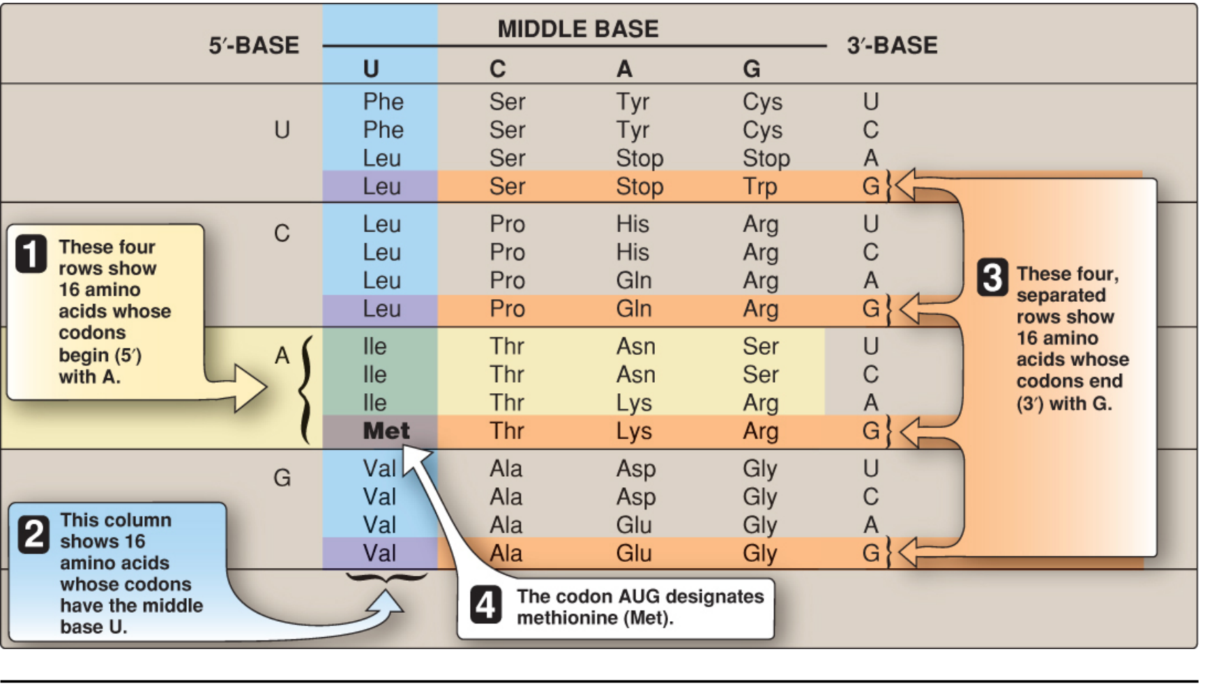
Types of Codons
| Type | Codons | Function |
|---|---|---|
| Start codon | AUG | Initiates translation; codes for Met (fMet in prokaryotes) |
| Sense codons | 60 others | Specify the 20 amino acids |
| Stop codons | UAA, UAG, UGA | Terminate translation (nonsense/termination codons) |
- AUG is the universal start codon - it both initiates translation AND codes for internal methionines within the protein.
- The three stop codons have mnemonics: "U Are Awful" (UAA), "U Are Gone" (UAG), "U Go Away" (UGA).
Properties (Characteristics) of the Genetic Code
1. Triplet (Three-letter)
- Each codon = exactly 3 nucleotides
- A sequence like
AGCUGGAAUCAUis read asAGC | UGG | AAU | CAU
2. Degenerate (Redundant)
- Multiple codons can specify the same amino acid (but one codon never codes for >1 amino acid)
- Example: Serine is coded by 6 codons: UCU, UCC, UCA, UCG, AGU, AGC
- Only Met (AUG) and Trp (UGG) have a single codon
- Degeneracy resides mostly in the 3rd (wobble) position of the codon
3. Unambiguous (Specific)
- A given codon always codes for the same amino acid - no ambiguity
- Degeneracy ≠ ambiguity: many codons → one amino acid (ok); one codon → many amino acids (never)
4. Nonoverlapping
- Each nucleotide belongs to only one codon
- The code is read sequentially, without any codon sharing nucleotides with the next
5. Commaless (Without Punctuation)
- No "spacers" between codons; read as a continuous run of triplets from start to stop
- Reading frame = the specific triplet grouping determined by the start codon AUG
6. Universal
- The code is essentially the same in all living organisms - from bacteria to humans
- This conservation implies the code evolved very early and any change would be lethal
- Exceptions: Mitochondria use a slightly different code:
- UGA → Trp (not Stop)
- AUA → Met (not Ile)
- AGA, AGG → Stop (not Arg) in mammalian mitochondria
7. Non-overlapping and Collinear
- The order of codons in mRNA is collinear with the order of amino acids in the protein (N-terminus to C-terminus corresponds to 5'-end to 3'-end of mRNA)
Codon-Anticodon Interaction
- The codon on mRNA is recognized by the anticodon of a specific tRNA
- Binding is antiparallel and follows Watson-Crick complementarity (mostly)
- Each tRNA carries a specific amino acid determined by the anticodon it holds
Wobble Hypothesis (Crick, 1966)
- The 3rd base of the codon (= 5th base of the anticodon) base-pairs less strictly - this is the "wobble position"
- The first (5') base of the anticodon can "wobble" and pair with more than one base at the 3' end of the codon
- Inosine (I) in the anticodon can pair with U, C, or A at the wobble position
- Result: fewer than 61 tRNA species are needed to read all 61 sense codons
- Explains why degeneracy is concentrated at the 3rd base of most codons
Mutations and Codons
| Mutation Type | Effect on Codon | Consequence |
|---|---|---|
| Silent / Synonymous | Same amino acid coded | No change in protein (due to degeneracy, usually at 3rd position) |
| Missense | Different amino acid coded | Altered protein (may or may not affect function) |
| Nonsense | Amino acid codon → Stop codon | Premature termination; truncated protein |
| Frameshift | Insertion/deletion shifts reading frame | All downstream codons altered; usually nonfunctional protein |
Codon Usage Bias
- Not all synonymous codons are used equally; usage varies between species and tissues
- tRNA levels generally mirror codon usage - abundant codons are decoded by abundant tRNAs
- Important in recombinant protein expression (codon optimization)
Summary
| Feature | Detail |
|---|---|
| Unit | 3 nucleotides (triplet) |
| Total codons | 64 |
| Sense codons | 61 (encode 20 amino acids) |
| Stop codons | 3 (UAA, UAG, UGA) |
| Start codon | AUG (Met) |
| Key property | Degenerate but unambiguous |
| Read direction | 5' → 3' on mRNA |
| Universal exception | Mitochondrial code |
Sources: Biochemistry 8th ed (Lippincott Illustrated Reviews); Harper's Illustrated Biochemistry 32nd ed; Emery's Elements of Medical Genetics and Genomics; Basic Medical Biochemistry 6th ed
This is a shared conversation. Sign in to Orris to start your own chat.