
Stomatitis
Reading File
Stomatitis refers to inflammation of the oral mucosa, which can involve the lips, cheeks, gums, tongue, and palate. It presents as pain, redness, ulceration, or white patches in the mouth and has many causes including infections, autoimmune conditions, nutritional deficiencies, and drug reactions.
Searching sources...
Finding Sources
Searching PubMed
stomatitis oral mucositis
Good hits. Let me read the most comprehensive sections in parallel.
Reading File
Reading File
Reading File
Now let me pull the aphthous stomatitis, nicotinic stomatitis, and bacterial stomatitis sections in parallel for comprehensive coverage.
Reading File
Reading File
Reading File
Let me also fetch the clinical image for aphthous ulcer and read the bacterial stomatitis section.
Loading Image
Reading File
Finding Sources
Reading File
Reading File
I now have comprehensive material from multiple authoritative textbooks. Let me compile the full answer.
Stomatitis
Stomatitis is inflammation of the oral mucosa - the lining of the mouth including the lips, cheeks, gums, tongue, floor of the mouth, and palate. It is a broad clinical term covering a range of conditions from infectious ulcers to drug reactions and nutritional deficiencies.
Classification and Causes
Stomatitis can be organized by etiology:
1. Infectious Stomatitis
Viral (most common overall)
- Herpetic gingivostomatitis (HSV-1/2): Primary HSV infection causes an extremely painful gingivostomatitis with high fever. Lesions appear as vesicles on the buccal mucosa and oropharynx (see image below). Recurrent HSV only involves keratinized mucosa (hard palate, attached gingiva) in immunocompetent hosts - an important distinguishing feature.
- Herpangina: Caused by coxsackievirus A16 and enterovirus 71. Vesicular lesions on the tonsils and soft palate in children 6 months to 10 years old, accompanied by fever and dysphagia. Heals spontaneously in 3-5 days.
- Hand-foot-and-mouth disease: Also enteroviral (coxsackievirus A16 most common). Oral vesicles that evolve into ulcers, often with simultaneous skin exanthem. Mainly affects children <5 years old.
- Epstein-Barr virus / CMV: Exudative pharyngitis with malaise, fatigue, and fever.
Bacterial
- Common pathogens: Streptococci, Staphylococci, Gonococci
- Tuberculosis can cause a painful, deep, irregular ulcer on the dorsum of the tongue, palate, or buccal mucosa.
Fungal
- Candida (oral candidiasis / thrush): Occurs as a superinfection, especially during chemotherapy-induced neutropenia or in immunocompromised states.
2. Recurrent Aphthous Stomatitis (RAS)
Also called canker sores or recurrent oral aphthae, this is the most clinically common form.
Epidemiology: Affects >20% of the population. Usually begins in the 2nd or 3rd decade; severity decreases with age.
Pathogenesis: Multifactorial and largely unknown, but T-cell-mediated immunological damage to the mucosal epithelial basal layer is central. It is not caused by HSV despite the "herpetiform" variant name.
Clinical subtypes (Table):
| Type | Size | Duration | Location | Scarring | Notes |
|---|---|---|---|---|---|
| Minor (most common) | <10 mm | 2-3 weeks | Non-keratinized mucosa only | No | <10 ulcers at a time; front of mouth |
| Major | >10 mm | Up to 3 months | Keratinized or non-keratinized | Possible | 1-3 ulcers; towards back of mouth |
| Herpetiform (least common) | <5 mm | Up to 2 weeks | Both types of mucosa | No | Up to 100 ulcers; coalesce into large areas |
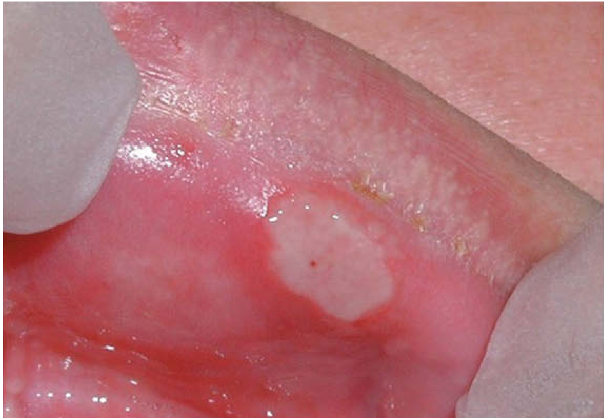
Associated systemic conditions to screen for:
- Behcet disease
- Inflammatory bowel disease (Crohn's, ulcerative colitis), celiac disease
- Cyclic neutropenia
- HIV infection
- PFAPA syndrome (periodic fever, aphthous stomatitis, pharyngitis, adenitis)
- Nutritional deficiencies: iron (ferritin), folate, vitamin B12 - importantly, these can be deficient even with a normal blood count, so a haematinic screen and coeliac screen should be performed in all RAS patients.
- Medications: mTOR inhibitors (e.g., everolimus)
Triggers: Stress, foods (nuts, citrus, acidic foods), sodium lauryl sulphate (SLS) in toothpaste, benzoate, chocolate, cinnamon.
"Complex aphthosis" = nearly constant disease with ≥3 oral ulcers ± genital ulcers - always requires a search for an underlying systemic disease.
3. Contact Stomatitis
An allergic contact reaction caused by:
- Dental materials: metals in fillings, gold caps/crowns, acrylic monomers, epoxy resins (~30% of patients with oral symptoms have relevant allergens)
- Food additives: flavorings (cinnamic aldehyde), antioxidants
- Dental/hygiene products: chewing gum (hexylresorcinol, thymol, oil of cinnamon, mint), toothpastes
Clinical signs: Bright erythema of the tongue and buccal mucosa with scattered erosions; angular cheilitis; oral lichenoid lesions (especially with metal sensitization).
4. Stomatitis Nicotina (Smoker's Keratosis)
Seen predominantly in heavy-smoking middle-aged men (and also in habitual hot-beverage drinkers). Heat appears to be the causative mechanism.
Appearance: Milky-white, slightly umbilicated, asymptomatic papules on the palate. The ductal ostia appear as red pinpoints. Intervening mucosa becomes white and thickened, desquamating to leave raw, beefy-red areas.
Severity correlates with pipe smoking and reverse smoking (the most heat-intense forms). Treatment: Abstain from tobacco and hot liquids.
5. Chemotherapy- and Radiotherapy-Induced Stomatitis (Mucositis)
Occurs in >40% of chemotherapy patients and is a major dose-limiting toxicity, especially when chemo is combined with radiotherapy.
Two mechanisms:
- Direct cytotoxic effect on oral epithelial cells (high mitotic index)
- Indirect effect via superimposed infections (Candida, HSV) and hemorrhage from bone marrow suppression (at the nadir of WBC count)
Key drugs causing mucositis: Anthracyclines (doxorubicin), 5-FU, methotrexate, busulfan, gemcitabine, irinotecan, vincristine, vinblastine, taxanes, capecitabine.
Prevention and management:
- Dental evaluation before starting chemotherapy
- Soft toothbrush + sodium bicarbonate rinses
- Prophylactic antivirals (acyclovir) and antifungals (fluconazole)
- Colony-stimulating factors to shorten neutropenia
- Topical anesthetics and anti-inflammatory agents
- Systemic analgesics when needed
- Palifermin (keratinocyte growth factor) in selected patients - note: can cause white tongue coating and transient keratoses
6. Stomatitis as Part of Systemic Disease
Many systemic conditions present with oral ulceration:
| Category | Examples |
|---|---|
| Skin diseases | Erosive lichen planus, pemphigus, pemphigoid, erythema multiforme, dermatitis herpetiformis, epidermolysis bullosa |
| GI diseases | Crohn's disease, ulcerative colitis, celiac disease |
| Blood disorders | Leukemia, neutropenia, haematinic deficiency |
| Malignancy | Squamous cell carcinoma, lymphoma |
| Iatrogenic | Antirheumatic drugs, chemotherapy, radiotherapy |
Treatment Summary
| Condition | Treatment |
|---|---|
| Aphthous (symptomatic) | Benzydamine rinse/spray, occlusive paste (Orabase), lignocaine gel - start at onset (before ulcer appears if possible, since immune damage precedes the ulcer) |
| Aphthous (immunomodulatory) | Topical corticosteroids; systemic immunotherapy by specialist only |
| Herpetic gingivostomatitis | Oral acyclovir 80 mg/kg/day in 4 divided doses x 5-7 days (in children, max 3200 mg/day); start within 72-96 hours |
| Herpangina / HFMD | Supportive - antipyretics, systemic analgesics; diphenhydramine + Maalox rinse for local pain |
| Contact stomatitis | Identify and remove the allergen; patch testing |
| Nicotinic stomatitis | Stop tobacco and hot liquids |
| Chemotherapy mucositis | Oral hygiene, prophylactic antifungals/antivirals, topical anesthetics, analgesics, palifermin |
| Nutritional deficiency RAS | Replace iron, folate, or B12 as indicated |
Key Differentiating Points
- HSV recurrence in immunocompetent patients only affects keratinized mucosa (hard palate, attached gingiva) - this distinguishes it from RAS, which affects non-keratinized mucosa.
- Herpetiform aphthae can look identical to herpetic gingivostomatitis but are HSV-negative.
- RAS ulcers have significant peripheral erythema and pain at onset; trauma ulcers have more ragged edges and less erythema; fixed drug eruptions and bullous diseases are less painful at onset.
- Any persistent ulcer not healing in 2-3 weeks should be biopsied to exclude squamous cell carcinoma.
Sources: Dermatology 2-Volume Set 5e; Tintinalli's Emergency Medicine; Andrews' Diseases of the Skin; Scott-Brown's Otorhinolaryngology Head & Neck Surgery; Cummings Otolaryngology; Lee's Essential Otolaryngology
This is a shared conversation. Sign in to Orris to start your own chat.