
Cervical radiculopathy
I'll start by reading the medical library skill, then search for comprehensive information on cervical radiculopathy.
Reading File
Now let me search the library and PubMed in parallel.
Finding Sources
Searching PubMed
"cervical radiculopathy"[MeSH Terms] AND treatment
Excellent - found dedicated sections in multiple textbooks. Let me read the key sources in parallel.
Reading File
Reading File
Finding Sources
Searching PubMed
"cervical radiculopathy" AND management
Excellent. Now let me read the main Bradley & Daroff section and Harrison's section in parallel for the full clinical detail.
Reading File
Reading File
Reading File
Let me read the treatment section from Bradley & Daroff and also pull the Tintinalli treatment section and the Frameworks dermatome table.
Reading File
Reading File
I now have comprehensive information to write a detailed response. Let me also quickly fetch the relevant cervical spine MRI image for visual clarity.
Loading Image
I now have all the information needed for a comprehensive response.
Cervical Radiculopathy
Definition and Pathophysiology
Cervical radiculopathy is a clinical syndrome caused by compression or inflammation of one or more cervical nerve roots. The two main mechanisms are:
- Disk herniation - more common in patients under 45. An acute soft disk protrudes posterolaterally into the neural foramen.
- Neuroforaminal stenosis from spondylosis - more common with increasing age. Osteophyte formation, facet joint arthropathy, and uncovertebral joint hypertrophy progressively narrow the foramen.
Both ischemia of the compressed root and inflammatory mediators (proinflammatory cytokines from degenerative disks) contribute to nerve root injury. The C5-C6 and C6-C7 levels are the most mobile segments and undergo the greatest degenerative change, making C6 and C7 the most commonly affected roots. The most frequently affected root is C7, followed by C6. Clinically evident C8 and root involvement above C5 is less common.
- Harrison's Principles of Internal Medicine 22E, p. 174
- Bradley and Daroff's Neurology in Clinical Practice, p. 2568
Clinical Presentation
Symptoms often appear suddenly, though most cases lack an identifiable traumatic precipitant.
Core symptoms:
- Pain originating in the neck radiating down the arm (radicular arm pain)
- Dysesthesias, paresthesias, numbness in a dermatomal distribution
- Limb weakness in the affected myotome
- Subscapular or interscapular pain (especially C6, C7, C8, T1)
- Pain worsens with Valsalva maneuver or coughing
- Neck pain alone (especially at C4 and above)
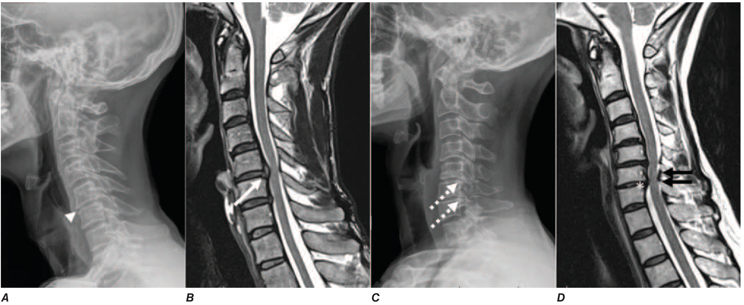
Root-Level Localization Table
| Root | Level | Sensory Changes | Motor Weakness | Reflex Lost |
|---|---|---|---|---|
| C5 | C4-C5 | Shoulder, lateral arm | Deltoid, biceps, brachioradialis | Biceps, supinator |
| C6 | C5-C6 | First two digits, lateral distal forearm | Brachioradialis, wrist extensors | Biceps, brachioradialis (may be inverted) |
| C7 | C6-C7 | Index, middle, ring fingers | Triceps, wrist flexors, pronators | Triceps |
| C8 | C7-T1 | 4th-5th digits, medial forearm | Intrinsic hand muscles | Finger flexors |
Note: "Inverted" biceps reflex (absent biceps reflex with paradoxical finger flexor contraction) suggests coexisting myelopathy at C5-C6.
- Bradley and Daroff's Neurology in Clinical Practice, p. 2569
Physical Examination
Key maneuvers:
- Spurling test (Spurling maneuver): Neck extension + ipsilateral rotation + axial compression reproduces or worsens arm pain. Highly specific for cervical radiculopathy. A 2025 systematic review and meta-analysis (PMID 39938056) confirmed its diagnostic value for subacute and chronic cervical radiculopathy.
- Shoulder abduction relief sign: Placing the ipsilateral hand on the head relieves pain (reduces root tension).
- Neck distraction test: Manual axial traction of the head reduces radicular symptoms.
Diagnostic Testing
Imaging
- Cervical plain films: Little value in diagnosing or excluding radiculopathy.
- MRI cervical spine: Study of choice - identifies nerve root compression in the neural foramina and assesses for myelopathy. Must be interpreted cautiously, as degenerative changes are common in asymptomatic individuals.
- CT myelography: Sometimes more sensitive than MRI; preferred in patients with MRI incompatibility (pacemakers, spinal cord stimulators), severe claustrophobia, or prior hardware from cervical fusion. Better for distinguishing osteophytes from soft disk herniation and for detecting lateral foraminal pathology missed by MRI.
Electrodiagnostics (EMG/NCS)
Useful in difficult diagnostic cases and to distinguish radiculopathy from brachial plexopathy or peripheral neuropathy.
-
Key NCS pattern: Reduced amplitude of compound muscle action potential (CMAP) with preserved sensory nerve action potential (SNAP). This dissociation occurs because the dorsal root ganglion (DRG) lies distal to the site of intraspinal compression - the sensory axon remains in continuity with its cell body, so antidromic sensory conduction is preserved.
-
Needle EMG: Denervation (fibrillations, positive sharp waves) in muscles of the affected myotome, including paraspinal muscles.
-
Bradley and Daroff's Neurology, p. 2569-2570
Differential Diagnosis
Key conditions to exclude:
-
Brachial plexopathy (Parsonage-Turner syndrome / brachial neuritis) - acute onset severe pain, patchy neurologic signs, EMG confirms
-
Thoracic outlet syndrome - vague shoulder pain, C8/T1 distribution, positional provocation
-
Peripheral nerve entrapment (carpal tunnel, cubital tunnel) - distal sensory symptoms, NCS distinguishes
-
Shoulder pathology (rotator cuff disease, calcific tendinitis) - may coexist; local anesthetic injection helps differentiate
-
Cervical myelopathy - bilateral symptoms, hyperreflexia below the lesion, Babinski sign, bladder dysfunction
-
Firestein & Kelley's Textbook of Rheumatology, p. 8888-8904
Management
Conservative (First-Line)
The natural history is generally favorable - most patients improve with nonoperative management.
- Activity modification: Avoid postures and movements that exacerbate symptoms.
- Analgesics: NSAIDs, acetaminophen; short-course opioids if needed.
- Muscle relaxants: May help with associated cervical muscle spasm.
- Oral corticosteroids: A 7-10 day course of methylprednisolone or prednisone is commonly prescribed for acute radiculopathy, though efficacy has not been definitively demonstrated in controlled trials.
- Cervical collar: Soft or hard collar use is controversial - no clear evidence for or against.
- Physical therapy: Cervical traction, McKenzie exercises, postural correction. A 2025 systematic review (PMID 40576779) found both articular and neural mobilization techniques effective for managing cervical radicular pain, with neural mobilization showing network meta-analysis superiority.
Interventional
- Epidural steroid injections (ESI): Effective for chronic cervical radiculopathy when other treatments have failed. Transforaminal or interlaminar approach under fluoroscopic guidance.
- Selective nerve root block: Both diagnostic and therapeutic.
Surgical
Indicated when:
- Progressive neurological deficit (especially progressive motor weakness)
- Refractory, disabling pain unresponsive to conservative treatment (typically after 6-12 weeks)
- Acute or progressive signs of myelopathy
Surgical options:
- Anterior cervical discectomy and fusion (ACDF): Most common approach; addresses ventral pathology.
- Total disk arthroplasty (TDA): Alternative to fusion at single levels, preserves motion.
- Posterior cervical laminoforaminotomy: Effective for foraminal stenosis, preserves motion, avoids anterior approach.
Surgery improves symptoms faster than conservative management but long-term outcomes are similar in the absence of progressive neurological deficit.
- Tintinalli's Emergency Medicine, p. 1925
- Rheumatology 2-Volume Set (Elsevier 2022), p. 1574-1581
- Bradley and Daroff's Neurology, p. 2570-2571
Indications for Urgent/Emergent Referral
Admit or urgently refer if:
- Progressive upper extremity weakness (especially C7 distribution)
- Acute or progressive signs of myelopathy (bilateral weakness, gait disturbance, hyperreflexia, Babinski, bladder/bowel involvement)
- Intractable radicular pain unresponsive to treatment
- Red flags suggesting malignancy or infection (constitutional symptoms, history of cancer, fever, weight loss)
Key Points Summary
| Feature | Detail |
|---|---|
| Most common root | C7 > C6 |
| Most common level | C5-C6, C6-C7 |
| Mechanism (young) | Disk herniation |
| Mechanism (older) | Spondylotic foraminal stenosis |
| Best imaging | MRI cervical spine |
| EMG hallmark | Reduced CMAP, preserved SNAP |
| Spurling sign | Specific (not sensitive) for diagnosis |
| First-line Rx | NSAIDs, PT, activity modification |
| Surgery threshold | Progressive deficit or refractory pain |
What to do when no abnormalities in MRI
When MRI is normal in a patient with suspected cervical radiculopathy, the key next steps are: (1) EMG/NCS to confirm radiculopathy and exclude mimics, (2) reconsider the diagnosis entirely, (3) consider dynamic or positional MRI, and (4) treat based on clinical picture if everything else fits.
Searching sources...
Finding Sources
Finding Sources
Searching PubMed
"cervical radiculopathy" AND "normal MRI" AND electrodiagnostic
Reading File
Reading File
Finding Sources
Finding Sources
Finding Sources
I now have all the key source material. Here is the comprehensive answer:
Normal MRI in Suspected Cervical Radiculopathy: What to Do
A normal MRI does not rule out cervical radiculopathy. This is a well-recognized clinical scenario and there is a structured approach to it.
Why MRI Can Be Normal
Understanding the limitations of MRI in this context is essential before deciding the next steps.
1. Demyelination without axonal loss
The most important mechanism: nerve root compression may cause intermittent ischemia or mechanical deformation resulting in isolated root demyelination - the patient experiences classical radicular symptoms (pain, paresthesias) but there is no structural distortion of the root visible on MRI and no axonal loss detectable on EMG. This is a pure demyelinating/conduction-block lesion.
2. Purely lateral/foraminal pathology
MRI can miss compression that lies lateral to the subarachnoid space. CT myelography depends on intrathecal contrast filling around the nerve root - if compression is entirely extraforaminal or at the far-lateral zone, it is invisible on both standard MRI and CT myelography. A standard supine MRI may underestimate foraminal narrowing that opens when upright.
3. False-negative rate of MRI technique
MRI quality is degraded by:
- Prior cervical hardware/fusion implants (metal artifact)
- Motion artifact in anxious or uncooperative patients
- Field strength (1.5 T vs. 3 T)
- Protocol gaps (missed dedicated coronal foraminal cuts)
4. MRI was done too soon or at wrong level
Early scans or scans done before the patient is appropriately positioned and examined can miss findings.
5. True functional/inflammatory radiculopathy
Inflammation of a nerve root (chemical radiculitis from nucleus pulposus contact, without mechanical compression) may not produce any visible structural abnormality on routine MRI.
- Bradley and Daroff's Neurology in Clinical Practice, p. 2570
Step-by-Step Approach
Step 1 - Proceed to EMG/NCS (Most Important Next Step)
Electrodiagnostic (EDX) testing is the key investigation when MRI is normal but radiculopathy is suspected clinically. It serves two purposes simultaneously: confirms radiculopathy and excludes mimics.
What to look for:
| Finding | Significance |
|---|---|
| Reduced CMAP amplitude, preserved SNAP | Confirms intraspinal root lesion (DRG is distal to compression, sensory cell body intact) |
| Fibrillation potentials / positive sharp waves in paraspinal muscles | Active denervation - confirms radiculopathy |
| Abnormal paraspinal EMG but normal limb EMG | Root lesion proximal to plexus |
| Normal paraspinal EMG, abnormal limb | Suggests plexopathy (DRG distal to lesion) |
| Normal EMG throughout | Demyelinating radiculopathy, early injury, or wrong diagnosis |
Critical timing caveat: EMG is of low yield in the hyperacute phase. Wallerian degeneration must complete first - this takes 5-6 days for motor fibers and 8-9 days for sensory fibers. Needle EMG sensitivity for radiculopathy is only 50-71% even when timed correctly. A normal EMG does not exclude radiculopathy - it simply means there is no axonal loss.
"Root compression resulting in intermittent ischemia or mechanical deformation may result in isolated root demyelination without secondary axonal loss. In this scenario, a patient may experience classical radicular symptoms without any objective evidence of the disease."
- Bradley and Daroff's Neurology in Clinical Practice, p. 2570
Step 2 - Consider Alternative Imaging
If EMG is also non-diagnostic and clinical suspicion remains high:
-
CT myelography: Sometimes more sensitive than MRI for bony foraminal stenosis, osteophyte-mediated compression, and lateral pathology. Particularly useful if prior cervical hardware is degrading MRI quality. Better at distinguishing calcified vs. soft disk herniation.
-
Flexion-extension or dynamic views: May reveal instability contributing to intermittent root compression that is not evident on static MRI.
-
Higher-field MRI (3 T) or dedicated foraminal protocol: If the original scan was 1.5 T or lacked dedicated foraminal imaging, upgrading the technique is worthwhile.
-
MRI brachial plexus: If the clinical picture is more consistent with plexopathy than radiculopathy (patchy, multi-root distribution; no paraspinal EMG changes), dedicated plexus MRI to exclude structural plexus lesion.
-
Goldman-Cecil Medicine, Ancillary Testing section
-
Bradley and Daroff's Neurology, Diagnostic Testing
Step 3 - Revisit the Differential Diagnosis
A normal MRI is a strong prompt to reconsider whether this is actually radiculopathy. Common mimics:
| Condition | Key Distinguishing Features |
|---|---|
| Brachial neuritis (Parsonage-Turner) | Acute severe pain followed by weakness/atrophy; patchy multi-nerve distribution; paraspinal EMG normal; plexus MRI shows T2 signal in muscles |
| Thoracic outlet syndrome | Positional symptoms; C8/T1 distribution; vascular signs |
| Carpal tunnel / cubital tunnel | Distal sensory pattern; NCS diagnostic |
| Shoulder pathology | Local shoulder tenderness; provocative shoulder tests; normal cervical maneuvers |
| Peripheral neuropathy | Stocking-glove; bilateral; NCS shows sensory abnormalities |
| MS / demyelinating disease | Multiple lesions; brain/cord MRI; cerebellar signs; visual symptoms |
| Motor neuron disease (ALS) | No sensory symptoms; upper + lower motor neuron signs; no pain |
The cervical paraspinal needle EMG is the critical test to separate radiculopathy from plexopathy:
-
Radiculopathy: fibrillations present in paraspinal muscles (proximal to DRG)
-
Plexopathy: paraspinal muscles normal (lesion is distal to DRG)
-
Bradley and Daroff's Neurology, p. 2614-2615
Step 4 - Treat Clinically If Diagnosis Fits
If the clinical picture is convincingly radiculopathic (dermatomal pain, positive Spurling test, appropriate myotomal weakness, consistent reflex change) and imaging/EDX are non-diagnostic:
- Proceed with conservative management - there is no harm in treating the clinical syndrome. Most cervical radiculopathy improves in 4-8 weeks regardless of treatment.
- NSAIDs, short-course oral steroids (7-10 days), physical therapy, activity modification.
- Epidural steroid injection can be both diagnostic and therapeutic - reproduction of symptoms + relief confirms the level.
"Patients with a typical clinical presentation and little or no neurological deficit usually can be managed with these noninvasive approaches without imaging or EDX studies."
- Bradley and Daroff's Neurology in Clinical Practice, p. 2571
Step 5 - Red Flags Requiring Urgent Escalation
Even with a normal MRI, escalate urgently if:
- Progressive weakness - myelopathy or motor neuron disease must be excluded regardless of MRI
- Constitutional symptoms (fever, weight loss, night sweats) - spinal infection or malignancy; MRI can have up to a 17% false-negative rate for spinal metastases
- Bilateral symptoms or long-tract signs - do not accept a normal standard MRI; request gadolinium-enhanced MRI and specialist review
- Bladder/bowel involvement - emergency evaluation
Summary Algorithm
Cervical Radiculopathy Suspected
           ↓
       MRI NORMAL
           ↓
    ┌──────────────────────────────────────────────┐
    │ 1. Timing? → Wait ≥2 weeks from onset for EMG│
    │ 2. EMG/NCS → Confirm root level, exclude      │
    │    plexopathy/peripheral nerve                │
    │ 3. Reconsider differential diagnosis          │
    │ 4. If MRI suboptimal → CT myelography         │
    │    or higher-field/protocol MRI               │
    │ 5. Treat conservatively if clinical fit       │
    │ 6. Epidural steroid: diagnostic + therapeutic │
    │ 7. Red flags? → Urgent specialist referral    │
    └──────────────────────────────────────────────┘
The key principle is that concordant clinical, radiological, and electrophysiological data are required before committing to any intervention - a normal MRI shifts the burden to EMG and careful clinical reassessment, not to surgery.
Symptoms - heaviness after eating even after meals..... especially after protein fodds.... regurgitation.... irritation on lying.... feeling of gas struck inside....a kind of movibg pain..
Reading File
This is a clinical case scenario. The symptoms - postprandial heaviness (especially with protein), regurgitation, irritation on lying down, trapped gas sensation, and moving/colicky pain - point strongly toward GERD, gastroparesis, or a functional upper GI disorder. Let me search the library comprehensively.
Searching sources...
Finding Sources
Finding Sources
Finding Sources
Reading File
Finding Sources
Finding Sources
Reading File
Reading File
Finding Sources
Reading File
Reading File
Finding Sources
Reading File
Finding Sources
I now have comprehensive information. Let me compile a thorough, clinically organized response.
Symptom Analysis: Postprandial Heaviness, Regurgitation, Positional Irritation, Trapped Gas, and Moving Pain
Clinical Impression
This symptom cluster maps most closely to Gastroesophageal Reflux Disease (GERD) with likely co-existing functional dyspepsia or gastroparesis features. Here is how each symptom maps:
| Your Symptom | Clinical Term | Points Toward |
|---|---|---|
| Heaviness after eating | Postprandial fullness / early satiety | Functional dyspepsia, gastroparesis |
| Worse with protein foods | Delayed gastric emptying (protein empties slower than carbs) | Gastroparesis |
| Regurgitation | Acid/food regurgitation | GERD |
| Irritation on lying down | Recumbent reflux worsening | GERD (classic) |
| Gas stuck inside | Bloating / intestinal gas trapping | Functional dyspepsia, GERD, IBS overlap |
| Moving/colicky pain | Visceral hypersensitivity, intestinal spasm | Functional dyspepsia, IBS overlap |
Most Likely Diagnosis: GERD + Functional Dyspepsia (Overlap)
GERD
The cardinal symptoms of GERD are heartburn and acid regurgitation. Symptoms typically occur after meals and characteristically increase when a patient is recumbent - exactly as you describe.
"Classic symptoms of GERD are heartburn, defined as a retrosternal burning discomfort, and acid regurgitation. Symptoms often occur after meals and may increase when a patient is recumbent."
- Yamada's Textbook of Gastroenterology, 7th Ed.
Regurgitation in GERD is the effortless return of esophageal or gastric contents into the chest or pharynx, without nausea or retching. Patients note a sour or burning fluid in the throat. Bending over or any maneuver increasing intra-abdominal pressure can trigger it.
The worsening on lying down happens because the lower esophageal sphincter (LES) is the only barrier against reflux in the upright position - when you lie flat, gravity no longer helps keep gastric contents down.
Functional Dyspepsia (Postprandial Distress Syndrome)
The Rome IV criteria define a specific subtype called Postprandial Distress Syndrome (PDS), which includes:
- Bothersome postprandial fullness after normal-sized meals
- Early satiety preventing finishing a meal
- Often accompanied by bloating and belching
This is exactly the "heaviness after meals" pattern you describe. Dyspepsia is described as symptoms originating from the gastroduodenal area - "postprandial heaviness, early satiety, and epigastric pain or burning."
- Yamada's Textbook of Gastroenterology (Rome III/IV definition)
Why Protein Foods Specifically?
Protein is the slowest macronutrient to leave the stomach. Gastric emptying order: carbohydrates > fats > proteins. If there is any degree of gastroparesis (delayed gastric emptying) or impaired gastric accommodation, protein meals will produce the most symptoms - fullness, bloating, and prolonged heaviness. High-protein meals also increase gastric acid secretion, worsening reflux.
Second Diagnosis to Consider: Gastroparesis
If symptoms are chronic and prominent, delayed gastric emptying (gastroparesis) deserves consideration. Key features:
- Postprandial fullness, early satiety, bloating
- Nausea (present in ~95% of gastroparesis patients)
- Abdominal pain as a predominant symptom in ~20%
- The "moving pain" may represent intestinal peristaltic contractions against a distended, slow-emptying stomach
The severity of early satiety and postprandial fullness in gastroparesis is associated with bodyweight, quality of life, and the degree of gastric emptying delay.
- Yamada's Textbook of Gastroenterology, gastroparesis section
Red Flags - Rule These Out First
See a doctor urgently if you have any of the following alongside your symptoms:
- Dysphagia (difficulty swallowing) or food getting stuck
- Unintentional weight loss
- Vomiting blood or black/tarry stools
- Pain that wakes you from sleep or is severe and constant
- Age >45 with new onset symptoms (warrants endoscopy to exclude malignancy)
- Anemia or persistent fatigue
What to Investigate
A stepwise workup is appropriate:
-
Upper GI endoscopy (OGD/EGD) - first and most important test. Rules out:
- Esophagitis, erosions (confirms GERD)
- Peptic ulcer disease
- H. pylori infection (biopsies)
- Gastric outlet obstruction
- Malignancy
-
H. pylori testing - Urea breath test or stool antigen test. H. pylori causes dyspepsia and contributes to GERD-like symptoms in many patients.
-
Gastric emptying scintigraphy - Gold standard for diagnosing gastroparesis if endoscopy is normal but symptoms persist. A solid-phase meal (usually egg) is labeled with a radiotracer and gastric emptying is measured at 1, 2, and 4 hours.
-
24-hour ambulatory pH-impedance monitoring - If endoscopy is negative but GERD is strongly suspected clinically. Detects both acid and non-acid reflux events, correlates them with symptoms.
Management
Lifestyle and Dietary Changes (Start Here)
These are effective and should be done regardless of which diagnosis is confirmed:
| Change | Reason |
|---|---|
| Eat smaller, more frequent meals | Reduces gastric distension and LES pressure |
| Avoid lying down for 2-3 hours after meals | Allows gravity to aid emptying before recumbency |
| Elevate head of bed by 15-20 cm | Reduces nocturnal reflux significantly |
| Reduce protein portion size per meal | Lessens gastric emptying burden |
| Avoid high-fat foods, carbonated drinks, caffeine, alcohol | All worsen LES tone and gas production |
| Chew food thoroughly, eat slowly | Reduces swallowed air (a main cause of "trapped gas") |
| Avoid tight clothing around waist | Reduces intra-abdominal pressure |
| Walk slowly after meals | Promotes gastric emptying |
| Do not eat within 3 hours of bedtime | Prevents recumbent reflux |
Medical Treatment
For GERD/acid symptoms:
- Proton pump inhibitors (PPIs): Omeprazole 20 mg or pantoprazole 40 mg once daily, 30-45 minutes before breakfast. PPIs are the most effective first-line agents for GERD and erosive esophagitis.
- H2-receptor antagonists (ranitidine/famotidine): Less potent than PPIs; useful for mild symptoms or on-demand use.
- Antacids: For immediate relief of heartburn; not for long-term use.
For postprandial heaviness and gastroparesis-type symptoms:
- Prokinetics (e.g., domperidone, metoclopramide): Promote gastric emptying and reduce postprandial fullness. Used when gastroparesis is confirmed or strongly suspected.
- Simethicone (gas-relief): Helps with the trapped gas/bloating sensation.
- Digestive enzyme supplements: May help reduce postprandial heaviness if there is a component of enzyme insufficiency.
If H. pylori positive:
- Triple therapy (PPI + amoxicillin + clarithromycin for 14 days) dramatically improves dyspeptic symptoms in H. pylori-positive patients.
"Treatment of GERD is multifaceted with lifestyle modification and use of antacids and over-the-counter H2 receptor antagonists or proton pump inhibitors (PPI). Lifestyle modifications include smoking cessation..."
- Berek & Novak's Gynecology (GERD treatment)
Summary
Your symptoms are very consistent with GERD overlapping with functional dyspepsia, and gastroparesis should be considered given the specific pattern of protein-food intolerance. This is a common, manageable condition. The priority steps are:
- Get an upper GI endoscopy to confirm diagnosis and exclude serious pathology
- Test for H. pylori
- Start lifestyle changes immediately (smaller meals, no lying down after eating, elevate head of bed)
- Consider a 2-4 week trial of a PPI (discuss with your doctor) while awaiting investigation
- Return to your doctor if red flag symptoms (dysphagia, weight loss, blood) develop
Note: This is medical information for educational purposes. Please see a doctor for diagnosis and a personalized treatment plan - these symptoms warrant proper clinical evaluation.
Burping after drinking water ..
Burping after drinking water is almost always caused by swallowing air along with the water (aerophagia). In the context of your other symptoms (GERD, postprandial heaviness, trapped gas), it fits into a clear pattern.
Searching sources...
Finding Sources
Finding Sources
Reading File
Reading File
This is very well explained in the textbooks. Here is the complete answer:
Burping After Drinking Water
What Is Happening
Burping after drinking water is not a disease in itself - it is a sign of aerophagia (air swallowing). When you drink water, you inevitably swallow small amounts of air along with it. The stomach then vents this air upward through the lower esophageal sphincter (LES) - that is the burp.
In the context of your earlier symptoms (GERD, postprandial heaviness, regurgitation, trapped gas), this is not coincidental. It is part of the same underlying cycle.
The Vicious Cycle (Why It Keeps Happening)
This is the key mechanism, explained clearly in Sleisenger & Fordtran's - the gold standard GI textbook:
"Repeated exposure of the squamous epithelium of the distal esophagus to gastric juice causes inflammation. The patient compensates by increased swallowing, allowing saliva to bathe the injured mucosa and alleviate the discomfort. Increased swallowing results in aerophagia, bloating, and repetitive belching. The distension induced by aerophagia leads to further exposure and repetitive injury."
- Mulholland & Greenfield's Surgery
So here is the cycle playing out in your case:
GERD / Esophageal irritation
         ↓
You feel discomfort → swallow more frequently (including when drinking water)
         ↓
Extra air enters stomach (aerophagia)
         ↓
Stomach distends → LES relaxes transiently
         ↓
Burp (and possibly more reflux)
         ↓
More esophageal irritation → more swallowing
         ↓
Cycle repeats
Two Types of Belching
The textbook distinguishes two mechanisms:
| Type | Mechanism | Significance |
|---|---|---|
| Gastric belching | Air swallowed and sitting in stomach is vented upward | Normal, occasional - everyone does this |
| Supragastric belching | Air rapidly sucked into esophagus and immediately expelled before even reaching the stomach | Behavioral/learned, often driven by anxiety or GI discomfort; can trigger more reflux |
Burping after every sip of water suggests the supragastric type - you are swallowing air reflexively, possibly because of underlying esophageal discomfort, and your body is expelling it immediately.
"Repetitive eructation results from inadvertent and compulsive aspiration of air into the hypopharynx and esophagus, most of which is immediately expelled before reaching the stomach... Supragastric belching can elicit regurgitation of reflux episodes."
- Sleisenger and Fordtran's GI and Liver Disease
Why Water Specifically Triggers It
Water is not the problem - how you drink it is:
- Drinking quickly or gulping water forces air down with each swallow
- Cold water causes a brief LES reflex relaxation, releasing trapped gas
- Water temporarily dilutes the acid pocket sitting at the gastroesophageal junction, causing a reflex LES relaxation and belch
- Drinking while anxious or tense increases unconscious air swallowing
How This Fits Your Full Symptom Picture
All your symptoms are connected:
| Symptom | Mechanism |
|---|---|
| Heaviness after meals | Delayed gastric emptying / functional dyspepsia |
| Worse with proteins | Proteins empty slowest; increase gastric acid |
| Regurgitation | Weak/relaxed LES, GERD |
| Irritation on lying | Gravity no longer helps; acid refluxes freely |
| Gas trapped inside | Air passes into intestine from aerophagia; not expelled as burp |
| Moving/colicky pain | Gas distension moving through gut; visceral hypersensitivity |
| Burping after water | Aerophagia from increased swallowing + LES relaxation triggered by GERD discomfort |
This is a single unified diagnosis: GERD-driven aerophagia and functional dyspepsia overlap. Treating the root cause (GERD + dyspepsia) will reduce the burping.
What to Do About It
Immediate drinking habits to change:
- Sip water slowly - small sips, pause between each
- Do not gulp or drink through a straw - both markedly increase air swallowing
- Avoid carbonated water or fizzy drinks entirely - these are literally dissolved gas
- Room-temperature water is better tolerated than cold (cold triggers more LES relaxation)
- Sit upright while drinking - never drink while lying or slouching
- Avoid talking while drinking
Treat the underlying GERD:
Treating GERD breaks the main driver of the cycle. As esophageal irritation settles, the reflex to swallow more frequently diminishes, and aerophagia reduces on its own.
- PPI (omeprazole/pantoprazole) - reduces acid, allows esophageal healing
- Smaller, slower meals
- No eating/drinking close to bedtime
- Head-of-bed elevation
For the burping itself:
"Treatment of repetitive eructation begins with providing a clear-cut pathophysiologic explanation... air swallowing rather than gas production is the problem. Patients should be instructed to refrain from belching; holding a pencil between the teeth during episodes of repetitive belching may help a patient become aware of swallowing and stop the cycle."
- Sleisenger and Fordtran's GI and Liver Disease
This may sound surprising, but suppressing the urge to burp actually breaks the supragastric cycle - the esophagus slowly reabsorbs the air, the urge passes, and you avoid the LES relaxation that causes more reflux.
Bottom Line
Burping after water is not a new or separate problem - it is the same GERD/aerophagia cycle expressing itself. Sip slowly, treat the GERD, and this will settle. If it persists despite treatment, ambulatory pH-impedance monitoring can objectively detect and classify the type of belching and confirm whether it is driving your reflux episodes.
This is a shared conversation. Sign in to Orris to start your own chat.