
Give mbbs notes on molar pregnancy
"hydatidiform mole"[MeSH Terms]
hydatidiform mole ultrasound snowstorm or gross specimen grape-like vesicles
complete hydatidiform mole
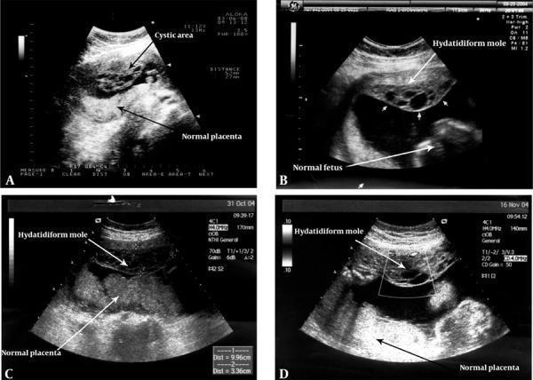
This obstetric ultrasound series (A-D) captures a rare twin pregnancy consisting of a complete hydatidiform mole and a coexisting viable fetus, spanning from 15 to 28 weeks of gestation. Image A (15 weeks) reveals a distinct cystic area at the anterior uterine wall characterized by multiple small, sonolucent vesicles, clearly separated from a homogeneous normal placenta. Image B (18 weeks) illustrates the characteristic heterogeneous, 'grape-like' vesicular echotexture of the hydatidiform mole situated superiorly, while a viable fetus with distinct anatomical outlines is visible inferiorly. Images C (24 weeks) and D (28 weeks) demonstrate the progression of the condition, showing the persistent, complex multicystic structure of the mole alongside the developing normal placenta and fetus. The series emphasizes the diagnostic hallmark of this condition: the presence of two distinct placental tissues—one with normal echogenicity and one exhibiting the disorganized, hydropic villi typical of molar pregnancy. This visual progression is critical for differentiating between a partial mole and a twin pregnancy with a complete mole and a coexistent live fetus.
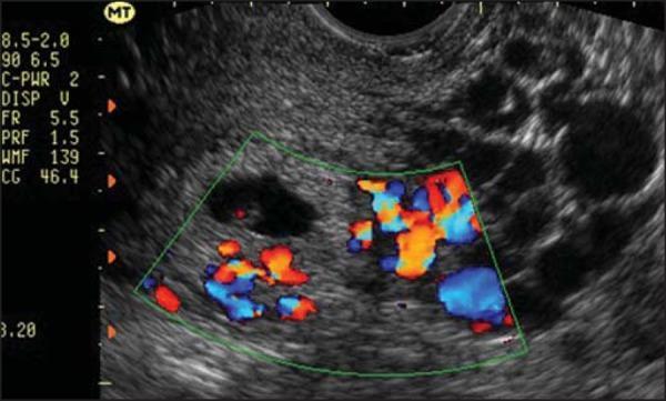
This diagnostic image is a transvaginal ultrasound featuring a sagittal view of the uterus, demonstrating a partial hydatidiform mole. The image shows a heterogeneous intrauterine mass with a classic 'snowstorm' or 'bunch of grapes' appearance, characterized by multiple small, anechoic (black) cystic vesicles representing hydropic villi. A distinct anechoic gestational sac containing a small fetal pole or embryo is visible, which is the key diagnostic feature distinguishing a partial mole from a complete hydatidiform mole. A Color Doppler overlay is applied to a central region of interest, showing prominent vascularity (red and blue signals) within the deciduous and placental tissue, indicating active blood flow. The surrounding myometrium appears intact. This image is used in obstetrics and gynecology to illustrate gestational trophoblastic disease (GTD) and the importance of high-resolution ultrasound in differentiating between complete and partial molar pregnancies based on the presence of fetal tissue and placental morphology.
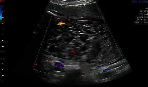
Diagnostic ultrasound image featuring a color Doppler study of a molar placenta, characteristic of a complete hydatidiform mole. The grayscale background reveals a heterogeneous, multicystic placental mass often referred to as a 'snowstorm' or 'swiss cheese' pattern. This is comprised of numerous small, anechoic vesicular structures representing hydropic villi. Superimposed color Doppler signals (red and blue) demonstrate active blood flow within the septations and around the cystic spaces, indicating significant vascularity of the molar tissue. This imaging modality is essential for distinguishing gestational trophoblastic disease from other uterine masses, such as degenerating fibroids or chorioangiomas. The clinical context involves a rare case of a twin pregnancy with a coexisting complete hydatidiform mole, where the diagnostic focus is on the distinct morphology of the abnormal third placental mass and its associated high vascularity.
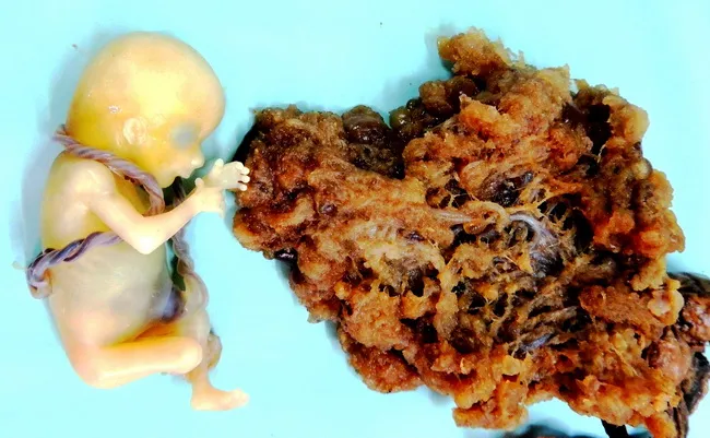
Gross pathology photograph of gestational tissue from a 30-year-old woman at approximately 14 weeks gestation. The image demonstrates placental tissue with admixture of vesicular hydropic villi and areas of normal villous architecture, accompanied by focal blood clots. A live fetus with intact umbilical cord is present adjacent to the placental tissue, consistent with partial hydatidiform mole in which a triploid conceptus coexists with molar villi. The case description notes paternal duplication or dispermic fertilization as the cytogenetic basis for triploidy (commonly 69,XXY) and highlights distinguishing features of partial mole. Incomplete moles can progress to invasion in less than 5% of cases, whereas complete hydatidiform moles show higher risk of persistent trophoblastic disease and choriocarcinoma. The gross morphology in this specimen--mixed vesicular and solid villous components with hemorrhagic clots--reflects hydropic change and trophoblastic proliferation occurring within placental tissue. Clinically, the presentation includes amenorrhea, vaginal bleeding, and uterine enlargement; ultrasound may reveal abnormal placentation with an intrauterine fetus. Differential considerations include complete mole, choriocarcinoma, invasive mole, and other GTD entities. Management typically involves termination with subsequent surveillance for persistent gestational trophoblastic disease. Ancillary notes emphasize molecular cytogenetics for definitive classification when available.
Molar Pregnancy (Hydatidiform Mole): MBBS Notes
1. Definition
2. Risk factors
- Extremes of maternal age (under 20 and over 35, with the highest risk over 40).
- A previous molar pregnancy (about 1% recurrence).
- Asian ethnicity and low socioeconomic status or dietary deficiency (low carotene or animal fat).
- Familial recurrent mole is rare. It is caused by NLRP7 mutations on 19q13.4, with biparental complete moles (Berek).
3. Complete vs partial mole (high-yield table)
| Feature | Complete mole | Partial mole |
|---|---|---|
| Karyotype | 46,XX (about 90%), 46,XY (about 10%) | Triploid, 69,XXY / XXX / XYY |
| Origin | All paternal (androgenetic). Empty ovum fertilised by one haploid sperm that duplicates (or 2 sperms). Mitochondrial DNA is maternal. | Haploid ovum fertilised by two sperms (dispermy), so 2 paternal + 1 maternal set |
| Fetus / embryo | Absent (rarely present) | Often present, with stigmata of triploidy (growth restriction, syndactyly, hydrocephalus) |
| Villi | All villi swollen, diffuse trophoblastic hyperplasia | Focal swelling, scalloped villi, stromal trophoblastic inclusions |
| p57 (KIP2) immunostain | Negative (paternally imprinted gene) | Positive |
| Presentation | Classic: bleeding, large-for-dates uterus, high hCG | Like incomplete or missed abortion |
| hCG | Markedly raised | Mildly raised or normal |
| Risk of persistent GTN | 15-20% (invasion about 15%, metastasis about 4%) | 1-4% |
| Choriocarcinoma | About 2.5% | Very rare |
4. Pathology
- Hydropic, avascular villi with central cisterns.
- Circumferential trophoblastic hyperplasia (cytotrophoblast and syncytiotrophoblast).
- Cellular atypia.
5. Clinical features
- Vaginal bleeding is the most common symptom (about 46% today, against 97% in older series). The classic discharge is prune-juice or brownish. The bleeding is intermittent and may be heavy.
- Uterus large for dates is seen in only about 28-50%. The uterus feels doughy, with no fetal parts or heart sounds.
- Hyperemesis gravidarum (about 8% today, historically 25%), due to high hCG.
- Early-onset preeclampsia, before 20 weeks (about 27% historically). Think of mole whenever preeclampsia appears this early. Eclampsia is rare.
- Hyperthyroidism (about 7%): tachycardia, tremor, warm skin. It occurs with very high hCG, and anaesthesia or surgery may precipitate thyroid storm.
- Anaemia (now only about 5%).
- Trophoblastic embolisation, causing respiratory distress, is rare.
- Theca lutein cysts, which may twist, rupture or bleed.
- Passage of vesicles per vaginam is a pathognomonic sign.
6. Diagnosis
- Ultrasound (transvaginal): the investigation of choice.
- Complete mole shows a "snowstorm" or "bunch of grapes" pattern: a heterogeneous mass with multiple small anechoic cysts and no fetus. Doppler shows vascularity.
- Partial mole shows a thickened placenta with cystic spaces and a gestational sac or fetal parts.
- Serum β-hCG: markedly raised, often over 100,000 mIU/mL in complete mole.
- Histopathology of all evacuated products is mandatory. Add p57 and ploidy when needed.
- Baseline work-up:
- Blood group and Rh.
- Full blood count.
- Thyroid function tests and renal and liver function tests.
- Chest X-ray (lung metastases).
- Coagulation profile if indicated.
7. Management
- Start an oxytocin infusion before or at the start of the procedure.
- Dilate the cervix and use a large cannula (12 mm).
- Follow with gentle sharp curettage.
- Give anti-D immunoglobulin to Rh-negative women, because trophoblast expresses RhD.
- Cross-match blood and have it available.
- Medical induction with prostaglandins or misoprostol and surgical hysterotomy are avoided. They increase bleeding and the risk of persistent disease.
8. Follow-up (essential)
- Serial serum β-hCG weekly until it is normal for 3 consecutive weeks. Then check monthly for 6 months for a complete mole. For a partial mole, many protocols stop after confirmed normalisation. Follow your local protocol.
- Contraception: use reliable contraception (combined oral contraceptive pills or barrier methods) for about 6 months. Avoid an IUCD until hCG is normal. The reason is to avoid a pregnancy that would make the hCG rise uninterpretable.
- Use an early ultrasound in future pregnancies. Send the products of conception for histology after any later miscarriage.
- High-risk features for persistent disease (Berek, p. 2503):
- hCG over 100,000 mIU/mL.
- Uterus large for dates.
- Theca lutein cysts of 6 cm or more.
- Age over 40.
9. Gestational trophoblastic neoplasia (post-molar)
- hCG plateau (4 values over 3 weeks).
- hCG rise of more than 10% (3 values over 2 weeks).
- hCG still detectable at 6 months.
- Histological choriocarcinoma.
10. Quick exam points
- A complete mole is 46,XX, all paternal, p57 negative, with no fetus.
- A partial mole is 69,XXY, has a fetus, is p57 positive, and has a low malignant risk.
- The characteristic sonogram shows a "snowstorm" or "bunch of grapes" pattern.
- Early preeclampsia (before 20 weeks), hyperemesis and hyperthyroidism all point to a mole, and are due to high hCG.
- Treatment is suction evacuation, then weekly hCG until normal, then monthly follow-up, with contraception.
- Anti-D is given to Rh-negative women.
- Hysterectomy does not eliminate the need for hCG follow-up.