
Renal Cell Carcinoma
Reading File
Renal cell carcinoma (RCC) is the most common primary malignancy of the kidney in adults, arising from renal tubular epithelium. It classically presents with hematuria, flank pain, and a palpable mass (the "classic triad"), though most cases are now found incidentally on imaging.
Searching sources...
Finding Sources
Searching PubMed
"renal cell carcinoma"[MeSH Terms] AND treatment
Good results across multiple textbooks. Let me read the key sections in parallel.
Reading File
Reading File
Reading File
Good content from Robbins and Goldman-Cecil. Now let me get the clinical/management sections from Campbell-Walsh (the premier urology reference) and continue reading the Goldman-Cecil content.
Reading File
Reading File
Reading File
I have excellent content on pathology, epidemiology, and classification. Now let me get the clinical presentation, staging, and management sections.
Reading File
Reading File
Good clinical content. Let me get the treatment/staging section from Goldman-Cecil and fetch relevant histological images from Robbins.
Reading File
Loading Image
Excellent. I now have comprehensive content covering all major aspects of RCC. Let me compile the full answer.
Renal Cell Carcinoma (RCC)
Definition and Overview
Renal cell carcinoma is not a single entity but a group of distinguishable epithelial neoplasms arising from the renal tubular epithelium, each with a strong relationship between its morphologic and genetic features. It represents 80-85% of all primary malignant renal neoplasms and 2-3% of all adult cancers, accounting for roughly 65,000-75,000 new diagnoses per year in the United States and approximately 14,000 deaths annually.
- 6th most common cancer in men, 9th in women
- Male:female ratio of ~2:1 to 3:1
- Peak incidence in the 6th-7th decade
- Highest incidence in Black Americans; lowest in Asians/Pacific Islanders
- Only 4-6% of cases are familial; the vast majority are sporadic
Risk Factors
| Risk Factor | Notes |
|---|---|
| Cigarette smoking | Most established modifiable risk |
| Obesity | Risk rises with increasing BMI |
| Hypertension | Independent risk factor |
| Diuretic use | Hard to separate from hypertension risk |
| Chronic dialysis / Acquired polycystic disease | 30-fold increased risk |
| Horseshoe kidney, polycystic kidney disease | Pre-existing renal injury |
| Occupational cadmium exposure | Less common |
| Genetic syndromes (VHL, HLRCC, BHD, SDH) | Familial forms |
Classification and Pathology
RCC is classified by morphology, growth pattern, and molecular genetics. The three main subtypes:
1. Clear Cell Carcinoma (65% of RCC; 90% of metastases)
- Derived from the proximal convoluted tubule
- Grossly: solitary, well-circumscribed, golden yellow due to cytoplasmic lipid
- Histologically: cells with clear cytoplasm arranged in nests surrounded by delicate vasculature
- Molecular hallmark: loss/inactivation of both VHL alleles on chromosome 3p25
- VHL normally causes oxygen-dependent degradation of HIFs (hypoxia-inducible factors)
- Loss of VHL --> HIF stabilization even in normoxia --> upregulation of VEGF --> tumor vascularization
- Also involves loss-of-function mutations in histone methylation regulators (epigenomic changes)
- Frequently invades the renal vein (and can extend into the inferior vena cava)
- Most virulent subtype
2. Papillary RCC (10-15%; largely indolent)
- Characteristic papillary growth pattern
- Frequently multifocal and bilateral
- Often presents as early-stage tumor
- Molecular hallmark: increased MET signaling (chromosome 7q)
- Sporadic: increased MET copy number or somatic activating MET mutations
- Familial: germline activating MET mutations
- Hypointense on T2-weighted MRI; hypoenhancing on all post-contrast phases
- Lower metastatic potential than clear cell
3. Chromophobe RCC (~5%; indolent)
- Neoplastic cells have eosinophilic cytoplasm with prominent cell membranes (like plant cells)
- Avid enhancement in corticomedullary phase on CT/MRI, but less than clear cell
- Better prognosis than clear cell or papillary
- Associated with Birt-Hogg-Dube (BHD) syndrome
Other Subtypes
- Collecting duct carcinoma: very rare, aggressive
- Oncocytoma: benign (10% of all renal tumors), mahogany-brown color, central stellate scar; cannot be reliably distinguished from RCC on imaging
- Sarcomatoid differentiation: can occur in any subtype; indicates aggressive biology and poor prognosis
Hereditary Syndromes
| Syndrome | Gene / Locus | RCC Subtype | Features |
|---|---|---|---|
| Von Hippel-Lindau (VHL) | VHL, 3p25 (AD) | Clear cell (bilateral, multiple) | Hemangioblastomas (cerebellum/retina), pheochromocytoma, pancreatic cysts |
| Hereditary Papillary RCC | MET, 7q (AD) | Papillary type 1 (bilateral, multiple) | No extrarenal manifestations |
| Hereditary Leiomyomatosis RCC (HLRCC) | Fumarate hydratase (FH), 1q42-43 (AD) | Papillary type 2 (solitary, aggressive) | Cutaneous + uterine leiomyomas; collecting duct carcinoma possible |
| Birt-Hogg-Dube (BHD) | Folliculin (FLCN), 17p (AD) | Chromophobe / oncocytic (bilateral) | Fibrofolliculomas, pulmonary cysts, spontaneous pneumothorax |
| SDH-RCC | SDHA/B/C/D, SDHAF2 | SDH-deficient RCC | Pheochromocytoma, paraganglioma; early onset (<40 yrs) |
Clinical Presentation
Most tumors (~50%) are now detected incidentally on CT or ultrasound performed for unrelated reasons. The classic triad of hematuria + flank pain + palpable mass is now seen in <10% of patients and usually indicates advanced disease.
Common symptoms and signs:
| Finding | Frequency |
|---|---|
| Anemia | 52% |
| Hepatic dysfunction (Stauffer syndrome) | 32% |
| Weight loss | 23% |
| Hypoalbuminemia | 20% |
| Malaise | 19% |
| Hypercalcemia | 13% |
| Anorexia | 11% |
| Thrombocytosis | 9% |
| Fever / Night sweats | 8% |
| Erythrocytosis | 4% |
| Hypertension | 3% |
Paraneoplastic syndromes are a hallmark of RCC:
- Hypercalcemia (~20%): from PTH-rP, IL-6-driven osteoclast activation, or bony mets
- Erythrocytosis: ectopic erythropoietin production
- Hypertension: excess renin secretion
- Stauffer syndrome: reversible hepatic dysfunction without hepatic metastases (resolves after nephrectomy)
- Fever of unknown origin: from cytokine (IL-6, IL-1) secretion
RCC is also known for venous extension - tumor thrombus can grow into the renal vein and up through the inferior vena cava into the right atrium, without necessarily carrying a worse prognosis stage-for-stage.
Imaging
CT (multiphasic contrast-enhanced) is the standard for characterization:
| Subtype | Enhancement Pattern |
|---|---|
| Clear cell | Avid corticomedullary enhancement, becomes hypoenhancing in nephrographic phase |
| Chromophobe | Avid enhancement, but less than clear cell |
| Papillary | Hypoenhancing on all post-contrast phases; hypointense on T2 MRI |
MRI is preferred when CT contrast is contraindicated (renal insufficiency) and is more sensitive for detecting enhancement within cystic lesions (subtraction imaging).
Bosniak Classification is used for cystic lesions:
- Category I/II: benign, no follow-up needed
- Category IIF: follow-up required
- Category III: surgical exploration (51% malignant)
- Category IV: surgical resection (>90% malignant)
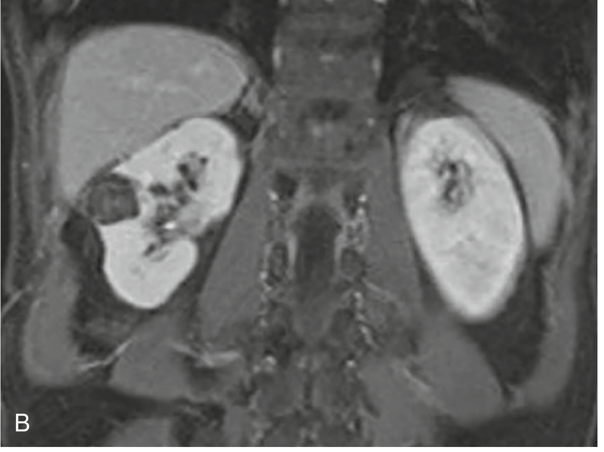
Below is a coronal MRI showing bilateral renal masses in a patient with bilateral RCC (as seen in VHL disease):
Staging (AJCC 8th Edition)
| Stage | T | N | M | Description |
|---|---|---|---|---|
| I | T1 | N0 | M0 | Tumor ≤7 cm, confined to kidney |
| II | T2 | N0 | M0 | Tumor >7 cm, confined to kidney |
| III | T1-2 | N1 | M0 | Regional lymph node involvement |
| III | T3a-c | N0-1 | M0 | Perinephric/renal vein/IVC extension below diaphragm |
| IV | T4 | Any N | M0 | Beyond Gerota fascia / adrenal invasion |
| IV | Any T | Any N | M1 | Distant metastasis |
- T3a: invasion of renal sinus fat or perinephric fat
- T3b: tumor thrombus in renal vein or IVC below diaphragm
- T3c: tumor thrombus in IVC above diaphragm or into right atrium
- ~30% of patients present with metastatic (Stage IV) disease
Common metastatic sites: lung (most common), bone, liver, brain, adrenal glands, and contralateral kidney.
Treatment
Localized Disease (Stages I-III)
Surgery is the only curative modality. RCC is resistant to conventional radiation therapy and cytotoxic chemotherapy.
- Partial nephrectomy (nephron-sparing): preferred for T1 tumors (≤7 cm) and when kidney preservation is important (solitary kidney, bilateral tumors, impaired renal function)
- Radical nephrectomy: standard for larger or more locally advanced tumors; can be performed laparoscopically or open (flank, transperitoneal, or transthoracic approach); ipsilateral adrenal gland often removed; lymph node dissection is optional and controversial
Adjuvant therapy (for high-risk patients post-nephrectomy):
- Sunitinib (VEGF-targeted TKI) - adjuvant use in high-risk patients
- Pembrolizumab (anti-PD-1 checkpoint inhibitor) - superior disease-free survival in high-risk patients after resection
Metastatic Disease (Stage IV)
Approximately 30% present with metastases; another 20-30% relapse after surgery.
Current first-line systemic therapy is combination immunotherapy + targeted therapy:
| Regimen | Notes |
|---|---|
| Nivolumab + Ipilimumab (anti-PD-1 + anti-CTLA-4) | Intermediate/poor risk; superior OS vs sunitinib |
| Nivolumab + Cabozantinib (anti-PD-1 + TKI) | Improved PFS and OS vs sunitinib |
| Pembrolizumab + Axitinib (anti-PD-1 + VEGFR TKI) | Superior PFS/OS vs sunitinib, all risk groups |
| Pembrolizumab + Lenvatinib (anti-PD-1 + TKI) | Superior PFS/OS vs sunitinib, all risk groups |
| Avelumab + Axitinib (anti-PD-L1 + TKI) | Option for favorable risk |
Key principle: Pembrolizumab + TKI (axitinib or lenvatinib) is recommended regardless of risk group. Nivolumab + ipilimumab is specifically recommended for intermediate/poor risk patients.
Surgery in metastatic disease:
- Cytoreductive nephrectomy: less commonly used in the current era of effective systemic therapy
- Metastasectomy: may extend survival and can cure selected patients (best candidates: disease-free interval >1 year, solitary metastatic site, lung metastases)
Radiation therapy: used for palliation of bone metastases and multiple brain metastases; stereotactic radiosurgery (SRS) for limited brain mets.
Prognosis
- Localized disease (Stage I-II): 5-year survival >80-90%
- Stage III: ~50-60%
- Metastatic (Stage IV): historically <10% at 5 years, but improving significantly with immunotherapy combinations
- Clear cell has the worst metastatic potential (90% of all RCC metastases)
- Chromophobe and papillary have lower metastatic rates but still require careful follow-up
- Sarcomatoid differentiation in any subtype is a poor prognostic indicator
Key High-Yield Points
- VHL gene (3p25) is the molecular hallmark of clear cell RCC - loss leads to HIF accumulation --> VEGF overexpression --> angiogenesis
- Clear cell RCC is prone to renal vein invasion and IVC thrombosis
- RCC is one of the great mimics - causes fever of unknown origin, hypercalcemia, erythrocytosis, and Stauffer syndrome
- Classic triad (hematuria + flank pain + mass) is now uncommon (<5-10%); most are found incidentally
- RCC is resistant to chemotherapy and conventional radiation - surgery for localized disease, immunotherapy combinations for metastatic
- Oncocytoma (benign) cannot be reliably distinguished from RCC on imaging - most are surgically resected
- VHL disease: bilateral clear cell RCC in 40-60% of affected individuals; autosomal dominant, 3p25
Sources: Robbins & Kumar Basic Pathology; Goldman-Cecil Medicine, 26th ed.; Campbell-Walsh-Wein Urology; National Kidney Foundation Primer on Kidney Diseases, 8th ed.
This is a shared conversation. Sign in to Orris to start your own chat.