
Give me detailed notes of EEG basics. Like a foundation for undertsadning and starting to learn EEG.
electroencephalography AND education
International Federation of Clinical Neurophysiology EEG recording standards electrode placement 10-20 system guideline
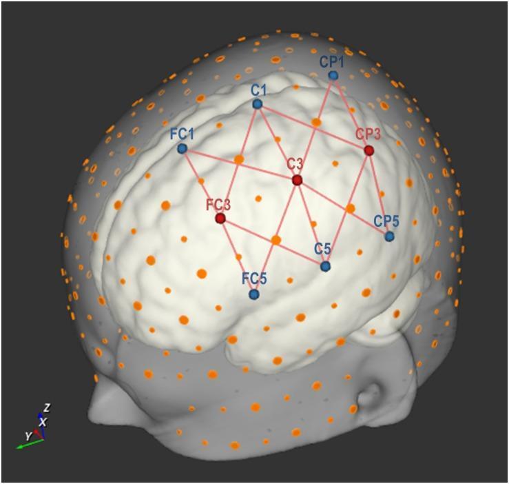
- What EEG measures - summed postsynaptic cortical electrical activity recorded from the scalp
- Waveform language - frequency, amplitude, morphology, polarity, field, synchrony, and reactivity
- Electrode placement - International 10-20 system and the meaning of labels such as Fp1, F3, C3, O1
- Technical fundamentals - referential versus bipolar montages, sensitivity, filters, impedance, sampling, and calibration
- Normal awake EEG - posterior dominant rhythm, beta activity, eye opening response, drowsiness
- Normal sleep EEG - vertex waves, sleep spindles, K-complexes, slow-wave sleep, REM
- Artifacts - eye movements, blink, muscle, ECG, sweat, electrode pop, movement, and 50/60-Hz interference
- Abnormal EEG terms - focal/generalized slowing, epileptiform discharges, periodic patterns, electrographic seizures
- Clinical interpretation - how to systematically read an EEG and correlate it with the patient
- Limits and applications - epilepsy, encephalopathy, coma/ICU monitoring, and why a normal EEG does not exclude epilepsy
Foundation overview
z means midline. Standardized placement matters because localization depends on comparing the distribution, field, phase reversal, and evolution of activity across channels. The ACNS electrode nomenclature guideline describes the standard system.- EEG signals and waveforms
- 10-20 electrodes and montages
- Normal awake and sleep EEG
- Artifacts and troubleshooting
- Epileptiform discharges and seizures
- A complete beginner-to-reader guide
Give me a complete beginner-to-reader EEG study guide
Which EEG module should I cover in detailed beginner-to-reader notes next?
- What EEG measures - summed postsynaptic cortical electrical activity recorded from the scalp
- Waveform language - frequency, amplitude, morphology, polarity, field, synchrony, and reactivity
- Electrode placement - International 10-20 system and the meaning of labels such as Fp1, F3, C3, O1
- Technical fundamentals - referential versus bipolar montages, sensitivity, filters, impedance, sampling, and calibration
- Normal awake EEG - posterior dominant rhythm, beta activity, eye opening response, drowsiness
- Normal sleep EEG - vertex waves, sleep spindles, K-complexes, slow-wave sleep, REM
- Artifacts - eye movements, blink, muscle, ECG, sweat, electrode pop, movement, and 50/60-Hz interference
- Abnormal EEG terms - focal/generalized slowing, epileptiform discharges, periodic patterns, electrographic seizures
- Clinical interpretation - how to systematically read an EEG and correlate it with the patient
- Limits and applications - epilepsy, encephalopathy, coma/ICU monitoring, and why a normal EEG does not exclude epilepsy
Foundation overview
z means midline. Standardized placement matters because localization depends on comparing the distribution, field, phase reversal, and evolution of activity across channels. The ACNS electrode nomenclature guideline describes the standard system.-
EEG signals and waveforms
-
10-20 electrodes and montages
-
Normal awake and sleep EEG
-
Artifacts and troubleshooting
-
Epileptiform discharges and seizures
-
A complete beginner-to-reader guideA complete EEG guide spans basic neurophysiology, technical recording, normal variants, and clinical interpretation. To make it useful rather than a very long surface-level note, here is the learning map.A complete beginner-to-reader EEG guide includes:
-
EEG signal foundations
What scalp EEG records, cortical generators, voltage, frequency, polarity, synchrony, and why deep sources may be missed. -
Electrodes and the 10-20 system
Scalp landmarks, electrode names, left-right and midline labels, referential electrodes, and localization basics. -
Montages and display settings
Bipolar and referential montages, phase reversal, sensitivity, time base, low-frequency and high-frequency filters, notch filter, impedance, and sampling. -
Normal background EEG
Posterior dominant rhythm, beta, theta, delta, symmetry, continuity, reactivity, and expected changes with eye opening and activation. -
Drowsiness and sleep
Normal transitions through N1, N2, N3, and REM: vertex waves, spindles, K-complexes, slow waves, and REM features. -
Artifacts and benign variants
Eye movements, muscle, ECG, sweat, movement, electrode pop, electrical interference, plus common normal variants that can mimic epileptiform activity. -
Abnormal background patterns
Focal slowing, generalized slowing, attenuation, asymmetry, discontinuity, periodic patterns, and their broad clinical implications. -
Epileptiform discharges and seizures
Spikes, sharp waves, spike-wave complexes, focal versus generalized patterns, seizure evolution, and the difference between interictal and ictal findings. -
Systematic EEG reading
A reproducible reading order, how to form an impression, and how to avoid overcalling normal activity or artifact as epilepsy. -
Clinical use and limitations
Role in epilepsy, altered consciousness, encephalopathy, ICU monitoring, and why a normal routine EEG does not rule out epilepsy.
All please
electroencephalography AND interpretation
ACNS guideline routine EEG technical standards filters sensitivity montages
international 10-20 EEG electrode placement scalp diagram labels Fp F C P O
https://www.acns.org/UserFiles/file/Guideline1-MinimumTechni…
EEG: Complete Beginner-to-Reader Study Guide
Purpose: Build the foundation to understand routine scalp EEG and begin reading it systematically. This is educational material, not a substitute for supervised clinical EEG training or formal reporting by a qualified electroencephalographer.
1. What is EEG?
- Horizontal axis: time
- Vertical axis: voltage, usually microvolts (µV)
- Each line: a recording channel, representing the voltage difference between electrodes or between one electrode and a reference
What scalp EEG detects well
- Superficial cortical electrical activity
- Generalized cerebral dysfunction
- State changes: wakefulness, drowsiness, sleep
- Many focal or generalized epileptiform discharges
- Electrographic seizures, especially with continuous video-EEG monitoring
What it detects poorly
- Very deep sources, such as mesial temporal, deep frontal, cingulate, or insular sources
- Very small cortical areas
- Brief, infrequent events during a short recording
- Structural lesions themselves
2. Core EEG language
| Feature | What it means | Questions to ask |
|---|---|---|
| Frequency | Cycles per second, Hz | Is it delta, theta, alpha, beta, or faster? |
| Amplitude | Height of waveform, µV | Is it low, normal, or high voltage? |
| Morphology | Shape | Sinusoidal, sharp, spiky, rhythmic, polymorphic? |
| Location | Where it is maximal | Frontal, temporal, central, parietal, occipital? |
| Distribution | How broadly it spreads | Focal, regional, unilateral, bilateral, generalized? |
| Symmetry | Left versus right comparison | Is the pattern similar on both sides? |
| Synchrony | Timing between regions | Are bilateral discharges simultaneous? |
| Reactivity | Change with stimulation or state | Does it change with eye opening, voice, touch, sleep? |
| Evolution | Meaningful change over time | Does frequency, amplitude, morphology, or location change? |
| Field | Spatial distribution across electrodes | Is there a physiologically believable scalp pattern? |
Frequency bands
| Band | Frequency | Usual context |
|---|---|---|
| Delta | <4 Hz | Normal in deep sleep; abnormal when prominent in an awake adult, especially if focal |
| Theta | 4-7 Hz | Normal in drowsiness and children; may reflect dysfunction if persistent while awake in adults |
| Alpha | 8-13 Hz | Posterior dominant rhythm in relaxed awake adults with eyes closed |
| Beta | >13 Hz | Low-amplitude fast activity, often frontocentral; increased by benzodiazepines and some sedatives |
| Gamma | >30 Hz | Often difficult to assess in routine scalp EEG because muscle artifact contaminates this range |
3. EEG polarity: why “up” and “down” can mislead you
- Negative at input 1 produces an upward deflection.
- Positive at input 1 produces a downward deflection.
- Where the waveform has its greatest amplitude
- How it distributes across neighboring electrodes
- Whether it reverses phase in a bipolar montage
- Whether it has a believable field
- Whether it evolves over time
4. The international 10-20 electrode system
- Nasion: bridge of the nose
- Inion: palpable prominence at the back of the skull
- Preauricular points: just anterior to each ear
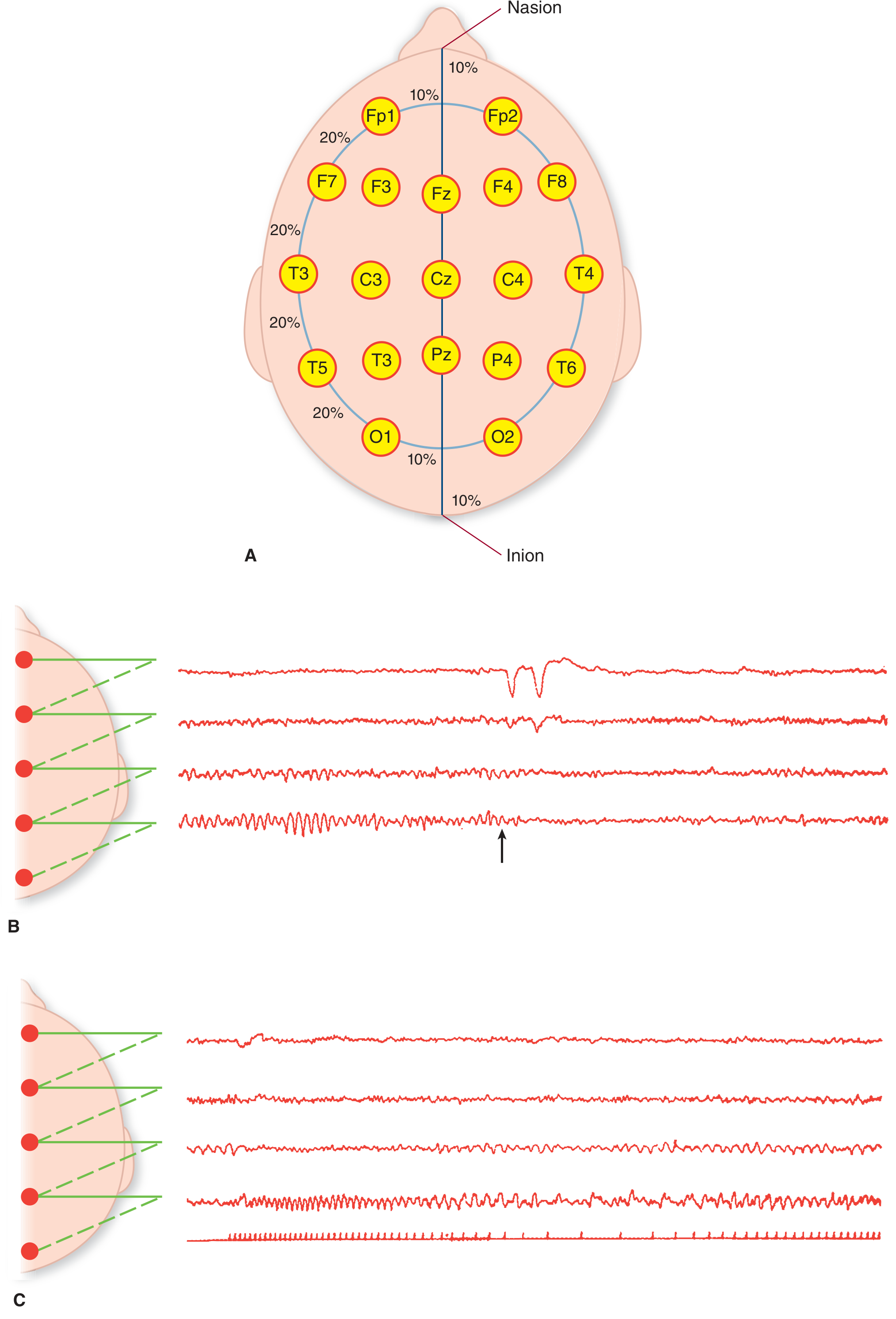
Electrode labels
| Letter | Region |
|---|---|
| Fp | Frontopolar |
| F | Frontal |
| C | Central |
| P | Parietal |
| O | Occipital |
| T | Temporal |
| A or M | Auricular or mastoid reference area |
| z | Midline |
Numbering
- Odd numbers: left hemisphere
- Even numbers: right hemisphere
- z: midline, for example Fz, Cz, Pz
- Fp1: left frontopolar
- F3: left frontal
- C3: left central
- P4: right parietal
- O2: right occipital
- F7: left anterior temporal/frontotemporal
- T7: left mid-temporal
- P7: left posterior temporal
- F8, T8, P8: corresponding right-sided sites
Important localization caution
5. What is a montage?
A. Bipolar montage
Fp1-F7
F7-T7
T7-P7
P7-O1
- Excellent for seeing local fields
- Helpful for recognizing phase reversal
- Helps distinguish focal activity from widespread artifact
B. Referential montage
F3-Avg
C3-Avg
P3-Avg
O1-Avg
- Helps identify areas of maximal amplitude
- Useful for generalized patterns
- Can make a focal discharge’s distribution easier to see
C. Why you must review several montages
6. Phase reversal
F7-T7      ↑
T7-P7      ↓
Key warning
Phase reversal = local maximum on the scalp display, not a final anatomical diagnosis.
7. EEG technical basics
Sensitivity or gain
- Lower µV/mm number = more sensitive display = waveform looks taller
- Higher µV/mm number = less sensitive display = waveform looks shorter
- At 5 µV/mm, a 50 µV wave spans 10 mm.
- At 10 µV/mm, the same 50 µV wave spans 5 mm.
Time base
- A faster display spreads waveforms out and helps inspect morphology.
- A slower display compresses time and helps identify rhythmicity, periodicity, and seizure evolution.
Filters
| Filter | Main purpose | Main danger |
|---|---|---|
| Low-frequency filter, also called high-pass | Reduces very slow drift, sweat artifact | Can attenuate or distort slow waves and long-duration components |
| High-frequency filter, also called low-pass | Reduces high-frequency noise and muscle artifact | Can blunt spikes and fast activity |
| Notch filter | Reduces 50/60-Hz line noise | Can mask an underlying technical issue and alter signal appearance |
First inspect the raw or minimally filtered signal. Do not solve every problem by applying stronger filters.
Impedance
- Excessive artifact
- Poor signal quality
- Electrode pops
- Increased 50/60-Hz interference
Sampling rate and digital EEG
8. Routine EEG protocol and activation procedures
Usual recording conditions
- Relaxed wakefulness, often eyes closed
- Eye opening and eye closure
- Drowsiness and, when possible, sleep
- ECG channel
- Video when available
- Documentation of medications, sleep deprivation, clinical events, and activation procedures
Hyperventilation
Intermittent photic stimulation
- Photic driving: normal posterior rhythmic response time-locked to flashes
- Photoparoxysmal response: epileptiform discharges provoked by flashes, sometimes generalized and sometimes associated with myoclonus or seizures
Sleep
9. Normal awake EEG
Posterior dominant rhythm, PDR
- Around 8-13 Hz
- Best seen posteriorly at O1 and O2
- Usually symmetric or only mildly asymmetric
- Most apparent when the person is relaxed with eyes closed
- Attenuates or blocks with eye opening and alerting
- Frequency
- Amplitude
- Symmetry
- Reactivity to eye opening
- Organization of the overall background
Beta activity
- Low-amplitude fast activity, usually frontocentral
- Often symmetric
- Can increase with benzodiazepines, barbiturates, and some sedating medicines
- May also reflect muscle artifact if it is irregular, very fast, and concentrated over temporal/frontal areas
Mu rhythm
- Arch-shaped or “comb-like”
- May look sharp
- Blocks with movement or intention to move the contralateral hand
- Can be mistaken for epileptiform activity by beginners
Lambda waves
10. Drowsiness and normal sleep EEG
Drowsiness, N1 sleep
- Posterior dominant rhythm becomes less organized and fades
- Generalized theta activity increases
- Slow rolling eye movements may appear
- Vertex sharp transients may occur
Vertex waves
- Brief sharp transients maximal at the vertex, often Cz
- Common in drowsiness and light sleep
- Can be asymmetric but should not have a consistent pathological field
N2 sleep
Sleep spindles
- Brief waxing and waning bursts
- Usually around 11-16 Hz
- Often maximal over central regions
- Bilateral and generally symmetric
K-complexes
- Large, biphasic waveform
- May occur spontaneously or after an auditory stimulus
- Often followed by a sleep spindle
- Normal in N2 sleep
N3 sleep
- High-amplitude delta activity
- Deep sleep
- Slow waves are broadly distributed and normal in this state
REM sleep
- Low-voltage mixed-frequency EEG, sometimes resembling wakefulness
- Rapid eye movements
- Reduced chin muscle tone on polysomnography
- Sawtooth waves may occur
A beginner safeguard
Is this a vertex wave, K-complex, spindle-related transient, or other normal sleep feature?
11. Artifacts: the first differential diagnosis
A. Eye blink and eye movement artifact
- Prominent in Fp1 and Fp2
- Large, slow frontal deflections
- Often symmetric with blinking
- Horizontal eye movements produce opposite polarity across left and right frontal electrodes
B. Muscle, EMG artifact
- Fast, irregular, low-amplitude or high-amplitude activity
- Common in frontal and temporal electrodes
- Seen with jaw clenching, talking, chewing, swallowing, frowning, or tension
- May obscure real cerebral activity
C. ECG artifact
- Repetitive waveform synchronized with heart rate
- Often greatest near ear, temporal, or lower electrodes
- Confirm by comparing with the ECG channel
D. Sweat artifact
- Very slow, drifting baseline movement
- Often frontal
- May improve as the patient cools or the electrode-skin interface is corrected
E. Electrode pop
- Abrupt, often high-amplitude transient
- Usually isolated to one electrode or a small number of channels
- May have a vertical or sharply discontinuous appearance
- Often no plausible cerebral field
F. Movement artifact
- Often large amplitude and irregular
- May involve many channels simultaneously
- Check video and technologist annotations
G. 50/60-Hz line noise
- Regular, monomorphic fast oscillation
- Usually widespread
- May occur in one region if a specific electrode is poorly connected
Artifact checklist
- Is it confined to one electrode?
- Does it have a credible field in adjacent electrodes?
- Does it correlate with video, ECG, EOG, EMG, breathing, or movement?
- Does it persist in another montage?
- Is it state-linked or stimulus-linked?
- Does its morphology look too abrupt, too perfectly regular, or technically implausible?
12. Normal variants that can mimic epileptiform activity
| Pattern | Typical clue |
|---|---|
| Mu rhythm | Central, arch-shaped, blocks with contralateral movement |
| Lambda waves | Occipital, during visual scanning with eyes open |
| Wicket rhythm | Temporal arch-like rhythm, no after-going slow wave, often in older adults |
| Rhythmic mid-temporal theta of drowsiness | Drowsiness-related, brief temporal theta bursts without evolution |
| Benign small sharp spikes | Low-amplitude, brief temporal transients in drowsiness/light sleep, usually no disruptive slow wave |
| 14-and-6-Hz positive bursts | Brief posterior temporal positive bursts, usually in youth |
| Breach rhythm | High-voltage, sharp-contoured fast activity near a skull defect; non-epileptic by itself |
Epileptiform discharges typically have a physiologic field and are often followed by a slow wave or background disruption. Benign variants commonly lack these features.
13. Abnormal background activity
Diffuse slowing
- Toxic-metabolic disturbance
- Sedative or anesthetic medications
- Systemic infection or organ failure
- Diffuse hypoxic-ischemic injury
- Neurodegenerative disease
- Reduced alertness or sleep, which must be excluded first
- Frequency and amount of slowing
- Continuity
- Reactivity
- Symmetry
- State dependence
- Presence of superimposed epileptiform activity
Focal slowing
- Structural lesion
- Postictal state
- Focal ischemia
- Hemorrhage
- Tumor
- Infection/inflammation
- Focal cortical dysfunction
Asymmetry
- Amplitude
- Frequency
- Organization
- Reactivity
- Continuity
Attenuation, suppression, and discontinuity
14. Epileptiform discharges
Common terms
| Term | Approximate duration | Description |
|---|---|---|
| Spike | <70 ms | Pointed transient with rapid rise and fall |
| Sharp wave | 70-200 ms | Pointed transient, broader than a spike |
| Spike-wave complex | Variable | Spike followed by a slow wave |
| Polyspike | Multiple spikes | Cluster of spikes, often followed by slow wave |
| Sharp-and-slow-wave complex | Variable | Sharp wave followed by a slow wave |
Features supporting an epileptiform discharge
- Distinctive sharp or spiky morphology
- Clear physiologic field across adjacent electrodes
- Voltage maximum in a plausible location
- Background disruption
- Often an after-going slow wave
- Consistency across montages
- Occurrence in a characteristic state, often drowsiness or sleep
- Clinical correlation with an epilepsy syndrome when applicable
Features arguing against an epileptiform discharge
- Only one electrode is involved
- No field in surrounding channels
- Perfectly stereotyped technical appearance
- Clear relation to eye movement, ECG, muscle, or movement
- Disappears or changes implausibly with montage
- Occurs only in a setting suggestive of artifact
Focal epileptiform discharges
- Left temporal sharp waves
- Right frontal spikes
- Centrotemporal spikes
Generalized epileptiform discharges
- Generalized spike-wave
- Polyspike-wave
- Generalized paroxysmal fast activity
15. What makes an electrographic seizure?
- Frequency
- Amplitude
- Morphology
- Spatial distribution
- Rhythmicity
Rhythmic theta in right temporal region
→ increases in amplitude
→ frequency changes
→ spreads to adjacent regions
→ slows and terminates
Think in terms of evolution
| Feature | More consistent with seizure | Less consistent with seizure |
|---|---|---|
| Duration | Sustained, usually tens of seconds or longer | Single brief transient |
| Evolution | Clear change over time | Static, unchanging pattern |
| Field | Plausible regional spread | One-electrode phenomenon |
| Clinical correlation | May have behavioral or motor change | No correlation does not exclude seizure, especially in ICU |
| Offset | Clear termination or postictal slowing | Abrupt technical disappearance |
16. A systematic method to read a routine EEG
Step 1: Confirm clinical context
- Age
- Indication
- Event description
- Epilepsy diagnosis and seizure types
- Medications, especially antiseizure drugs and sedatives
- Wakefulness, sleep deprivation, and sleep during the study
- Brain imaging and previous EEGs
- Recent seizure or postictal state
Step 2: Check technical quality
- Is the recording sufficiently long?
- Are all standard electrodes present?
- Is the ECG channel visible?
- Are video and event markers available?
- Is there excessive artifact?
- Are settings and montages appropriate?
- Are there loose or noisy electrodes?
Step 3: Identify state
- Awake
- Drowsy
- N1, N2, N3, or REM sleep
- Sedated
- Stuporous/comatose
Step 4: Describe the posterior dominant rhythm
- Frequency
- Symmetry
- Organization
- Reactivity to eye opening
- Whether it is appropriate for age and state
Step 5: Evaluate background organization
- Continuity
- Symmetry
- Amount and distribution of theta/delta
- Beta activity
- Reactivity
- Focal attenuation or excess fast activity
- Whether normal sleep elements are present when expected
Step 6: Review activation procedures
- Hyperventilation response
- Photic driving
- Photoparoxysmal response
- Eye opening/closure
- Sleep
Step 7: Search for focal abnormalities
- Focal slowing
- Asymmetry
- Attenuation
- Focal spikes or sharp waves
- Rhythmic focal activity
- A consistent maximum and field
Step 8: Search for generalized abnormalities
- Generalized slowing
- Generalized spike-wave or polyspike-wave
- Periodic or rhythmic patterns
- Lack of organization or reactivity
Step 9: Determine whether any event is a seizure
- Onset
- Evolution
- Spread
- Duration
- Offset
- Clinical/video correlate
- Post-event background change
Step 10: Write the impression
- Is the EEG normal or abnormal?
- If abnormal, what is the main abnormality?
- Is there epileptiform activity?
- Were electrographic seizures recorded?
- What is the most appropriate clinical interpretation, stated cautiously?
17. Template for a beginner’s structured EEG description
Background
The awake background is organized, symmetric, and continuous.
A posterior dominant rhythm of approximately __ Hz is present over the occipital regions and attenuates with eye opening.
Low-amplitude beta activity is seen frontocentrally.
Sleep
Drowsiness and N2 sleep are captured, with vertex waves, symmetric sleep spindles, and K-complexes.
Activation
Hyperventilation produces expected generalized slowing without epileptiform activation.
Photic stimulation produces symmetric photic driving without photoparoxysmal response.
Abnormalities
Intermittent polymorphic delta slowing is present over the left temporal region.
No definite epileptiform discharges are identified.
No electrographic seizures occur.
Impression
Abnormal EEG due to intermittent left temporal slowing, indicating focal cerebral dysfunction in this region. No epileptiform discharges or electrographic seizures are recorded.
18. Common beginner mistakes
-
Calling every sharp waveform a spike
Review morphology, field, after-going slow wave, state, and artifact correlation. -
Ignoring sleep stage
Vertex waves and K-complexes can be mistaken for epileptiform activity. -
Reading only one montage
Switch between bipolar and referential displays. -
Treating phase reversal as exact localization
It identifies a scalp voltage maximum, not necessarily the source. -
Ignoring the ECG channel
Cardiac artifact may look sharply periodic. -
Calling muscle artifact “fast activity”
Examine distribution and video. Jaw and temporalis EMG are common. -
Using filters to hide a problem
First identify the source of noise and inspect minimally filtered data. -
Equating a normal EEG with absence of epilepsy
Routine EEG has limited sampling time and may not capture interictal discharges. -
Equating interictal spikes with a seizure during the recording
Spikes indicate seizure tendency, while seizures show sustained evolving ictal activity. -
Ignoring the patient
EEG is interpreted with the event history, neurological examination, imaging, medications, age, and level of consciousness.
19. Clinical uses and limits of EEG
Major uses
- Evaluation of possible seizures and epilepsy
- Classification of focal versus generalized epileptiform abnormalities
- Detection of nonconvulsive seizures or status epilepticus
- Evaluation of altered mental status and encephalopathy
- Continuous EEG monitoring in critical care
- Sleep studies and polysomnography
- Supportive testing in selected conditions, including brain death protocols and Creutzfeldt-Jakob disease
Major limits
- Diagnose epilepsy solely from symptoms without electroclinical correlation
- Exclude epilepsy when normal
- Reliably define every seizure onset zone
- Replace MRI or CT for structural lesion localization
- Determine the cause of diffuse slowing without clinical correlation
20. Suggested learning sequence
Week 1: Signal and electrode basics
- Learn frequency bands
- Memorize 10-20 locations
- Understand left/right and midline naming
- Learn bipolar versus referential montage
- Practice identifying eye and muscle artifact
Week 2: Normal wake and sleep EEG
- Posterior dominant rhythm
- Eye opening response
- Beta activity
- Drowsiness
- Vertex waves
- Sleep spindles and K-complexes
- N3 delta and REM background
Week 3: Abnormal backgrounds and artifacts
- Focal versus generalized slowing
- Symmetry, continuity, reactivity
- Electrode pop, ECG, sweat, EMG, eye movement, line noise
- Common normal variants
Week 4: Epileptiform patterns and seizures
- Spikes, sharp waves, spike-wave
- Focal versus generalized discharges
- Field and phase reversal
- Ictal evolution
- Electroclinical correlation
Practice rule
- State
- Background/PDR
- Symmetry and reactivity
- Artifacts and activation response
- Focal, generalized, epileptiform, or ictal findings
21. One-page revision checklist
- I know the patient’s state.
- I have checked electrode quality and artifact.
- I reviewed more than one montage.
- I assessed PDR, symmetry, continuity, and reactivity.
- I looked at wakefulness, drowsiness, and sleep.
- I considered activation procedures.
- The waveform has a credible field.
- I considered normal variants.
- I distinguish interictal discharges from seizures.
- A suspected seizure demonstrates sustained evolution.
- My final interpretation is compatible with the clinical context.
Evidence update
2nd, 3rd and 4th please
electroencephalography AND artifact
ACNS routine clinical EEG guideline 10-20 electrodes montage sensitivity filters artifact
EEG electrode artifacts eye blink muscle ECG artifact waveform examples
EEG Study Notes: 2. Electrodes and Montages, 3. Normal Awake EEG, 4. Sleep and Artifacts
2. Electrodes and montages
A. The International 10-20 electrode system
Important anatomical landmarks
- Nasion: depression at the bridge of the nose
- Inion: bony prominence at the posterior skull
- Left and right preauricular points: immediately anterior to the ears
- The nasion-inion line
- The left-right preauricular line
- Head circumferences and parasagittal arcs
Meaning of electrode names
| Prefix | Approximate scalp region |
|---|---|
| Fp | Frontopolar |
| F | Frontal |
| C | Central |
| P | Parietal |
| O | Occipital |
| T | Temporal |
| A/M | Auricular/mastoid reference area |
| z | Midline |
Left, right, and midline
- Odd numbers: left side, for example F3, C3, O1
- Even numbers: right side, for example F4, C4, O2
- z: midline, for example Fz, Cz, Pz
High-yield electrode positions
| Electrode | Region | Practical relevance |
|---|---|---|
| Fp1, Fp2 | Frontopolar | Very sensitive to blink and eye-movement artifact |
| F3, F4 | Frontal | Frontal activity and frontal slowing |
| F7, F8 | Anterior temporal/frontotemporal | Eye movement and temporal/frontal abnormalities |
| T7, T8 | Mid-temporal | Important for temporal discharges |
| P7, P8 | Posterior temporal | Important in posterior temporal fields |
| C3, C4 | Central | Mu rhythm, sleep spindles, central spikes |
| Cz | Midline central | Vertex waves and sleep features |
| P3, P4 | Parietal | Parietal activity |
| O1, O2 | Occipital | Posterior dominant rhythm and photic responses |
Older versus current temporal terminology
| Older label | Current 10-20 label |
|---|---|
| T3 | T7 |
| T4 | T8 |
| T5 | P7 |
| T6 | P8 |
B. Electrodes, channels, and references
F7 - T7
Reference, ground, and electrode confusion
- Active/scalp electrode: electrode over the scalp region being recorded.
- Reference electrode: comparator for a referential channel.
- Ground electrode: improves electrical safety and reduces common electrical noise. It is not a recording reference.
C. What is a montage?
1. Bipolar montage
Fp1-F7
F7-T7
T7-P7
P7-O1
- Shows local spatial relationships clearly
- Helps identify phase reversal
- Helps determine whether activity has a credible field
- Useful for focal abnormalities
- A widespread pattern can be difficult to appreciate
- The apparent maximum may be less obvious than in a referential montage
2. Referential montage
F3-Avg
C3-Avg
P3-Avg
O1-Avg
- Average reference
- Linked ears or linked mastoids
- A single mastoid
- Cz
- Other laboratory-specific references
- Makes amplitude maxima easier to see
- Useful for generalized patterns
- Useful for comparing homologous right and left electrodes
- A contaminated or electrically active reference can create misleading activity across many channels
- Activity at the reference can make other electrodes appear falsely abnormal
3. Transverse bipolar montage
Fp1-Fp2
F7-F8
T7-T8
P7-P8
O1-O2
D. Phase reversal
F7-T7      upward
T7-P7      downward
The rule
A phase reversal indicates a local voltage maximum in that montage.
- Exact anatomical source
- Seizure onset zone
- Epileptogenic zone
- That the waveform is pathological
E. Field: one of the most important EEG concepts
- Appears in several anatomically neighboring electrodes
- Decreases gradually away from its maximum
- Is recognizable in several montages
- Has a plausible distribution over the scalp
- Appears in only one electrode
- Has an abrupt or implausible distribution
- Does not spread logically to neighboring electrodes
- Is linked to movement, ECG, eye movement, muscle, or loose electrode
- Changes dramatically or disappears in another montage
Example: plausible focal temporal field
F7-T7      moderate sharp wave
T7-P7      large sharp wave
P7-O1      smaller sharp wave
Example: likely electrode artifact
F7-REF     huge abrupt transient
F7-T7      huge abrupt transient
Fp1-F7     huge abrupt transient
Other channels: normal
F. Basic technical controls
Sensitivity or gain
- Lower number, such as 5 µV/mm: waveform appears taller
- Higher number, such as 10 µV/mm: waveform appears shorter
Lower µV per mm = more display sensitivity.
Time base
- Faster time base: waveform is spread out. Better for inspecting spikes or morphology.
- Slower time base: waveform is compressed. Better for identifying rhythmicity, periodic patterns, and seizure evolution.
Filters
| Filter | What it reduces | Important risk |
|---|---|---|
| Low-frequency filter/high-pass filter | Very slow drift and sweat artifact | May attenuate slow waves or distort long-duration components |
| High-frequency filter/low-pass filter | Muscle and high-frequency noise | May blunt spikes and fast activity |
| Notch filter | 50/60-Hz electrical interference | May hide a technical issue and alter waveform appearance |
3. Normal awake EEG
A. Start with state
- Alert and awake
- Relaxed with eyes closed
- Drowsy
- Asleep
- Sedated
- Encephalopathic
B. Posterior dominant rhythm
Typical features in a healthy awake adult
- Frequency approximately 8-13 Hz
- Best seen in posterior regions, especially O1 and O2
- More apparent with eyes closed
- Attenuates, blocks, or becomes less organized with eye opening
- Usually symmetric, though minor asymmetry can occur
- Often has a smooth or sinusoidal appearance
How to describe a PDR
- Frequency: for example, 10 Hz
- Location: posterior/occipital maximum
- Symmetry: symmetric or asymmetric
- Organization: well or poorly organized
- Reactivity: attenuates with eye opening or not
A symmetric, well-organized 10-Hz posterior dominant rhythm is present,
maximal in the occipital regions and attenuating appropriately with eye opening.
When a PDR may be abnormal
- PDR too slow for age and alertness
- Poorly formed or absent PDR in an alert adult
- Marked persistent asymmetry
- Lack of reactivity when the patient is clearly awake and cooperative
C. Beta activity
- Often maximal over frontal and central regions
- Usually symmetric
- May become prominent with benzodiazepines, barbiturates, and other sedative drugs
- Can be enhanced over a skull defect, called breach effect or breach rhythm
Beta versus muscle artifact
| Beta | Muscle artifact |
|---|---|
| More regular and lower amplitude | Irregular, often more chaotic |
| Often symmetric frontocentral | Often frontal/temporal and uneven |
| Cerebral distribution | Linked to jaw clenching, frowning, talking, chewing |
| Persists when muscles relax | Reduces when patient relaxes |
D. Mu rhythm
- Arched or comb-like morphology
- Often unilateral or asymmetric
- Can resemble sharp waves
- Attenuates with movement or intention to move the opposite hand
- Does not behave like a posterior alpha rhythm
Mu rhythm blocks with contralateral hand movement, whereas posterior dominant rhythm blocks with eye opening.
E. Eye opening and eye closure
- PDR becomes more evident posteriorly.
- PDR attenuates or blocks.
- Blink artifact may appear prominently at Fp1/Fp2.
F. Photic stimulation
Normal photic driving
- Posterior rhythmic activity
- Time-locked to flashes
- Often more visible at harmonics of the flash frequency
- Usually symmetric
Photoparoxysmal response
- Generalized or focal spikes, polyspikes, or spike-wave discharges triggered by flashing
- May persist beyond the flash train
- May correlate with myoclonus or a clinical event
G. Hyperventilation
- Generalized slowing
- Higher-amplitude theta and delta activity
- Greater effect in children and younger adults
4. Sleep and artifacts
A. Why sleep matters
- It changes normal EEG patterns in predictable ways.
- It increases the yield of epileptiform discharges in many patients.
- It helps distinguish state-related normal activity from pathology.
- It can reveal sleep-activated epileptiform abnormalities.
B. Drowsiness and N1 sleep
Drowsiness
- Attenuation and fragmentation of the PDR
- Increased generalized theta activity
- Slow rolling eye movements
- Reduced responsiveness
- Occasional vertex sharp transients
Slow rolling eye movements
- Large slow deflections
- Maximal in frontal leads
- Associated with drowsiness
- Seen in EOG channels, if recorded
C. Vertex waves
- Brief, sharp-looking waves
- Maximal at Cz or near the vertex
- Seen in drowsiness and light sleep
- Can be quite prominent in children
- May occur singly or in brief trains
D. N2 sleep: spindles and K-complexes
Sleep spindles
- Brief bursts around 11-16 Hz
- Waxing and waning shape
- Usually maximal over central regions
- Often bilateral and symmetric
- Can be asynchronous across the two sides, especially in younger individuals
K-complexes
- Large, biphasic waveform
- May occur spontaneously
- May be provoked by sound or other stimuli
- Often followed by a sleep spindle
- Normal feature of N2 sleep
K-complex versus epileptiform discharge
| K-complex | Epileptiform sharp wave |
|---|---|
| Seen in N2 sleep | May occur in wake or sleep |
| Large, biphasic, broadly distributed | Usually has a more localized or syndrome-specific field |
| May be stimulus-induced | Not usually consistently stimulus-induced |
| Often followed by sleep spindle | Often followed by after-going slow wave, not spindle |
| Part of normal sleep architecture | May disrupt background or recur in a focal field |
E. N3 sleep
- High-amplitude, generalized delta activity
- Slow waves occupying a substantial part of the epoch
- Reduced frequency of spindles and K-complexes compared with N2
Generalized delta can be normal in deep sleep but may be abnormal in an alert adult.
F. REM sleep
- Low-voltage mixed-frequency activity
- Relative reduction in synchronized slow activity
- Rapid eye movements
- Low chin muscle tone, seen on polysomnography
- Possible sawtooth waves
Artifacts
G. Definition
- Physiologic artifacts: generated by the patient, but not the brain
- Extraphysiologic artifacts: generated by electrodes, equipment, environment, or external electrical sources
H. Eye blink and vertical eye movement artifact
- Large slow potentials
- Maximal at Fp1 and Fp2
- Often symmetric with blinks
- Strongest frontally, decreasing toward posterior electrodes
- May be captured directly on vertical EOG channels
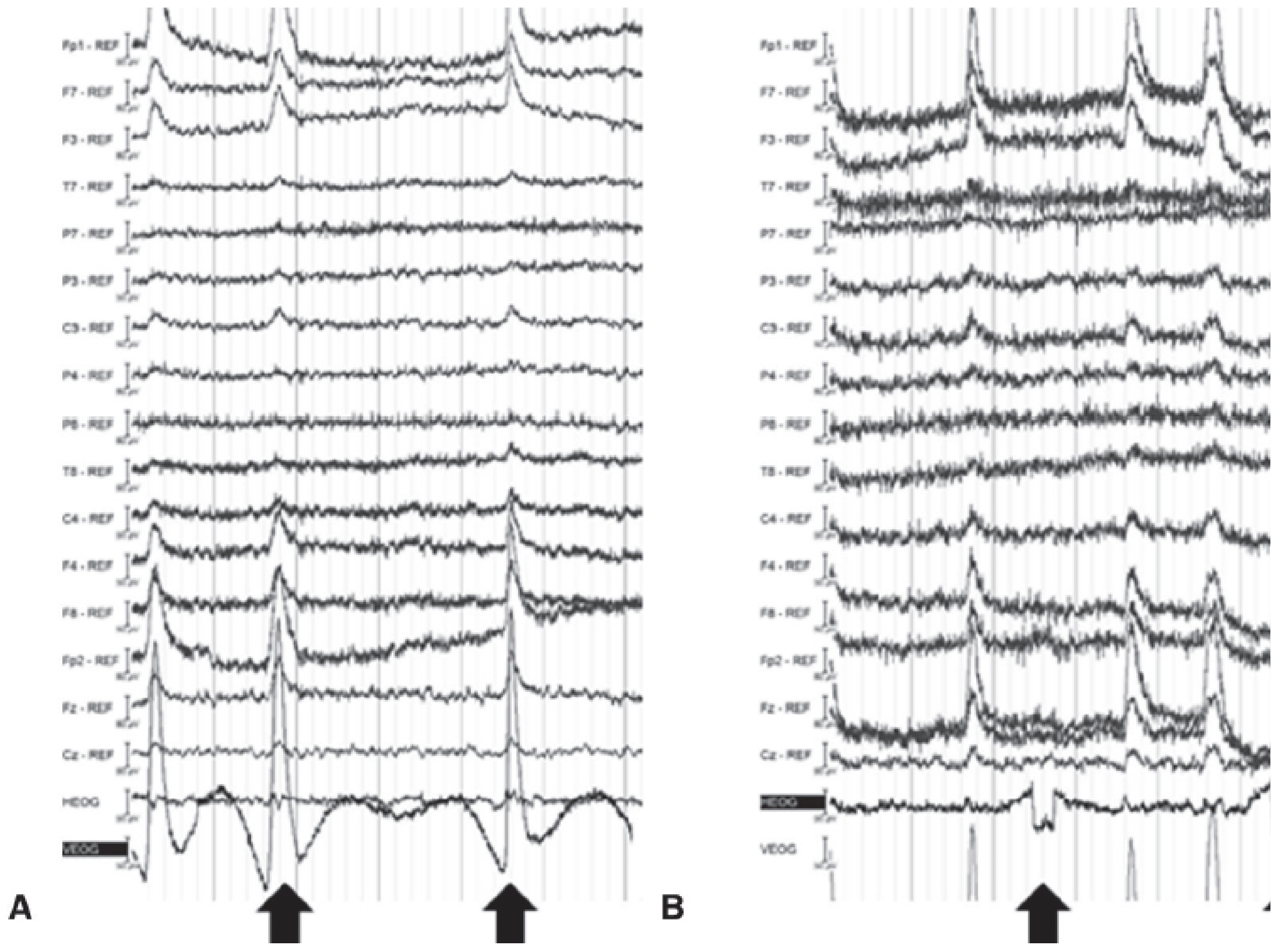
Recognition clue
I. Horizontal eye movement artifact
- F7 and F8 are often involved
- Left and right frontal electrodes can show opposite polarity
- The pattern may mimic a frontal phase reversal
- Video
- EOG channels
- Timing with gaze changes
- Symmetry/opposite polarity at lateral frontal sites
J. Muscle artifact, EMG
- Jaw clenching
- Chewing
- Talking
- Swallowing
- Frowning
- Forehead tension
- Shivering
- Fast activity
- Irregular and often chaotic morphology
- Often most prominent in frontal and temporal electrodes
- Can obscure true spikes or fast cerebral activity
- Usually improves when the patient relaxes
Practical response
- Relax the jaw
- Stop talking or chewing
- Loosen facial muscles
- Avoid reading or squinting
- Remain still
K. ECG artifact
- Repetitive sharp waveform
- Occurs at the heart rate
- Often more visible in inferior temporal, ear, or mastoid-related electrodes
- Time-locks to the ECG channel
Recognition rule
If a sharp transient recurs exactly with every QRS complex, it is ECG artifact unless there is strong evidence otherwise.
L. Sweat artifact
- Very slow baseline drift
- Often frontal
- Can mimic slow-wave activity
- Usually irregular and not a stable cerebral field
- Cool the patient if appropriate
- Check skin and electrode contact
- Allow time for sweating to reduce
- Use technical adjustments carefully
M. Electrode pop
- Abrupt, high-amplitude transient
- Often isolated to one electrode
- May have a vertical edge or “pop”
- No physiologic field
- Can recur irregularly
Recognition strategy
Fp1-F7     abrupt transient
F7-T7      abrupt transient
F7-REF     abrupt transient
Other channels: no corresponding event
N. Movement artifact
- Large irregular waveforms
- Widespread disturbance
- Electrode displacement
- Cable movement artifact
- Poor baseline stability
O. 50/60-Hz electrical interference
- Highly regular fast oscillation
- Often widespread
- Can be limited to one electrode if its impedance is poor
- May worsen with electrical equipment nearby
- Check electrode impedances and contact.
- Check cables and nearby electrical equipment.
- Inspect the unfiltered signal.
- Use a notch filter only when necessary and document its use.
P. Artifact-versus-brain checklist
- Does it have a believable field?
- Is it present in neighboring electrodes in a logical distribution?
- Does it persist in a different montage?
- Is it time-locked to eye movement, ECG, movement, or video?
- Does it occur in a single electrode only?
- Does it occur at 50/60 Hz or heart rate?
- Does the pattern change when the patient relaxes or moves?
- Could it be a normal sleep feature or normal variant?
High-yield revision table
| Topic | Remember |
|---|---|
| 10-20 system | Odd = left, even = right, z = midline |
| Channel | Difference in voltage between two inputs |
| Montage | Display arrangement, not a different recording |
| Bipolar montage | Best for local field and phase reversal |
| Referential montage | Useful for amplitude maxima and generalized activity |
| Phase reversal | Indicates local scalp voltage maximum, not exact seizure onset |
| PDR | Posterior, 8-13 Hz, eyes closed, attenuates with eye opening |
| Mu rhythm | Central, arch-shaped, blocks with contralateral hand movement |
| N2 sleep | Spindles and K-complexes |
| N3 sleep | High-amplitude delta is normal |
| Blink artifact | Frontal, especially Fp1/Fp2 |
| EMG artifact | Fast, irregular, frontal/temporal, improves with relaxation |
| ECG artifact | Time-locked to QRS |
| Electrode pop | Abrupt, one-electrode phenomenon, no proper field |
| Line noise | Regular 50/60-Hz activity |
Find recent guidelines for routine EEG recording
site:acns.org clinical EEG recording guideline routine EEG 2024 2025 2026 ACNS
IFCN routine EEG recording guideline standards 2023 2024 clinical EEG
electroencephalography AND practice guideline
PMID: 36762397
PMID: 28436800
PMID: 28436788
PMID: 28436792
https://acns.org/practice/guidelines
Most relevant recent guideline for routine EEG recording
Essential recommendations to learn
| Topic | Practical recommendation |
|---|---|
| Electrode placement | Use standardized electrode placement. The 10-20 system remains foundational; the IFCN standardized array supports 25 electrodes including inferior temporal electrodes for standard clinical recordings when feasible. |
| Recording duration | Record enough artifact-free EEG to assess the background and capture state changes. A longer routine recording improves detection of epileptiform abnormalities. |
| Sleep | Record sleep whenever feasible because it raises the yield of interictal epileptiform discharges. Sleep deprivation is generally preferred over sedation for sleep induction when appropriate. |
| Video | Use synchronized video whenever possible, especially when events, seizures, or unusual movements are a concern. |
| ECG channel | Include at least one ECG channel to identify cardiac artifact and to help correlate events. |
| Hyperventilation | Perform unless contraindicated. It is particularly useful for provoking generalized spike-wave discharges in absence epilepsy. Document effort and any reason for omission. |
| Intermittent photic stimulation | Perform according to a standardized frequency sequence unless contraindicated. Document photic driving, photoparoxysmal response, symptoms, and any clinical event. |
| Montages | Review EEG in more than one montage, particularly bipolar and referential reconstructions. Use additional electrodes or modified montages when temporal, anterior temporal, or mesial frontal localization is clinically important. |
| Technical quality | Record digitally, maintain good electrode contact, identify artifacts during acquisition, and avoid excessive filtering that can distort real cerebral signals. |
| Documentation | Record patient state, medications, activation procedures, sleep achieved, technical limitations, clinical events, and video correlation. |
Supporting standards still commonly used
-
ACNS Guideline 1: Minimum Technical Requirements for Performing Clinical EEG
This remains a main technical reference for routine scalp EEG. It supports:- Full standard electrode coverage
- Digital recording
- At least 20 minutes of artifact-free EEG, including activation procedures, as a minimum baseline
- Routine display sensitivity around 5-10 µV/mm
- Hyperventilation for at least 3 minutes, with recording before and after, when safe
- Inclusion of a single ECG channel
ACNS Guideline 1 . 2016 . PMID: 28436800
-
ACNS Guideline 3: Standard Montages in Clinical EEG
Useful for choosing bipolar and referential montages and for additional anterior temporal or mesial frontal coverage. ACNS Guideline 3 . 2016 . PMID: 28436788 -
ACNS Guideline 7: EEG Reporting
Recommends a structured routine EEG report with five sections:- History
- Technical description
- EEG description
- Impression
- Clinical correlation
Document activation procedures and state changes, and explain why hyperventilation or photic stimulation was omitted. ACNS Guideline 7 . 2016 . PMID: 28436792
Practical routine EEG checklist
- Patient identity, age, indication, antiseizure/sedating medicines, and last clinical event
- Awake, drowsy, and sleep states achieved
- Standardized 10-20 electrode placement, with extra electrodes where indicated
- ECG and, ideally, synchronized video
- Bipolar and referential montage review
- Background, posterior dominant rhythm, symmetry, continuity, and reactivity
- Hyperventilation and photic stimulation, or a documented reason not to perform them
- Artifact burden and technical limitations
- Interictal epileptiform discharges, seizures, and clinical correlation
- A concise standardized final impression
Compare IFCN-ILAE and ACNS EEG standards
https://pmc.ncbi.nlm.nih.gov/articles/PMC10006292/
PMID: 36762397
electroencephalography AND practice guideline
IFCN ILAE Routine and sleep EEG minimum recording standards 2023 recommendations routine duration electrodes sleep hyperventilation photic
IFCN-ILAE vs ACNS standards for routine EEG
Bottom line
| Feature | IFCN-ILAE | ACNS |
|---|---|---|
| Main routine EEG standard | 2023 routine and sleep EEG minimum recording standard | Core routine EEG series mainly 2016 |
| Organizations | International Federation of Clinical Neurophysiology + International League Against Epilepsy | American Clinical Neurophysiology Society |
| Geographic orientation | International, intended for broad global applicability | Primarily U.S. clinical practice |
| Scope | Indications, technical recording, routine and sleep duration, sleep induction, activation procedures | Technical requirements, electrode nomenclature, montage design, digital recording, pediatric EEG, reporting |
| Methodology | Systematic evidence review, GRADE, QUADAS-2, and modified Delphi consensus | Society guideline and expert consensus updates |
| Routine EEG duration | Suggests 20 minutes, excluding preparation, conditional recommendation | Minimum 20 minutes of artifact-free recording, including activation procedures |
| Sleep EEG duration | Suggests 30 minutes, excluding preparation | Sleep should be recorded whenever possible; no equivalent universal 30-minute sleep EEG specification in Guideline 1 |
| Electrode array | Suggests the 25-electrode IFCN array, including six inferior temporal electrodes, when feasible | Standard 21-electrode 10-20 system is required for routine clinical EEG; additional electrodes as clinically needed |
| Activation | Hyperventilation, photic stimulation, eye opening/closure suggested unless contraindicated | Hyperventilation and photic stimulation are routine unless contraindicated; gives more procedure-specific operational detail |
| Reporting | Focuses chiefly on recording standards | Has a dedicated structured reporting guideline |
| Video | Supports video where available and useful | Time-locked video is incorporated into modern routine EEG reporting practice but is not universally mandatory in the core technical guideline |
1. Scope and purpose
IFCN-ILAE 2023
- Indications
- Technical recording conditions
- Electrode application
- Recording duration
- Sleep induction
- Hyperventilation
- Intermittent photic stimulation
- Eye opening and eye closure
ACNS 2016 series
- Guideline 1: minimum technical requirements for clinical EEG
- Guideline 2: electrode nomenclature
- Guideline 3: standard montages
- Guideline 4: digital EEG recording
- Guideline 5: pediatric EEG
- Guideline 7: EEG reporting
2. Recording duration
| Standard | Routine EEG | Sleep EEG | Interpretation |
|---|---|---|---|
| IFCN-ILAE 2023 | 20 minutes, excluding preparation | 30 minutes, excluding preparation | Conditional recommendation due to limited evidence |
| ACNS 2016 | At least 20 minutes of artifact-free EEG, including activation procedures | Sleep whenever possible, especially in suspected/known seizure disorders | Longer studies increase yield |
Important practical difference
- IFCN-ILAE explicitly separates routine EEG from sleep EEG and specifies 30 minutes for sleep EEG.
- ACNS emphasizes that the recorded sample must be artifact-free, and activation procedures plus sleep often make a longer study necessary.
Obtain at least 20 minutes of artifact-free routine EEG, and extend the study to obtain drowsiness/sleep and complete activation procedures when clinically indicated.
3. Electrode number and placement
IFCN-ILAE
ACNS
- Anterior temporal/subtemporal electrodes
- Sphenoidal or other special temporal electrodes, depending on local practice
- Extra electrodes near a skull defect
- Expanded 10-10 coverage
Practical comparison
| Question | IFCN-ILAE approach | ACNS approach |
|---|---|---|
| Minimum routine array | 25-electrode array suggested when feasible | 21-electrode 10-20 array required for a standard routine study |
| Temporal coverage | Stronger emphasis on routine inferior temporal coverage | Add electrodes when localization is clinically important |
| Naming | 10-10 nomenclature integrated into the recommended array | 10-20 standard, with 10-10 extension accepted |
| Resource sensitivity | Explicitly recognizes feasibility and resource limitations | More prescriptive about comprehensive standard coverage |
4. Technical recording standards
Areas of agreement
- Digital EEG recording
- Standardized electrode placement
- Good electrode contact and artifact control
- Review in more than one montage
- Recording wakefulness and sleep when possible
- ECG recording
- Careful documentation of technical limitations
- Cautious and selective filtering
ACNS is more operational
- Typical routine display sensitivity: 5-10 µV/mm
- At least one ECG channel
- A review of an unfiltered system-reference segment is recommended before relying on a notch filter
- Explicit technical considerations for calibration, filters, and montage changes
IFCN-ILAE is more technology-aware and internationally framed
- Cup electrodes and head caps
- Need to verify impedance with caps
- Current limitations of dry-electrode EEG, including movement and sweat artifact
- MRI-compatible and needle electrode use in selected settings
- Preference for enhanced temporal coverage when feasible
5. Sleep recording and sleep induction
Shared principle
IFCN-ILAE 2023
- Routine versus sleep EEG duration
- Methods of sleep induction
- Sleep deprivation
- Balancing diagnostic benefit against patient practicality and safety
ACNS 2016
6. Hyperventilation
Agreement
ACNS: more detailed operational protocol
- At least 3 minutes of hyperventilation
- At least 1 minute of pre-hyperventilation EEG using the same montage
- At least 1 minute of EEG after hyperventilation
- Documentation of the quality of patient effort
- Recording the reason if hyperventilation is omitted
IFCN-ILAE: broader contraindication list and conditional recommendation
- Sickle cell disease or trait
- Moyamoya disease or syndrome
- Recent cerebrovascular event
- Vascular malformations/aneurysm
- Raised intracranial pressure
- Recent myocardial infarction
- Significant arrhythmia or severe cardiac disease
- Severe pulmonary disease
- Pregnancy
7. Intermittent photic stimulation
Areas of agreement
- Photic stimulation as a routine activation procedure unless contraindicated
- Documentation of the procedure and response
- Recording whether photic driving occurs
- Documentation of photoparoxysmal response, symptoms, myoclonus, or induced seizure
- Explicit documentation if omitted
Difference in emphasis
- ACNS gives practical environment and sequence details, such as dim lighting and performing photic stimulation before hyperventilation or after hyperventilation-related EEG changes have resolved.
- IFCN-ILAE emphasizes standardized activation protocols, eye-open/eye-closed baseline recording, and tailoring stimulation to clinical questions and patient safety.
8. Montages
ACNS
IFCN-ILAE
Practical conclusion
- Use IFCN-ILAE to decide the minimum electrode coverage and routine recording elements.
- Use ACNS to build the local montage library and display-review workflow.
9. Reporting
- History
- Technical description
- EEG description
- Impression
- Clinical correlation
- Number and placement of electrodes
- Video and ECG channels
- Montages and post hoc review settings
- Recording duration
- Wakefulness, drowsiness, and sleep
- Hyperventilation and photic stimulation
- Reason for omitting activation procedures
- Technical limitations and relevant artifacts
Recommended combined protocol
-
Primary recording standard: IFCN-ILAE 2023
Use it for minimum routine/sleep recording duration, modern electrode considerations, sleep strategy, and activation procedures. -
Baseline electrode coverage:
Use a complete 21-electrode 10-20 array at minimum. Add the IFCN inferior temporal electrodes, creating the 25-electrode array, when feasible, particularly for epilepsy evaluation. -
Technical acquisition: ACNS Guideline 1
Use it for digital acquisition, sensitivity, filters, ECG, calibration, artifact management, activation procedure details, and minimum artifact-free recording. -
Montage review: ACNS Guideline 3
Use bipolar and referential reconstructions. Include additional temporal or targeted electrodes when the clinical question requires improved localization. -
Structured report: ACNS Guideline 7
Use the five-part report format and document state, technical parameters, activation, artifacts, findings, and clinical correlation.
Exam-style summary
IFCN-ILAE 2023 is the newer international standard for minimum routine and sleep EEG recording. It recommends a 20-minute routine EEG, a 30-minute sleep EEG, activation procedures unless contraindicated, and a 25-electrode array with inferior temporal electrodes when feasible.ACNS 2016 is more detailed for day-to-day U.S. laboratory practice: it specifies at least 20 minutes of artifact-free recording, full 21-electrode 10-20 coverage, ECG, technical settings, detailed hyperventilation procedures, montage guidance, and standardized reporting.