
Para spinal muscles
Reading File
The paraspinal (erector spinae) muscles are the main extensors of the vertebral column, running along either side of the spine. They include three columns - iliocostalis (lateral), longissimus (intermediate), and spinalis (medial) - plus deeper muscles like the multifidus and semispinalis.
Searching sources...
Finding Sources
Finding Sources
Finding Sources
Finding Sources
Good, found relevant sources. Let me read the most detailed anatomical section from Fischer's Mastery of Surgery.
Reading File
Reading File
Reading File
Finding Sources
Finding Sources
Finding Sources
Searching the Web
paraspinal muscles anatomy origin insertion innervation function
Reading File
There's an anatomical illustration at line 1579. Let me fetch that image.
Loading Image
Now I have comprehensive information to provide a thorough answer on paraspinal muscles.
Paraspinal Muscles
The paraspinal muscles (also called intrinsic or deep back muscles) are the group of muscles running on either side of the vertebral column, enclosed within the thoracolumbar fascia. They are the primary extensors and stabilizers of the spine.
Layers of Back Musculature
The muscular support of the spine is organized into three layers (- Textbook of Family Medicine 9e):
| Layer | Type | Muscles |
|---|---|---|
| Superficial | Long, polysegmental | Erector spinae (chief extensors) |
| Middle | Short, polysegmental | Multifidus |
| Deep | Small, intersegmental | Rotatores, interspinales, intertransversarii |
1. Erector Spinae (Superficial Paraspinal Group)
The erector spinae is the largest paraspinal muscle group. It is divisible into three columns, from lateral to medial (- Fischer's Mastery of Surgery 8e):
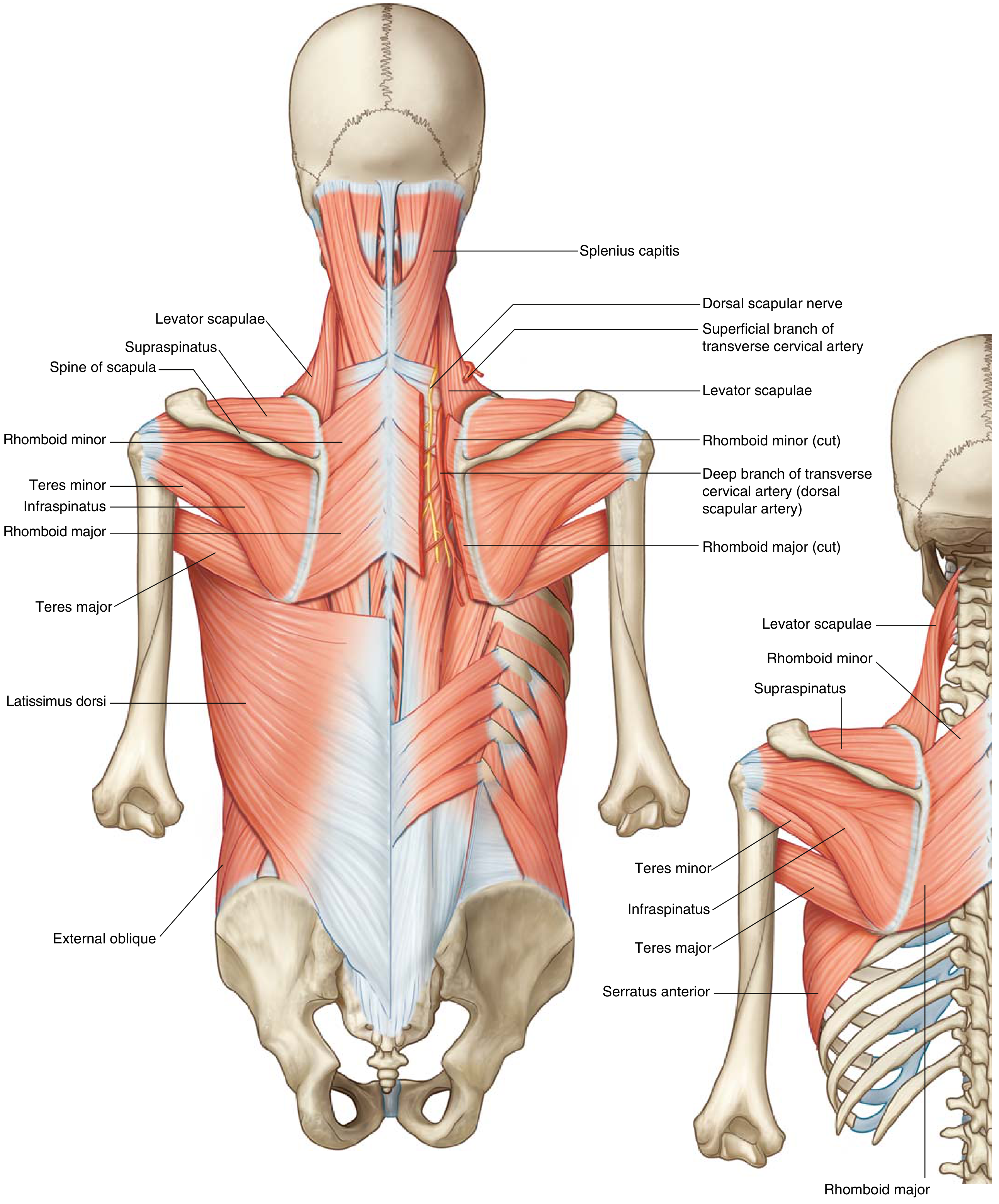
A. Iliocostalis (Lateral Column)
- Most lateral of the three erector spinae columns
- Three parts: iliocostalis lumborum, thoracis, cervicis
- Origin: Iliac crest, sacrum, lower ribs
- Insertion: Angles of ribs, transverse processes of cervical vertebrae
- Action: Extension, lateral flexion of vertebral column; assists respiration
B. Longissimus (Intermediate Column)
- Largest of the three columns; occupies the interval between spinalis and angles of lower ribs
- Three parts: longissimus thoracis, cervicis, capitis
- Origin: Transverse and articular processes of lumbar/thoracic/cervical vertebrae
- Insertion: Transverse processes of superior vertebrae, ribs (between tubercle and angle), mastoid process (capitis)
- Action: Extension and lateral flexion of spine; extends and rotates head (capitis)
C. Spinalis (Medial Column)
- Smallest of the three, adjacent to spinous processes
- Three parts: spinalis thoracis, cervicis, capitis
- Origin: Spinous processes of T11-L2 (thoracis); C7-T1 and nuchal ligament (cervicis)
- Insertion: Spinous processes of T2-T8 (thoracis); C2-C4 (cervicis); occipital bone (capitis)
- Action: Extension of spine; lateral flexion
2. Transversospinalis Group (Intermediate/Deep)
These muscles run obliquely from transverse processes (below) to spinous processes (above) - hence the name. Their main role is spinal stabilization.
A. Semispinalis
- Spans 4-6 vertebral levels
- Three parts: semispinalis capitis, cervicis, thoracis
- Origin: Transverse processes of T6-T10 (thoracis); T1-T6 (cervicis); articular processes C4-C7 + transverse processes T1-T6 (capitis)
- Insertion: Spinous processes C6-T4 (thoracis); spinous processes C2-C5 (cervicis); between nuchal lines of occipital bone (capitis)
- Innervation: Semispinalis capitis - greater occipital nerve (C2) and C3; others - posterior rami of adjacent spinal nerves
B. Multifidus
- Spans 2-4 vertebral levels
- Runs the full length of the spine, most prominent in the lumbar region
- Origin: Sacrum, posterior iliac spine, mammillary processes of lumbar vertebrae, transverse processes of thoracic vertebrae, articular processes of C4-C7
- Insertion: Spinous processes 2-4 levels above
- Function: Primary stabilizer of individual vertebral segments; especially important for lumbar spine stability
- Innervation: Medial branches of posterior rami of spinal nerves
C. Rotatores
- Shortest of the transversospinalis group; most developed in thoracic region
- Rotatores breves: Origin - transverse process; insertion - lamina/spinous process of vertebra 1 level above
- Rotatores longi: Origin - transverse process; insertion - 2 levels above
- Function: Bilateral = extension; unilateral = contralateral rotation
- Innervation: Medial branches of posterior rami of spinal nerves
3. Segmental (Deepest) Muscles
| Muscle | Location | Function |
|---|---|---|
| Interspinales | Between adjacent spinous processes | Fine extension control |
| Intertransversarii | Between adjacent transverse processes | Lateral flexion; stabilization |
Innervation - Key Principle
All deep back (paraspinal) muscles are innervated by the dorsal (posterior) primary rami of spinal nerves (- Miller's Review of Orthopaedics 9e). This segmental innervation is why paraspinal muscles can be used for electromyographic localization of nerve root lesions - each segmental level innervates the corresponding paraspinal muscle.
Blood Supply
- Dorsal branches of posterior intercostal arteries (thoracic region)
- Lumbar arteries (lumbar region)
- Lateral sacral arteries (sacral region)
Clinical Relevance
| Condition | Paraspinal Involvement |
|---|---|
| Lumbar strain | Erector spinae and multifidi most often strained; presents with painful paraspinal spasm (- Textbook of Family Medicine 9e) |
| Disc degeneration / LBP | Paraspinal atrophy (especially multifidus) is common and contributes to instability |
| Paraspinal EMG | Denervation in paraspinals localizes lesion to nerve root (proximal to dorsal ramus takeoff) |
| Erector spinae plane block | Regional anesthesia targeting the erector spinae fascia at the transverse process for chest/abdominal surgery (- Barash Clinical Anesthesia 9e) |
| Posterior spinal surgery | Subperiosteal dissection of paraspinal musculature off spinous processes is the standard approach (- Miller's Review of Orthopaedics 9e) |
| Paraspinal muscle atrophy on MRI | Loss of cross-sectional area, especially multifidus, is strongly associated with chronic low back pain and intervertebral disc degeneration |
Summary
The paraspinal muscles form a hierarchical system:
- Superficial (erector spinae): Power generators for spinal extension and lateral flexion
- Intermediate (transversospinalis - semispinalis, multifidus): Span multiple segments; control inter-segmental motion
- Deep (rotatores, interspinales, intertransversarii): Fine segmental stabilizers
All are innervated by dorsal rami of spinal nerves. The multifidus is the most studied clinically due to its role in lumbar stability and its atrophy in chronic low back pain.
This is a shared conversation. Sign in to Orris to start your own chat.