
Layers of skin
layers of skin epidermis dermis hypodermis diagram cross section
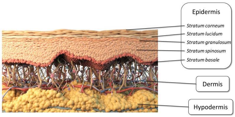
Anatomical diagram providing a detailed cross-sectional schematic of human skin layers, categorized into three primary regions: epidermis, dermis, and hypodermis. The epidermis is further subdivided into its five constituent layers, labeled from superficial to deep: stratum corneum (outermost barrier), stratum lucidum, stratum granulosum, stratum spinosum, and stratum basale (innermost germinative layer). The dermis is depicted with a complex network of connective tissue fibers, blood vessels (arterioles and venules), and neural elements. The hypodermis (subcutaneous layer) is shown at the base, characterized by yellow globular clusters representing adipose tissue. This illustration serves as an educational resource for understanding skin histology and is relevant to dermatology, pharmaceutics (specifically transdermal drug delivery), and clinical anatomy. The visual highlights the structural complexity of the skin barrier, particularly the dense stratum corneum which serves as the primary rate-limiting step for topical absorption.
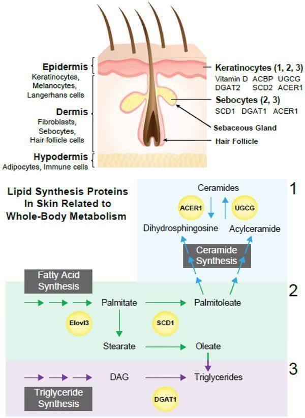
This medical illustration depicts the histological structure of human skin alongside metabolic pathways for lipid synthesis. The top section shows a cross-sectional anatomical diagram identifying three layers: the epidermis (containing keratinocytes, melanocytes, and Langerhans cells), the dermis (containing fibroblasts, sebocytes, and hair follicles), and the hypodermis (containing adipocytes and immune cells). The diagram maps specific enzymes and proteins to these cell types, such as Vitamin D, ACBP, UGCG, DGAT2, SCD2, and ACER1 in keratinocytes, and SCD1, DGAT1, and ACER1 in sebocytes. The bottom section outlines three distinct lipid synthesis pathways: (1) Ceramide synthesis involving ACER1 and UGCG enzymes; (2) Fatty acid synthesis showing the conversion of Palmitate and Stearate mediated by Elovl3 and SCD1; and (3) Triglyceride synthesis involving the conversion of Diacylglycerol (DAG) to triglycerides via DGAT1. This resource illustrates the integration of skin biology with whole-body lipid metabolism and dermatological physiology.
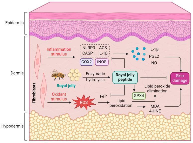
This medical schematic diagram illustrates the pathophysiology of skin damage and the protective mechanism of Royal Jelly Peptide (RJP) within a cross-section of human skin. The visual is divided into three primary layers: the stratified epidermis, the fibroblast-rich dermis, and the adipose-dense hypodermis. The central dermal region depicts two main pathways of injury. First, an inflammatory pathway triggered by stimulus leads to the activation of the NLRP3 inflammasome complex (including ASC and CASP1) and inflammatory enzymes COX2 and iNOS, resulting in the release of IL-1β, PGE2, and NO. Second, an oxidative stress pathway shows oxidant stimuli generating Reactive Oxygen Species (ROS), which, in the presence of Fe2+, drives lipid peroxidation to produce MDA and 4-HNE. Both pathways culminate in skin damage. The diagram highlights the therapeutic role of Royal Jelly Peptide, derived from enzymatic hydrolysis of royal jelly, which acts by inhibiting the NLRP3 complex and iron-dependent lipid peroxidation, while simultaneously promoting GPX4 activity for lipid peroxide elimination. This illustrates RJP's potential in dermatological health by modulating inflammation and oxidative homeostasis.
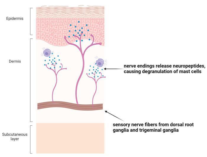
An anatomical diagram illustrating neuro-immune crosstalk within the skin layers, created for medical education. The cross-section depicts three primary layers: the epidermis (top, showing stratified keratinocytes), the dermis (middle, containing connective tissue and specialized cells), and the subcutaneous layer (bottom). The illustration highlights the pathway of sensory nerve fibers originating from the dorsal root ganglia and trigeminal ganglia. These branched nerve fibers extend through the dermis toward the dermo-epidermal junction. The diagram specifically demonstrates the release of neuropeptides (represented by small blue granules) from terminal nerve endings. This release is shown interacting with dermal mast cells, triggering their degranulation. This visual focuses on the signaling pathway between the nervous and immune systems, explaining how neural stimuli can amplify inflammatory responses and contribute to clinical symptoms such as pruritus (itching) in dermatological conditions.
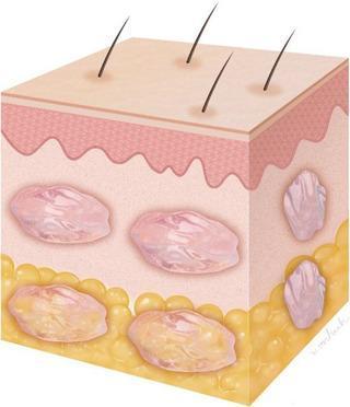
This anatomical diagram provides a 3D schematic representation of a skin biopsy-style section, illustrating a multi-layer injection technique for cosmetic or regenerative medicine. The diagram depicts three distinct integumentary layers: the epidermis, shown as a thin, undulating top layer with emerging terminal hairs; the dermis, a thicker mid-layer; and the subcutaneous layer (hypodermis) at the base, characterized by yellow globular adipose tissue. The focus of the illustration is the targeted administration of a therapeutic substance, such as polynucleotide (PN), which appears as pink boluses. The diagram demonstrates a dual-depth injection strategy: intradermal boluses located within the deep dermis and subdermal boluses positioned at the junction between the dermis and the superficial subcutaneous layer. This visual is designed to explain the clinical procedure for addressing skin depressions or volumetric loss through layered filler or biostimulator delivery. It serves as an educational tool for dermatology and aesthetic surgery to visualize precise anatomical placement of injectable substances.
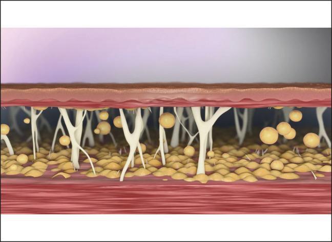
This medical illustration provides a cross-sectional view of the human integumentary system, focusing on the anatomy of the subcutaneous layer following a liposuction procedure. The diagram depicts the epidermis and dermis as thin, stratified upper layers. Beneath the dermis lies the hypodermis (subcutaneous tissue), characterized by prominent fibrous septae—white, branching connective tissue strands that tether the dermis to the underlying deep fascia and muscle tissue (shown at the bottom in red). Yellowish, spherical lobules representing adipocytes (fat cells) are interspersed within this structural framework. The image highlights a post-liposuction state where a significant portion of the fat volume has been removed, leaving behind the vertical and oblique connective tissue network. This visual emphasizes the relationship between the skin, the scaffolding of the subcutaneous connective tissue, and the residual fat, illustrating why additional treatments may be required to address tissue laxity when these supporting strands do not spontaneously contract after fat aspiration. The content is designed for intermediate to advanced learners in plastic surgery or dermatology to understand the structural mechanics of soft tissue contraction.
Layers of Skin
1. Epidermis (outermost layer)
Four major cell populations (Dermatology 5e):
- Keratinocytes - the dominant cell type; migrate upward from the basal layer and differentiate into the protective stratum corneum
- Melanocytes - produce melanin pigment; protect against UV radiation by transferring melanosomes to nearby keratinocytes (positioned above the nucleus)
- Langerhans cells - antigen-presenting cells; metabolize complex antigens and migrate to regional lymph nodes to regulate immunity
- Merkel cells - found in the basal layer; contain neuroendocrine peptides; thought to play a role in mechanoreceptor responses
The 5 sublayers of the epidermis (deep to superficial):
| Layer | Key Features |
|---|---|
| Stratum basale | Deepest, single cell layer; mitotically active stem cells; contains melanin pigment; expresses KRT5/KRT14 keratins |
| Stratum spinosum | Several cells thick; cells have spinous processes (intercellular bridges via desmosomes); expresses KRT1/KRT10 |
| Stratum granulosum | Cells contain keratohyalin granules (rich in filaggrin); cells begin to lose their nuclei |
| Stratum lucidum | Present only in thick skin (palms and soles); a pale, translucent band between the granulosum and corneum |
| Stratum corneum | Outermost; anucleate, keratinized "dead" cells (corneocytes); forms the primary physical and chemical barrier; prevents fluid loss and microbial invasion |
Under normal conditions, keratinocytes take about 2 weeks to exit the nucleated compartment, and another 2 weeks to transit through the stratum corneum. - Dermatology 5e
2. Dermis (middle layer)
| Sublayer | Composition | Notes |
|---|---|---|
| Papillary dermis (superficial) | Thin, loose, fine non-bundled collagen; abundant elastic fibers; capillaries | Projects upward as dermal papillae into the epidermis; capillaries here supply the avascular epidermis |
| Reticular dermis (deep) | Thick, compact, tightly interwoven collagen bundles | The bulk of the dermis; damage to this layer results in permanent scarring |
3. Hypodermis / Subcutaneous Layer (deepest layer)
- Thermal insulation
- Mechanical cushioning
- Energy storage
- Anchoring of the skin to underlying fascia and muscle
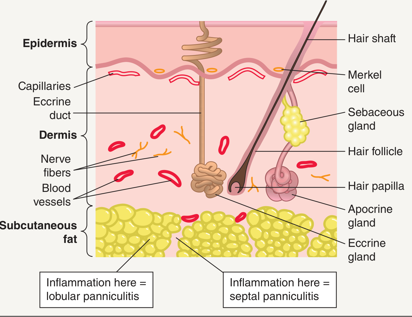
Summary Diagram
Disease of skin
Which area of skin diseases would you like to cover? · How detailed should the answer be?
common skin diseases overview dermatology rash lesions
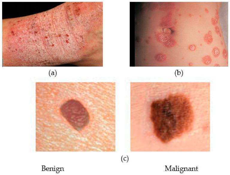
This composite clinical photograph illustrates common dermatological conditions for comparative medical education. Panel (a) shows atopic dermatitis (eczema) presenting as a diffuse, erythematous, and pruritic-appearing rash with visible excoriation, lichenification, and scaling on an extremity. Panel (b) depicts psoriasis, characterized by well-demarcated, erythematous papules and plaques with a silvery-white micaceous scale, distributed in a nummular (coin-shaped) pattern. Panel (c) provides a side-by-side comparison of melanocytic lesions: the 'Benign' lesion (likely a melanocytic nevus) exhibits a symmetrical oval shape, regular borders, and uniform reddish-brown pigmentation; the 'Malignant' lesion (malignant melanoma) demonstrates classic ABCDE warning signs, including asymmetry, irregular or notched borders, and variegated, dark brown-to-black pigmentation. The image serves as a visual guide for distinguishing between inflammatory skin diseases and neoplastic lesions, emphasizing morphology, border definition, and pigmentation patterns in clinical dermatology.
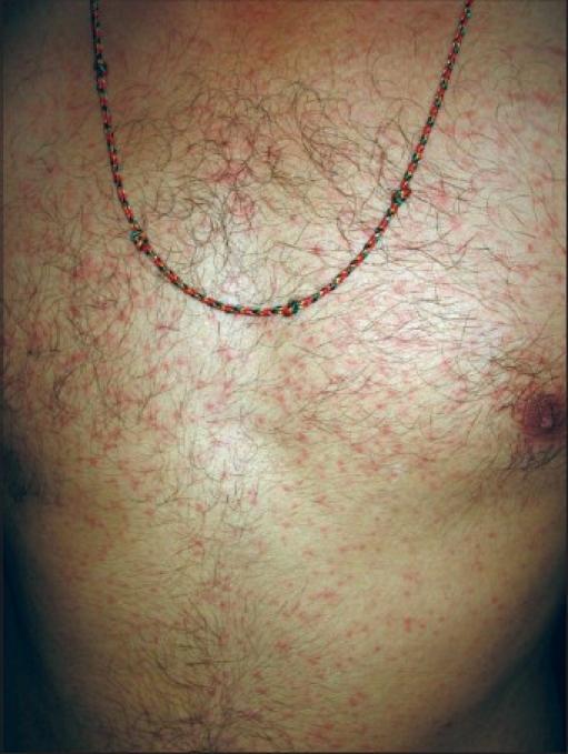
This clinical photograph shows the anterior trunk of an adult male patient demonstrating a generalized erythematous maculopapular exanthem. The skin lesions consist of numerous small, red macules and slightly elevated papules distributed symmetrically across the chest and abdomen. The rash is non-confluent in most areas, presenting as discrete spots against the background of normal skin. The anatomical region is notable for significant terminal hair growth and the presence of a decorative thread necklace. Clinically, this type of maculopapular rash is a common cutaneous finding in various systemic conditions, including viral exanthems, drug eruptions, or specific infectious diseases such as early-stage syphilis or Chikungunya fever, depending on the broader clinical context. The image serves as a teaching tool for identifying morphology and distribution patterns in dermatology and infectious disease specialties.
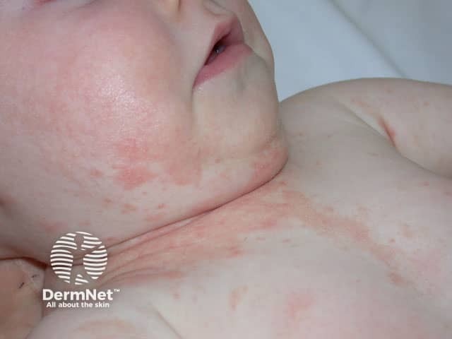
Clinical photograph of neonatal skin showing erythematous, patchy rash localized to the anterior neck folds and spread onto the upper chest. The image captures a close-up frontal view with high color fidelity, natural skin tone, and diffuse lighting. The lesions appear as pink to red, ill‑defined macules and patches without obvious vesicles, crusting, or purulent exudate. Skin elsewhere looks well hydrated, with mild transient erythema at the fold lines where creases may trap moisture. No fever or systemic signs are evident in the image, and there is no obvious edema. Pattern suggests involvement of flexural surfaces in infancy, a common presentation for inflammatory dermatitis. Differential diagnoses include irritant contact dermatitis from saliva, drool, or clothing friction; infantile atopic dermatitis (eczema) with predilection for cheeks, scalp, trunk, and neck folds; miliaria rubra (heat rash) in warm environments; seborrheic dermatitis with erythema; and less likely candidal intertrigo in skin folds. The image provides educational value for recognizing cutaneous inflammatory patterns in newborns and supports clinical correlation with history, timing, irritant exposure, and family atopy. Not a stand-alone diagnosis; should be integrated with physical examination and, if lesions persist or worsen, dermatology referral and follow-up. This description improves searchability for dermatology education resources.
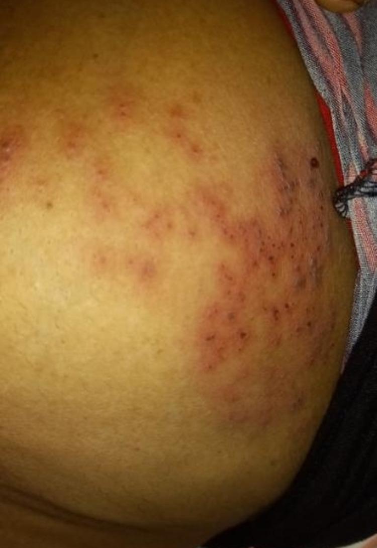
Clinical photograph of a patient's left chest wall demonstrating a localized, erythematous skin eruption. The lesions consist of multiple small, red papules and vesicles clustered in a dermatomal distribution corresponding to the T4 thoracic level. The rash is characterized by varying intensities of inflammation, ranging from light pink to dark red, set against a background of yellowish-brown skin. The distribution is unilateral and follows a clear neural pathway, which is highly suggestive of Herpes Zoster (shingles). This image serves as an educational example of dermatomal viral rashes and is relevant for medical training in infectious diseases and dermatology for identifying classic clinical presentations of varicella-zoster virus reactivation.
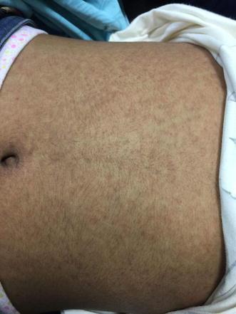
This clinical photograph displays an erythematous maculopapular rash on the abdomen of a patient. The eruption is characterized by a diffuse and symmetric distribution of numerous small, reddish, non-confluent lesions. Individual lesions are primarily macules and slight papules, mostly round to oval in shape and under 5mm in diameter. The rash covers a broad anatomical region, extending across the umbilical and epigastric areas without following a dermatomal or specific linear pattern. The background skin appears tanned, providing a visual contrast to the inflammatory erythematous spots. This morphology is common in systemic conditions such as viral exanthems, drug eruptions, or certain bacterial infections. The visual information serves as a classic educational example of a generalized maculopapular eruption, useful for clinical dermatology and infectious disease studies.
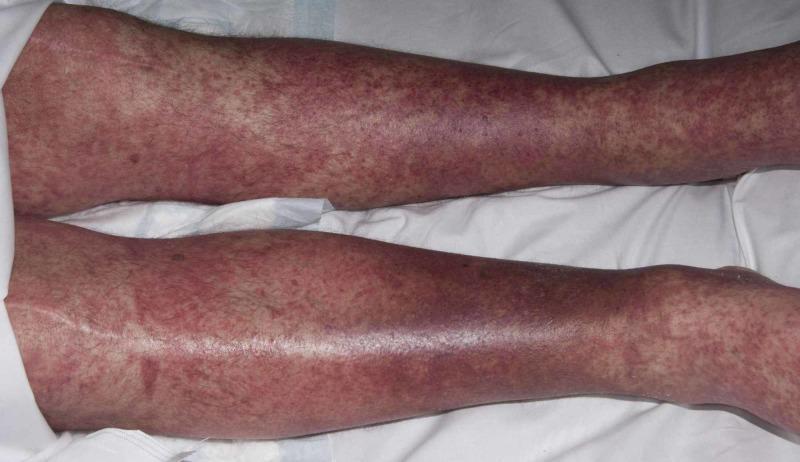
Clinical photograph of a patient's lower extremities demonstrating a widespread, symmetrical erythematous papular rash. The eruption is characterized by numerous small, reddish, raised lesions (papules) that are densely distributed across the anterior and lateral surfaces of both legs, extending from the thighs down to the ankles. In several regions, particularly over the shins and calves, the lesions show a tendency toward confluence, forming larger erythematous patches. The background skin exhibits generalized erythema without evidence of vesiculation, bullae, or overt necrosis. This morbilliform (measles-like) presentation is clinically significant in dermatology as a common manifestation of systemic drug eruptions, such as Drug Reaction with Eosinophilia and Systemic Symptoms (DRESS) syndrome. The image illustrates the classic cutaneous morphology of a hypersensitivity reaction, where the density and distribution of the maculopapular rash are key diagnostic features for evaluating adverse drug reactions and differentiating from other inflammatory or infectious dermatoses.