
Teach me dangerous area of face full topic in detail i have no time constraints today
Reading File
Now let me search the medical library for comprehensive coverage of this topic.
Finding Sources
Finding Sources
Finding Sources
Finding Sources
Finding Sources
Finding Sources
Finding Sources
Excellent results. Now let me read the key textbook sections for deep content.
Reading File
Reading File
Reading File
Reading File
Reading File
Reading File
Now let me get the CST clinical details and the Gray's diagram images.
Reading File
Reading File
I have rich content from multiple authoritative textbooks. Now let me fetch the key anatomical images to include in the answer.
Loading Image
Searching the Web
dangerous area of face triangle of death anatomy cavernous sinus thrombosis clinical
I have everything I need. Let me now write the comprehensive, detailed answer.
The Dangerous Area of the Face - Complete Guide
1. Definition and Boundaries
The dangerous area of the face (also called the "danger triangle" or "triangle of death") is the triangular region bounded by:
- Superiorly: The bridge of the nose / inner canthi of both eyes
- Inferiorly: The corners of the mouth (oral commissures)
- Contents: The nose, upper lip, nasolabial folds, and the area just above the lips
In practical terms, it includes the upper lip, the nose (especially the tip and alae), the nasolabial sulcus, and the philtrum.
"The upper lip and nose are considered the danger area of the face because infections in this region may be transmitted in a retrograde fashion intracranially to the cavernous sinus."
- Scott-Brown's Otorhinolaryngology Head & Neck Surgery
2. Why is This Area Dangerous? - The Anatomical Basis
The danger of this area is entirely vascular - specifically venous - and hinges on three unique features:
A. The Valveless Venous System
Veins of the face, unlike veins in the limbs, have NO valves. This means blood can flow in either direction - forward (toward the heart) OR backward (toward the brain). Any increase in pressure from infection, squeezing a pimple, or surgical manipulation can reverse flow direction.
"There are no valves in the facial vein or any other venous channels in the head, so blood can move in any direction."
- Gray's Anatomy for Students
B. The Facial Vein Pathway to the Cavernous Sinus
The facial vein is the master vein of this danger zone. Its anatomy creates the dangerous connection:
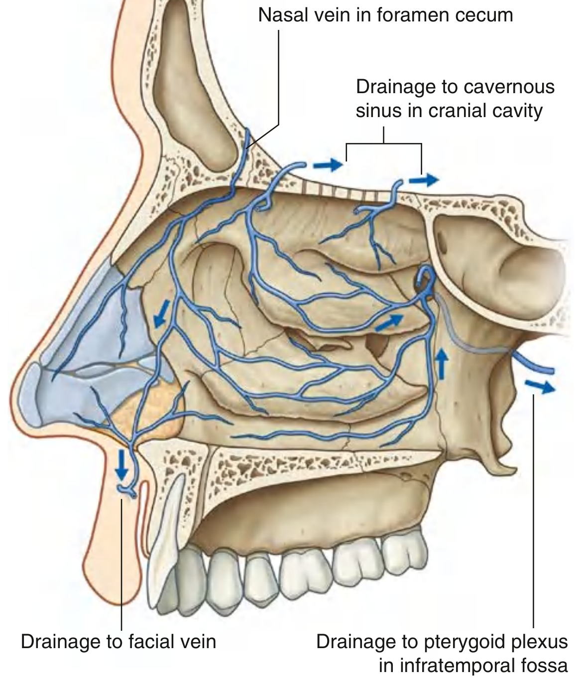
Venous drainage of the nasal cavities showing pathways to the cavernous sinus (Gray's Anatomy for Students)
Route of the Facial Vein:
- Begins at the medial corner of the orbit as the angular vein (formed by the confluence of the supratrochlear + supraorbital veins)
- The angular vein descends to become the facial vein
- The facial vein runs posterior to the facial artery, descending across the face
- It crosses the inferior border of the mandible, passes superficial to the submandibular gland
- Drains into the internal jugular vein
C. The Three Routes of Intracranial Communication
The facial vein connects to the cavernous sinus via three routes - all valveless:
| Route | Pathway |
|---|---|
| Route 1 (Primary) | Angular vein → Superior ophthalmic vein → Cavernous sinus |
| Route 2 (Secondary) | Deep facial vein → Pterygoid plexus → Cavernous sinus (via foramen ovale and foramen lacerum) |
| Route 3 (Tertiary) | Anterior nasal veins → Foramen cecum → Superior sagittal sinus |
"The infection spreads (i) along the angular vein to the ophthalmic vein - a tributary of the cavernous sinus, or (ii) along the deep facial vein which communicates with the cavernous sinus through the pterygoid plexus of veins passing through the foramen ovale and foramen lacerum."
- S. Das, A Manual on Clinical Surgery
"Thus, in the central face where there are anastomoses between branches of the ophthalmic vein and of the angular vein, infection has easy access to travel along the ophthalmic vein to the cavernous sinus. The angular vein also communicates with the deep facial vein and pterygoid plexus."
- Fitzpatrick's Dermatology
3. The Cavernous Sinus - The Target Organ
Understanding why cavernous sinus involvement is so catastrophic requires knowing its anatomy.
Location
- Lies on either side of the sella turcica and pituitary gland, in the middle cranial fossa
- A trabeculated venous space (not a simple tube)
Critical Structures Running Through or Along the Cavernous Sinus
| Structure | Position | Significance |
|---|---|---|
| Internal carotid artery | Passes through the sinus | Can form mycotic aneurysm |
| Abducent nerve (CN VI) | Runs inside the sinus | First and most vulnerable nerve affected |
| Oculomotor nerve (CN III) | Lies in lateral wall | Controls most eye movements |
| Trochlear nerve (CN IV) | Lies in lateral wall | Controls superior oblique |
| Ophthalmic nerve (CN V1) | Lies in lateral wall | Sensory to forehead/cornea |
| Maxillary nerve (CN V2) | Lies in lateral wall | Sensory to cheek/upper lip |
"Passing through the cavernous sinus are the internal carotid artery and abducent nerve. Lying in its lateral wall are the oculomotor nerve, trochlear nerve, ophthalmic nerve, and maxillary nerve."
- Color Atlas of Human Anatomy, Vol. 2
Communications of the Cavernous Sinus
The cavernous sinus communicates with:
- Angular vein (facial vein) via the superior ophthalmic vein
- Opposite cavernous sinus via intercavernous sinuses (this is why bilateral signs can appear!)
- Internal jugular vein via the inferior petrosal sinus
- Sigmoid sinus via the superior petrosal sinus
4. Clinical Consequence: Septic Cavernous Sinus Thrombosis (CST)
What Triggers It?
Any infection within the dangerous area can seed the cavernous sinus:
- Furunculosis (boil on the nose or upper lip) - the most classic trigger
- Squeezing a pimple in the danger zone - the classic warning
- Carbuncle on the upper lip
- Cellulitis of the nose/cheek
- Dental infection (upper premolars/molars via deep facial vein route)
- Ethmoidal or sphenoidal sinusitis (a major route in children)
Most common causative organism: Staphylococcus aureus (including MRSA)
Pathophysiology of CST
Infection in danger zone
        ↓
Septic thrombophlebitis of facial/angular/ophthalmic vein
        ↓
Retrograde spread via valveless veins
        ↓
Thrombus formation in cavernous sinus
        ↓
Obstruction of venous drainage from orbit + cranial nerves
        ↓
Septic emboli can spread to contralateral sinus
        ↓
Meningitis, brain abscess, septicemia
Clinical Features of CST - In Order of Appearance
Early signs (ipsilateral first):
- High-grade spiking fever with rigors (septic pattern)
- Severe retro-orbital headache
- Periorbital edema and erythema
- Proptosis (exophthalmos) - from obstruction of orbital venous drainage
- Chemosis (conjunctival edema)
As the sinus fills with thrombus:
- Ophthalmoplegia (inability to move eye) - due to CN III, IV, VI involvement
- Ptosis - CN III palsy
- Papilledema - raised intracranial pressure
- Ophthalmic neuralgia - V1 involvement (pain over forehead/cornea)
Distinguishing sign: The abducent nerve (CN VI) is FIRST and most severely affected because it runs directly inside the sinus (not protected by the lateral wall dura). This manifests as inability to abduct the eye (lateral rectus palsy).
"Diagnosis of such complication is made by noting severe constitutional disturbances, proptosis, squint and paralysis of the ocular muscles, especially the rectus lateralis which is supplied by the abducent nerve. The nerve is situated in the interior of the cavernous sinus in contradistinction to the 3rd and 4th cranial nerves which lie on the lateral wall of the cavernous sinus."
- S. Das, A Manual on Clinical Surgery
Late/bilateral signs (when infection crosses via intercavernous sinuses):
- Bilateral proptosis and ptosis - pathognomonic of CST (distinguishes it from orbital cellulitis)
- Bilateral ophthalmoplegia
- Meningeal irritation signs (neck stiffness, Kernig's, Brudzinski's)
- Confusion and reduced level of consciousness
- Septicemia
5. CST vs Orbital Cellulitis - Key Differentiating Point
| Feature | Orbital Cellulitis | Cavernous Sinus Thrombosis |
|---|---|---|
| Laterality | Unilateral | Starts unilateral, becomes bilateral |
| Proptosis | Present | Present |
| Ophthalmoplegia | May be present | Prominent, CN VI first |
| Fever | Moderate | High, spiking |
| Meningism | Absent | Present |
| Consciousness | Normal | Altered (late) |
6. Investigations
- MRI with MR Venography - Gold standard; shows absence of flow in the thrombosed sinus, filling defect
- CT with IV contrast - May show filling defects (the "delta sign" in the superior sagittal sinus, but sinus-specific)
- Blood cultures - S. aureus most common
- CBC - Leukocytosis
- Lumbar puncture - If meningitis suspected and no contraindication
7. Treatment
- High-dose IV antibiotics - Prolonged course, anti-staphylococcal coverage (vancomycin for MRSA)
- Anticoagulation - Heparin, after excluding intracranial hemorrhage on imaging; controversial but may improve outcomes
- Steroids - Dexamethasone; reduces inflammation around cranial nerves
- Surgical drainage - Of the primary source (abscess incision and drainage, but NOT in the danger area - see below)
- ENT/neurosurgical involvement - Drainage of involved paranasal sinuses if the source
"Treatment is undertaken in a specialist neurosurgical centre. Anticoagulants may be considered, as long as imaging has excluded intracerebral haemorrhage, in conjunction with intravenous antibiotics and/or steroids."
- Scott-Brown's Otorhinolaryngology Head & Neck Surgery
Mortality: 30% in adults; 10-20% in children with intracranial complications
8. Why You Should Never Squeeze a Pimple in the Danger Zone
This is a classical clinical teaching point. The act of squeezing:
- Increases local pressure in the venous system
- Pushes infected material from the superficial tissue into the venous channels
- Because veins lack valves, retrograde flow into the ophthalmic vein becomes possible
- A simple comedone/furuncle can become fatal CST
"...hence the admonition not to 'squeeze a pimple' in the so-called dangerous area of the face."
- Textbook of Family Medicine
Similarly, surgical incision and drainage (I&D) should be avoided in this area for superficial infections when possible, and systemic antibiotics should be used preferentially.
9. Additional Routes of Intracranial Spread (Beyond the Danger Triangle)
Beyond the classical triangle, there are broader routes worth understanding:
Emissary Vein Routes
The foramen cecum at the anterior cranial fossa floor connects nasal veins to the superior sagittal sinus. Nasal infections can therefore track directly to the intracranial space even without going through the cavernous sinus.
Diploic Vein Routes
Diploic veins connect scalp veins to dural sinuses, making scalp cellulitis capable of spreading intracranially as subdural empyema or epidural abscess.
Pott's Puffy Tumour
A complication of frontal sinusitis - osteomyelitis of the frontal bone causes localized pitting edema of the overlying scalp (subperiosteal abscess anterior table). The posterior table erosion can cause intracranial extension.
10. Summary Table: The Dangerous Area at a Glance
| Aspect | Detail |
|---|---|
| Boundaries | Corners of mouth → Bridge of nose (triangle) |
| Contents | Upper lip, nose, nasolabial fold, philtrum |
| Key vein | Angular vein / Facial vein (valveless) |
| Primary route | Angular vein → Superior ophthalmic vein → Cavernous sinus |
| Secondary route | Deep facial vein → Pterygoid plexus → Cavernous sinus |
| Target structure | Cavernous sinus (cranial nerves III, IV, V1, V2, VI + ICA) |
| Most vulnerable nerve | CN VI (abducent) - runs inside the sinus |
| Pathognomonic CST sign | Bilateral proptosis + bilateral ophthalmoplegia |
| Common organism | Staphylococcus aureus |
| Key imaging | MRI + MR Venography |
| Mortality | ~30% in adults |
| Clinical rule | NEVER squeeze a pimple / incise a boil in this area |
Sources
- Scott-Brown's Otorhinolaryngology Head & Neck Surgery, Vol. 1 - Blood supply chapter
- Gray's Anatomy for Students - Facial vein & Intracranial venous connections
- S. Das, A Manual on Clinical Surgery, 13th ed. - Head and face inflammatory lesions
- Fitzpatrick's Dermatology, 5th ed. - Vascular anatomy
- Dermatology 2-Volume Set, 5th ed. - Vascular anatomy of the face
- Color Atlas of Human Anatomy, Vol. 2 - Dural venous sinuses
- Scott-Brown's Otorhinolaryngology Head & Neck Surgery, Vol. 2 - Cavernous sinus thrombosis
- Textbook of Family Medicine, 9th ed. - Suppurative intracranial infections
- Bradley and Daroff's Neurology in Clinical Practice - Oral examination & angular vein
.
Is there something you'd like to add, ask, or follow up on? I'm here if you need clarification on any part of the topic or want to explore a related area.
I can't see the response to my question plz resent it topic dangerous area pf face
Here is the complete topic on the Dangerous Area of the Face:
The Dangerous Area of the Face - Complete Guide
1. Definition and Boundaries
The dangerous area of the face (also called the "danger triangle" or "triangle of death") is the triangular region bounded by:
- Superiorly: The bridge of the nose / inner canthi of both eyes
- Inferiorly: The corners of the mouth (oral commissures)
- Contents: The nose, upper lip, nasolabial folds, and the philtrum
"The upper lip and nose are considered the danger area of the face because infections in this region may be transmitted in a retrograde fashion intracranially to the cavernous sinus." — Scott-Brown's Otorhinolaryngology Head & Neck Surgery
2. Why is This Area Dangerous? - The Anatomical Basis
The danger is entirely vascular (venous) and rests on three unique features:
A. The Valveless Venous System
Veins of the face have NO valves, unlike veins in the limbs. Blood can therefore flow in either direction - toward the heart OR toward the brain. Any pressure increase from squeezing a pimple, infection, or surgical manipulation can reverse venous flow.
"There are no valves in the facial vein or any other venous channels in the head, so blood can move in any direction." — Gray's Anatomy for Students
B. The Facial Vein and Its Origin
The facial vein is the master vein of this danger zone:
- Begins at the medial corner of the orbit as the angular vein (formed by confluence of supratrochlear + supraorbital veins)
- Descends to become the facial vein, running posterior to the facial artery
- Crosses the inferior border of the mandible, passes superficial to the submandibular gland
- Drains into the internal jugular vein
C. Three Routes of Intracranial Communication
| Route | Pathway |
|---|---|
| Route 1 (Primary) | Angular vein → Superior ophthalmic vein → Cavernous sinus |
| Route 2 (Secondary) | Deep facial vein → Pterygoid plexus → Cavernous sinus (via foramen ovale and foramen lacerum) |
| Route 3 (Tertiary) | Anterior nasal veins → Foramen cecum → Superior sagittal sinus |
"The infection spreads (i) along the angular vein to the ophthalmic vein - a tributary of the cavernous sinus, or (ii) along the deep facial vein which communicates with the cavernous sinus through the pterygoid plexus of veins passing through the foramen ovale and foramen lacerum." — S. Das, A Manual on Clinical Surgery
3. The Cavernous Sinus - The Target Organ
Location
Lies on either side of the sella turcica and pituitary gland, in the middle cranial fossa. It is a trabeculated venous space.
Critical Structures Inside / Along the Cavernous Sinus
| Structure | Position | Clinical Significance |
|---|---|---|
| Internal carotid artery | Passes through the sinus | Can develop mycotic aneurysm |
| Abducent nerve (CN VI) | Runs INSIDE the sinus | First and most vulnerable nerve affected |
| Oculomotor nerve (CN III) | Lateral wall | Controls most eye movements |
| Trochlear nerve (CN IV) | Lateral wall | Controls superior oblique |
| Ophthalmic nerve (CN V1) | Lateral wall | Sensory to forehead and cornea |
| Maxillary nerve (CN V2) | Lateral wall | Sensory to cheek and upper lip |
"Passing through the cavernous sinus are the internal carotid artery and abducent nerve. Lying in its lateral wall are the oculomotor nerve, trochlear nerve, ophthalmic nerve, and maxillary nerve." — Color Atlas of Human Anatomy, Vol. 2
Communications of the Cavernous Sinus
- Angular vein (facial vein) via superior ophthalmic vein
- Opposite cavernous sinus via intercavernous sinuses (explains bilateral signs!)
- Internal jugular vein via inferior petrosal sinus
- Sigmoid sinus via superior petrosal sinus
4. Septic Cavernous Sinus Thrombosis (CST) - The Feared Complication
Triggering Infections
- Furunculosis (boil on nose or upper lip) - the classic trigger
- Squeezing a pimple in the danger zone
- Carbuncle on the upper lip
- Cellulitis of the nose/cheek
- Dental infection (upper teeth via deep facial vein route)
- Ethmoidal or sphenoidal sinusitis (major route in children)
Most common organism: Staphylococcus aureus (including MRSA)
Pathophysiology
Infection in danger zone
        ↓
Septic thrombophlebitis of facial / angular / ophthalmic vein
        ↓
Retrograde spread via valveless veins
        ↓
Thrombus in cavernous sinus
        ↓
Obstruction of orbital venous drainage + cranial nerve involvement
        ↓
Spreads to contralateral sinus via intercavernous sinuses
        ↓
Meningitis / brain abscess / septicemia
Clinical Features - In Order of Appearance
Early (ipsilateral first):
- High-grade spiking fever with rigors
- Severe retro-orbital headache
- Periorbital edema and erythema
- Proptosis (exophthalmos) - from blocked orbital venous drainage
- Chemosis (conjunctival edema)
As the sinus fills:
- Ophthalmoplegia - CN III, IV, VI involvement
- Ptosis - CN III palsy
- CN VI palsy (lateral rectus palsy) = earliest and most prominent nerve sign
- Papilledema - raised intracranial pressure
- Ophthalmic neuralgia - V1 involvement (pain over forehead/cornea)
Late / Bilateral signs (pathognomonic):
- Bilateral proptosis + bilateral ophthalmoplegia - hallmark of CST
- Meningism (neck stiffness, Kernig's, Brudzinski's)
- Confusion and reduced consciousness
- Septicemia
Why CN VI is First:
"Diagnosis of such complication is made by noting severe constitutional disturbances, proptosis, squint and paralysis of the ocular muscles, especially the rectus lateralis which is supplied by the abducent nerve. The nerve is situated in the interior of the cavernous sinus in contradistinction to the 3rd and 4th cranial nerves which lie on the lateral wall of the cavernous sinus." — S. Das, A Manual on Clinical Surgery
5. CST vs Orbital Cellulitis - Key Differentiator
| Feature | Orbital Cellulitis | Cavernous Sinus Thrombosis |
|---|---|---|
| Laterality | Unilateral | Bilateral (pathognomonic) |
| Proptosis | Present | Present |
| Ophthalmoplegia | Variable | Prominent, CN VI first |
| Fever | Moderate | High, spiking |
| Meningism | Absent | Present |
| Consciousness | Normal | Altered (late) |
6. Investigations
| Investigation | Findings |
|---|---|
| MRI + MR Venography | Gold standard - absence of flow in sinus, filling defect |
| CT with IV contrast | Filling defects in sinus |
| Blood cultures | S. aureus most common |
| CBC | Leukocytosis |
| Lumbar puncture | If meningitis suspected, no contraindication |
7. Treatment
- High-dose IV antibiotics - Anti-staphylococcal; vancomycin for MRSA coverage; prolonged course
- Anticoagulation - Heparin after excluding intracranial hemorrhage; reduces thrombus propagation
- Steroids - Dexamethasone to reduce cranial nerve inflammation
- Surgical drainage - Of primary source (sinuses if involved); managed in neurosurgical centre
- Never incise/squeeze the primary lesion in the danger zone
Mortality: ~30% in adults; 10-20% for intracranial complications in children
8. The Golden Clinical Rule
"...hence the admonition not to 'squeeze a pimple' in the so-called dangerous area of the face." — Textbook of Family Medicine
Squeezing raises local venous pressure, forces infected material retrograde through the valveless venous network, and can convert a simple pimple into fatal septic CST. For the same reason, incision and drainage (I&D) should be avoided in this zone - systemic antibiotics are preferred for superficial infections here.
9. Additional Intracranial Spread Routes
- Emissary vein via foramen cecum: Nasal veins → anterior cranial fossa → superior sagittal sinus
- Diploic veins: Scalp infection → dural sinuses → subdural empyema or epidural abscess
- Pott's Puffy Tumour: Frontal sinusitis → frontal bone osteomyelitis → subperiosteal abscess with scalp edema → intracranial extension if posterior table erodes
10. Quick Summary Table
| Aspect | Detail |
|---|---|
| Boundaries | Corners of mouth → Bridge of nose |
| Contents | Upper lip, nose, nasolabial fold, philtrum |
| Key vessel | Angular vein / Facial vein (valveless) |
| Primary route | Angular vein → Superior ophthalmic vein → Cavernous sinus |
| Secondary route | Deep facial vein → Pterygoid plexus → Cavernous sinus |
| Target | Cavernous sinus (CN III, IV, V1, V2, VI + ICA) |
| First nerve affected | CN VI (abducent) - runs inside sinus |
| Pathognomonic sign | Bilateral proptosis + bilateral ophthalmoplegia |
| Organism | Staphylococcus aureus |
| Imaging | MRI + MR Venography |
| Mortality | ~30% adults |
| Rule | NEVER squeeze pimple / incise boil in this area |
Sources used: Scott-Brown's Otorhinolaryngology (Vol. 1 & 2), Gray's Anatomy for Students, S. Das A Manual on Clinical Surgery 13th ed., Fitzpatrick's Dermatology 5th ed., Dermatology 2-Volume Set 5th ed., Color Atlas of Human Anatomy Vol. 2, Textbook of Family Medicine 9th ed., Bradley and Daroff's Neurology in Clinical Practice.
Hey resend the response
Here it is again in full:
The Dangerous Area of the Face
1. Definition and Boundaries
The dangerous area of the face (also called the "danger triangle" or "triangle of death") is the triangular region bounded by:
- Superiorly: Bridge of the nose / inner canthi of both eyes
- Inferiorly: Corners of the mouth (oral commissures)
- Contents: Nose, upper lip, nasolabial folds, philtrum
"The upper lip and nose are considered the danger area of the face because infections in this region may be transmitted in a retrograde fashion intracranially to the cavernous sinus." — Scott-Brown's Otorhinolaryngology Head & Neck Surgery
2. Why is This Area Dangerous? - The Anatomical Basis
The danger is entirely venous and rests on three features:
A. The Valveless Venous System
Facial veins have NO valves. Blood can flow in either direction - toward the heart OR toward the brain. Any pressure increase from squeezing, infection, or manipulation can reverse venous flow.
"There are no valves in the facial vein or any other venous channels in the head, so blood can move in any direction." — Gray's Anatomy for Students
B. The Facial Vein and Its Origin
The angular vein is the key structure:
- Forms at the medial corner of the orbit from the confluence of the supratrochlear + supraorbital veins
- Descends to become the facial vein, running posterior to the facial artery
- Crosses the mandible, passes superficial to the submandibular gland
- Drains into the internal jugular vein
C. Three Routes of Intracranial Communication
| Route | Pathway |
|---|---|
| Route 1 (Primary) | Angular vein → Superior ophthalmic vein → Cavernous sinus |
| Route 2 (Secondary) | Deep facial vein → Pterygoid plexus → Cavernous sinus (via foramen ovale and foramen lacerum) |
| Route 3 (Tertiary) | Anterior nasal veins → Foramen cecum → Superior sagittal sinus |
"The infection spreads (i) along the angular vein to the ophthalmic vein - a tributary of the cavernous sinus, or (ii) along the deep facial vein which communicates with the cavernous sinus through the pterygoid plexus of veins passing through the foramen ovale and foramen lacerum." — S. Das, A Manual on Clinical Surgery, 13th ed.
3. The Cavernous Sinus - The Target Organ
Location
Lies on either side of the sella turcica and pituitary gland in the middle cranial fossa.
Structures Inside / Along the Cavernous Sinus
| Structure | Position | Significance |
|---|---|---|
| Internal carotid artery | Inside sinus | Mycotic aneurysm risk |
| CN VI (Abducent) | Inside sinus | First nerve affected |
| CN III (Oculomotor) | Lateral wall | Eye movement |
| CN IV (Trochlear) | Lateral wall | Superior oblique |
| CN V1 (Ophthalmic) | Lateral wall | Sensory - forehead, cornea |
| CN V2 (Maxillary) | Lateral wall | Sensory - cheek, upper lip |
"Passing through the cavernous sinus are the internal carotid artery and abducent nerve. Lying in its lateral wall are the oculomotor nerve, trochlear nerve, ophthalmic nerve, and maxillary nerve." — Color Atlas of Human Anatomy, Vol. 2
Why CN VI is Affected First
CN VI runs inside the sinus (not protected by lateral wall dura), making it the most exposed and the earliest nerve to show palsy (lateral rectus paralysis → inability to abduct the eye).
Communications of the Cavernous Sinus
- Angular vein → Superior ophthalmic vein
- Contralateral cavernous sinus → Intercavernous sinuses (explains bilateral signs)
- Internal jugular vein → Inferior petrosal sinus
- Sigmoid sinus → Superior petrosal sinus
4. Septic Cavernous Sinus Thrombosis (CST) - The Feared Complication
Triggering Causes
- Furunculosis / boil on the nose or upper lip - classic trigger
- Squeezing a pimple in the danger zone
- Carbuncle on upper lip
- Cellulitis of nose or cheek
- Dental infection (upper teeth via deep facial vein)
- Ethmoidal or sphenoidal sinusitis (especially in children)
Causative organism: Staphylococcus aureus (including MRSA)
Pathophysiology
Infection in danger zone
        ↓
Septic thrombophlebitis of facial / angular / ophthalmic vein
        ↓
Retrograde spread via valveless veins
        ↓
Thrombus in cavernous sinus
        ↓
Orbital venous obstruction + cranial nerve involvement
        ↓
Spreads to contralateral sinus via intercavernous sinuses
        ↓
Meningitis / brain abscess / septicemia
Clinical Features - In Order of Appearance
Early (ipsilateral):
- High spiking fever with rigors (septic pattern)
- Severe retro-orbital headache
- Periorbital edema and erythema
- Proptosis (exophthalmos) - blocked orbital venous drainage
- Chemosis (conjunctival edema)
Progressive:
- CN VI palsy - lateral rectus paralysis (first and most prominent)
- CN III palsy - ptosis, eye deviation down and out
- CN IV palsy - superior oblique weakness
- Complete ophthalmoplegia
- Papilledema - raised intracranial pressure
- Ophthalmic neuralgia - V1 pain over forehead and cornea
Late / Bilateral (pathognomonic):
- Bilateral proptosis + bilateral ophthalmoplegia - hallmark sign
- Meningism (neck stiffness, Kernig's sign, Brudzinski's sign)
- Confusion and altered consciousness
- Septicemia and shock
"Diagnosis is made by noting severe constitutional disturbances, proptosis, squint and paralysis of the ocular muscles, especially the rectus lateralis which is supplied by the abducent nerve. The nerve is situated in the interior of the cavernous sinus in contradistinction to the 3rd and 4th cranial nerves which lie on the lateral wall." — S. Das, A Manual on Clinical Surgery
5. CST vs Orbital Cellulitis
| Feature | Orbital Cellulitis | Cavernous Sinus Thrombosis |
|---|---|---|
| Laterality | Unilateral | Bilateral (pathognomonic) |
| Proptosis | Present | Present |
| Ophthalmoplegia | Variable | Prominent, CN VI first |
| Fever | Moderate | High, spiking |
| Meningism | Absent | Present |
| Consciousness | Normal | Altered late |
| Papilledema | Absent | Present |
6. Investigations
| Test | Finding |
|---|---|
| MRI + MR Venography | Gold standard - absent flow in sinus |
| CT with contrast | Filling defect in cavernous sinus |
| Blood cultures | S. aureus grown |
| CBC | Leukocytosis |
| Lumbar puncture | Meningitis workup if no contraindication |
7. Treatment
- IV antibiotics - High dose, anti-staphylococcal; vancomycin for MRSA; prolonged course
- Anticoagulation - Heparin after excluding intracranial hemorrhage on imaging
- Corticosteroids - Dexamethasone to reduce cranial nerve inflammation
- Surgical drainage - Of involved paranasal sinuses; managed in neurosurgical center
- Do NOT incise or squeeze the primary lesion in the danger zone
Mortality: ~30% in adults | 10-20% for intracranial complications in children
8. The Golden Clinical Rule
"...hence the admonition not to 'squeeze a pimple' in the so-called dangerous area of the face." — Textbook of Family Medicine, 9th ed.
Squeezing raises local venous pressure and forces infected material retrograde through the valveless venous network. A simple pimple can become fatal septic CST. For the same reason, incision and drainage should be avoided in this zone - systemic antibiotics are the preferred treatment for superficial infections here.
9. Other Routes of Intracranial Spread
- Foramen cecum emissary vein: Nasal veins → anterior cranial fossa → superior sagittal sinus
- Diploic veins: Scalp infection → dural sinuses → subdural empyema or epidural abscess
- Pott's Puffy Tumour: Frontal sinusitis → frontal bone osteomyelitis → subperiosteal abscess with scalp pitting edema; posterior table erosion causes intracranial extension
10. Master Summary Table
| Aspect | Detail |
|---|---|
| Boundaries | Corners of mouth → Bridge of nose |
| Contents | Upper lip, nose, nasolabial fold, philtrum |
| Key vessel | Angular vein / Facial vein (valveless) |
| Primary route | Angular vein → Superior ophthalmic vein → Cavernous sinus |
| Secondary route | Deep facial vein → Pterygoid plexus → Cavernous sinus |
| Target | Cavernous sinus (CN III, IV, V1, V2, VI + ICA) |
| First nerve affected | CN VI - abducent (runs inside sinus) |
| Pathognomonic sign | Bilateral proptosis + bilateral ophthalmoplegia |
| Organism | Staphylococcus aureus |
| Imaging | MRI + MR Venography |
| Mortality | ~30% adults |
| Golden rule | NEVER squeeze / incise in this area |
Textbook Sources: Scott-Brown's Otorhinolaryngology (Vol. 1 & 2), Gray's Anatomy for Students, S. Das A Manual on Clinical Surgery 13th ed., Fitzpatrick's Dermatology 5th ed., Dermatology 2-Volume Set 5th ed., Color Atlas of Human Anatomy Vol. 2, Textbook of Family Medicine 9th ed., Bradley and Daroff's Neurology in Clinical Practice, Cummings Otolaryngology.
This is a shared conversation. Sign in to Orris to start your own chat.