
Rickets
Reading File
Now I'll search the medical library and PubMed in parallel for comprehensive coverage.
Finding Sources
Searching PubMed
rickets vitamin D deficiency
Good. Now let me read the most comprehensive sections in parallel.
Reading File
Reading File
Reading File
Excellent content. Let me now get more detail including the radiology, hereditary forms, and fetch key images in parallel.
Reading File
Reading File
Loading Image
Now let me fetch the radiological image and also read the Brenner & Rector section on hereditary hypophosphatemic rickets.
Loading Image
Reading File
Finding Sources
I now have comprehensive content to write a thorough answer. Let me also quickly read the XLH section from Rockwood for treatment detail.
Reading File
Finding Sources
I have comprehensive information from multiple authoritative textbooks. Here is the full answer:
Rickets
Rickets is a metabolic bone disease of childhood caused by inadequate mineralization of the growth plate and osteoid, resulting from insufficient calcium, phosphate, or vitamin D. When the same process occurs in adults after the growth plates close, it is called osteomalacia.
Pathophysiology
The fundamental defect is a reduced calcium-phosphate product in the extracellular fluid surrounding developing growth plates. Normally, hypertrophied chondrocytes in the growth plate undergo orderly apoptosis and mineralization, creating an organized scaffold for bone formation. When Ca²⁺ × PO₄³⁻ product falls, this chondrocyte apoptosis is disrupted - the result is a disorganised, expanded, hypomineralised growth plate, which produces the classic radiological changes.
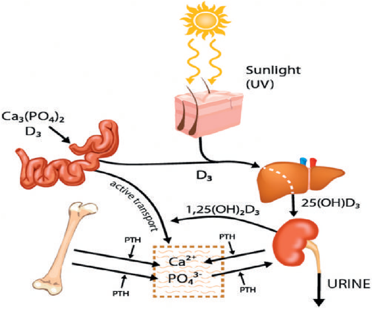
Vitamin D activation pathway. A disruption anywhere - diet, sun exposure, liver, kidney, receptor - can cause rickets. (Grainger & Allison's Diagnostic Radiology)
Vitamin D is activated by two-step hydroxylation:
- Liver: Vitamin D → 25-hydroxyvitamin D₃ (calcidiol)
- Kidney: 25(OH)D₃ → 1,25-dihydroxyvitamin D₃ (calcitriol, the active form)
Calcitriol acts via the vitamin D receptor (VDR) to enhance GI absorption of calcium and phosphorus. Without vitamin D, only 10-15% of dietary calcium and 60% of phosphorus are absorbed.
Classification & Causes
Rickets can be caused by a disturbance at almost any point along the Ca/PO₄/Vitamin D pathway:
1. Nutritional / Vitamin D Deficiency Rickets (most common)
- Inadequate dietary intake of vitamin D or calcium, or insufficient sun exposure
- Serum vitamin D (25-OH-D) is low
- Responds promptly to vitamin D supplementation
- Radiographic changes can be florid but improve rapidly with treatment
- Still among the most common childhood diseases in developing countries; routine milk fortification has reduced incidence in the West
2. Vitamin D-Dependent Rickets (VDDR) - Autosomal Recessive
Both types have normal vitamin D intake and serum levels, but errors in activation or receptor interaction:
| Type | Defect | Onset | Features |
|---|---|---|---|
| Type I | Deficiency of renal 1α-hydroxylase (can't convert 25-OH-D → 1,25-OH₂-D) | <2 years | Severe bony changes, hypocalcaemic tetany/seizures; responds to calcitriol, not vitamin D |
| Type II | Inactivating mutations in VDR (end-organ resistance) | <1 year | Alopecia, poor dentition; deficient response to both vitamin D and calcitriol |
3. Vitamin D-Resistant (Hypophosphataemic) Rickets
- Impaired proximal renal tubular reabsorption of phosphate → renal phosphate wasting
- Serum vitamin D levels are normal or elevated
- Subtypes:
- X-linked hypophosphataemic rickets (XLH) - most common; dominant mutation in the PHEX gene (phosphate-regulating endopeptidase); excess FGF23 drives phosphate wasting
- Autosomal dominant hypophosphataemic rickets (ADHR) - FGF23 gain-of-function mutations
- Hereditary hypophosphataemic rickets with hypercalciuria (HHRH) - mutations in SLC34A3 (sodium-phosphate co-transporter 2C); presents with rickets, short stature, hypercalciuria
- Fanconi syndrome (multiple proximal tubular defects)
- Untreated XLH → severe bowing deformities, short stature, dental abscesses
4. Renal Osteodystrophy
- Chronic renal failure → failure of second-step activation of vitamin D (kidney can't make calcitriol)
- Phosphate retention + hypocalcaemia → secondary hyperparathyroidism
- Elevated serum phosphate and ALP; normal or low serum calcium
5. Tumour-induced (Oncogenic) Rickets
- FGF23-secreting tumours (usually mesenchymal) → phosphaturia and hypophosphataemia
- Treatment requires resection of the tumour
Clinical Features
| Feature | Description |
|---|---|
| Short stature | Growth retardation |
| Limb bowing | Usually genu varum (bow-legs); weight-bearing deformity |
| Bone pain | Diffuse aching |
| Rachitic rosary | Enlargement/beading of costochondral junctions |
| Craniotabes | Soft skull with "ping-pong ball" feel (infants) |
| Frontal bossing | Prominent forehead |
| Harrison's sulcus | Groove along lower chest margin (diaphragm pulling on soft ribs) |
| Dental defects | Delayed dentition, enamel hypoplasia |
| Hypocalcaemic features | Tetany, seizures (in VDDR type I especially) |
| Ligamentous laxity | |
| Muscle weakness / hypotonia |
Radiology
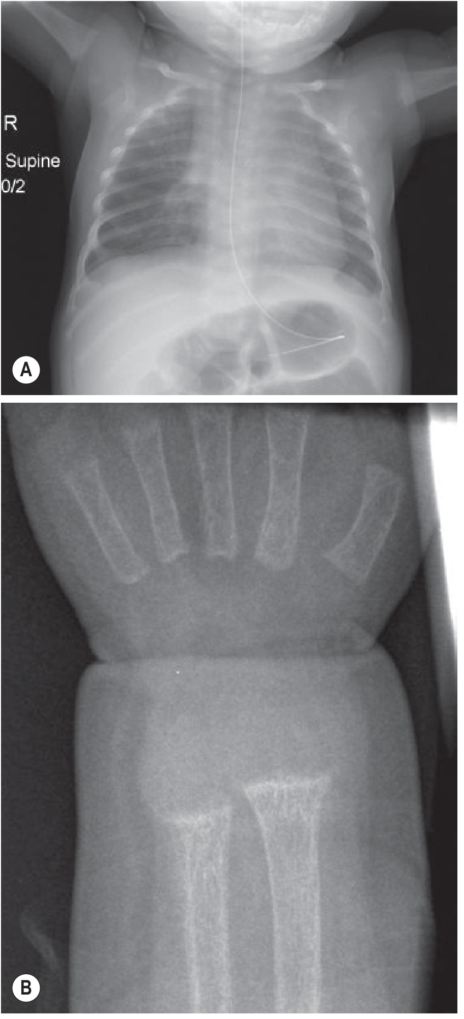
Severe nutritional rickets. (A) Marked bone density reduction, widening of costochondral junctions (rachitic rosary), cupping and fraying of proximal humeral metaphyses. (B) Cupped, frayed wrist metaphyses - the classic plain film sign. (Grainger & Allison's Diagnostic Radiology)
Classic radiological findings:
- Widened, flared, and cupped metaphyses (most visible at distal radius/ulna and proximal tibia)
- Frayed or brush-border metaphyseal margins
- Physeal widening (growth plate appears broadened)
- Generalised osteopenia (reduced bone density)
- Indistinct epiphyseal margins
- Looser zones (pseudofractures) - radiolucent bands perpendicular to cortex, especially at pubic rami, medial femoral necks, posterior ulna, and ribs
- Bowing of long bones (especially femur, tibia)
- Rachitic rosary (rib expansion at costochondral junctions)
- Dorsal kyphosis ("cat back"), flattened skull
- Histologically: widened osteoid seams, "Swiss cheese trabeculae," enlarged maturation zone of growth plate, poorly defined zone of provisional calcification
Investigations / Laboratory Findings
| Test | Nutritional Rickets | XLH | VDDR Type I | VDDR Type II |
|---|---|---|---|---|
| Serum Ca²⁺ | Low | Normal | Low | Low |
| Serum PO₄³⁻ | Low/Normal | Low | Low | Low |
| 25-OH-D | Low | Normal | Normal/Low | Normal |
| 1,25-(OH)₂-D | Low | Low/Normal | Low | High |
| ALP | Elevated | Elevated | Elevated | Elevated |
| PTH | Elevated (2° HPT) | Normal/Low | Elevated | Elevated |
| Urine phosphate | Normal | High (phosphaturia) | Normal | Normal |
Treatment
Principle: treat the underlying cause.
Nutritional rickets
- Vitamin D₃ (cholecalciferol) supplementation - preferred over D₂ (ergocalciferol) as it is more biologically potent with better absorption
- Children with normal GI absorption: 2,000-5,000 IU/day × 6-12 weeks (Harriet Lane)
- Calcium supplementation if dietary intake is low
- Serum ALP may initially rise as mineralisation increases, then falls to normal as bone heals
- Rapid radiological and clinical improvement expected
Malabsorption
- Children: 10,000-25,000 IU/day; adults: 10,000-300,000 IU/day (Harriet Lane)
VDDR Type I
- Calcitriol (1,25-(OH)₂D₃) directly, bypassing the defective 1α-hydroxylase
- Calcium supplementation
VDDR Type II
- High-dose calcitriol; some cases refractory (VDR mutation)
- IV calcium infusions have been used in severe cases
X-linked Hypophosphataemic Rickets (XLH)
- Traditional: phosphate supplements (multiple daily doses) + calcitriol or 1α-hydroxyvitamin D
- Goal: compensate for renal phosphate losses and overcome impaired 1,25-(OH)₂D synthesis
- Burosumab (anti-FGF23 monoclonal antibody): now licensed for XLH in children and adults in multiple countries
- Corrects hypophosphataemia, promotes fracture healing, improves stiffness and pain
- A 2024 meta-analysis (PMID 39211452) confirms burosumab as a promising treatment for paediatric XLH
Renal osteodystrophy
- Active vitamin D analogues (calcitriol, alfacalcidol)
- Phosphate binders
- Manage secondary hyperparathyroidism
Surgical
- Orthopedic surgery may be necessary for severe bowing deformities in children
Monitoring
- Serum Ca²⁺, PO₄³⁻, 25-OH-D (target ≥20 ng/mL in children), and ALP
- Ca × PO₄ product should be kept <70 mg/dL to avoid ectopic calcification/nephrocalcinosis
- Watch for signs of hypercalcaemia during supplementation: weakness, polyuria, nephrocalcinosis
Key Summary
Rickets = failure to mineralise the growth plate in children. The adult equivalent is osteomalacia. The common pathway is an insufficient Ca²⁺ × PO₄³⁻ product around chondrocytes. Most cases are nutritional and highly preventable; hereditary forms (XLH, VDDR) require targeted therapy including, increasingly, burosumab for XLH.
Sources: Grainger & Allison's Diagnostic Radiology; Ganong's Review of Medical Physiology (26th ed); Miller's Review of Orthopaedics (9th ed); Rockwood & Green's Fractures in Adults (10th ed); Brenner and Rector's The Kidney; Harriet Lane Handbook (23rd ed)
This is a shared conversation. Sign in to Orris to start your own chat.