
Bening tumors of vocal.cord
benign tumors of vocal cord larynx
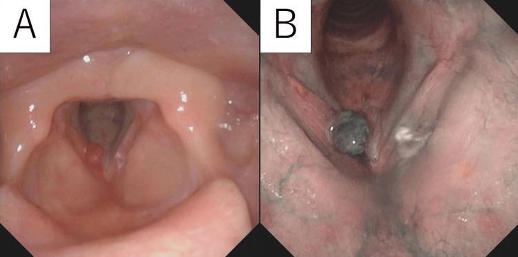
Side-by-side endoscopy images (laryngoscopy) of the larynx comparing White Light (WL) and Narrow-Band Imaging (NBI) modalities for a vocal cord tumor. Image A (WL) shows a pale red, globular, nodular mass with a smooth surface, located on the anterior third of the right vocal cord near the anterior commissure. The contralateral vocal cord appears normal. Image B (NBI) enhances the vascular detail, showing the same mass as dark green with significant internal vascularity and prominent subepithelial vessels in the surrounding mucosa. This comparison demonstrates the utility of NBI in highlighting neovascularization and lesion borders for surgical planning of laryngeal pathologies, such as a cavernous hemangioma. The clinical context involves a patient with chronic hoarseness, and the visual evidence guides the differential diagnosis between benign polyps and vascular tumors.
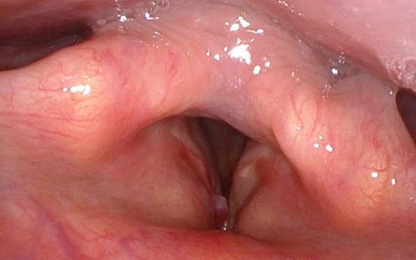
This diagnostic endoscopic image, obtained via fibrolaryngoscopy, provides a superior view of the larynx, focusing on the glottis. A distinct, pedunculated polypoid lesion is visible on the anterior third of the right vocal cord, situated near the anterior commissure. The lesion exhibits a smooth surface with prominent vascularization, characteristic of an angiomatous appearance. While the mass partially encroaches upon the glottic aperture, the airway space remains largely patent. The contralateral (left) vocal cord appears healthy with normal mucosal color and texture. There is no evidence of diffuse inflammation or significant structural distortion of the surrounding laryngeal tissues. This clinical photograph is used in otolaryngology to identify benign and malignant laryngeal masses, such as inflammatory myofibroblastic tumors or angiomatous polyps, and to assess their impact on vocal fold mobility and airway patency.
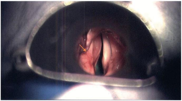
This clinical photograph shows a direct laryngoscopy endoscopic view of the human larynx, oriented with the anterior commissure at the top of the frame. The image illustrates a significant reddish, submucosal mass on the left side (indicated by an orange arrow), localized to the left vallecula and supraglottic region. The lesion exhibits a smooth, bulging morphology and extends toward the subepithelial area of the left vocal cord, resulting in visible anatomical asymmetry and partial narrowing of the glottic aperture (airway obstruction). In contrast, the right vocal cord and adjacent supraglottic structures appear relatively normal. This visual serves as a primary clinical example of a laryngeal mass—pathologically confirmed in this case as a schwannoma—demonstrating how localized benign tumors can displace laryngeal architecture and impinge upon the airway. This material is suitable for medical education in Otolaryngology regarding the endoscopic assessment of laryngeal tumors and differential diagnosis of airway obstruction.
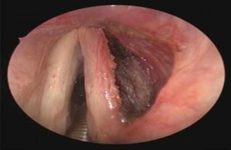
This endoscopic clinical image provides a postoperative view of the larynx following a transoral CO2 laser excision of a deep vocal cord lesion, specifically identified as an adult rhabdomyoma. The view focuses on the glottis, where the left vocal cord appears intact with smooth, pale pink mucosa. In contrast, the right vocal cord shows significant tissue disruption where the microflap technique was utilized. A prominent surgical defect or 'minus' is visible within the right thyroarytenoid muscle area. This surgical cavity exhibits irregular margins and a dark, hemorrhagic base typical of immediate postoperative healing. The surrounding laryngeal mucosa shows reactive erythema and mild edema. This visual demonstrates the immediate anatomical result of a conservative endolaryngeal enucleation of a benign laryngeal tumor, emphasizing the preservation of the superficial mucosal layers while revealing the underlying muscular excision site intended for healing by secondary intention.
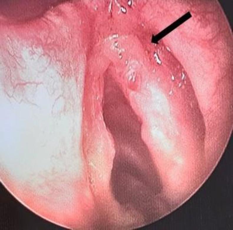
This diagnostic endoscopic image, obtained via direct laryngoscopy, demonstrates the anatomical structures of the larynx with a focus on the true vocal folds. A prominent, irregular polypoidal lesion is visible on the anterior half of the right vocal cord (indicated by a black arrow), appearing as a localized, protruding mass that disrupts the smooth glottic margin. The surrounding laryngeal mucosa exhibits a pink, healthy appearance without evidence of generalized edema or significant inflammatory erythema. The contralateral (left) vocal cord appears normal in morphology and surface texture. Clinically, such lesions are common causes of persistent or intermittent hoarseness and require surgical excision and histopathological analysis to differentiate between benign entities like laryngeal myxoma or vocal polyps and malignant pathologies. This visual is representative of Otolaryngology (ENT) diagnostic imaging for upper airway evaluation.
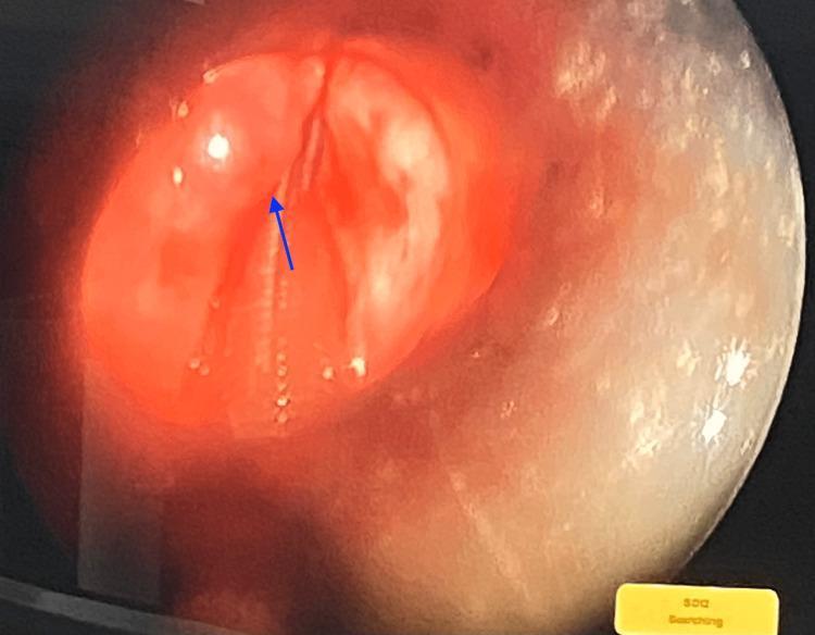
This clinical photograph displays a laryngeal view obtained via micro-laryngoscopy. The image provides an endoscopic perspective of the glottic opening, showing the true vocal cords (vocal folds) and the surrounding laryngeal mucosa. A blue arrow highlights a discrete, rounded mass located on the surface of the vocal cord. The mass appears well-circumscribed and pedunculated or bulging, characteristic of a benign-appearing laryngeal polyp or small nodule. The surrounding mucosa appears intact without obvious signs of severe acute inflammation or malignancy. The image demonstrates the surgical site for an endoscopic excision, a common procedure in otolaryngology for treating vocal cord lesions causing symptoms such as hoarseness (dysphonia). The lighting and magnification are consistent with micro-laryngoscopy, intended for detailed visualization and precise surgical intervention on the delicate structures of the larynx.
Benign Tumors of the Vocal Cord
Endoscopic Views: Vocal Cord Lesions
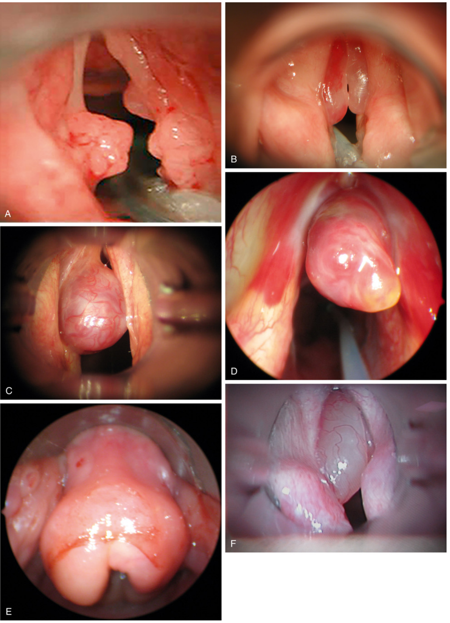
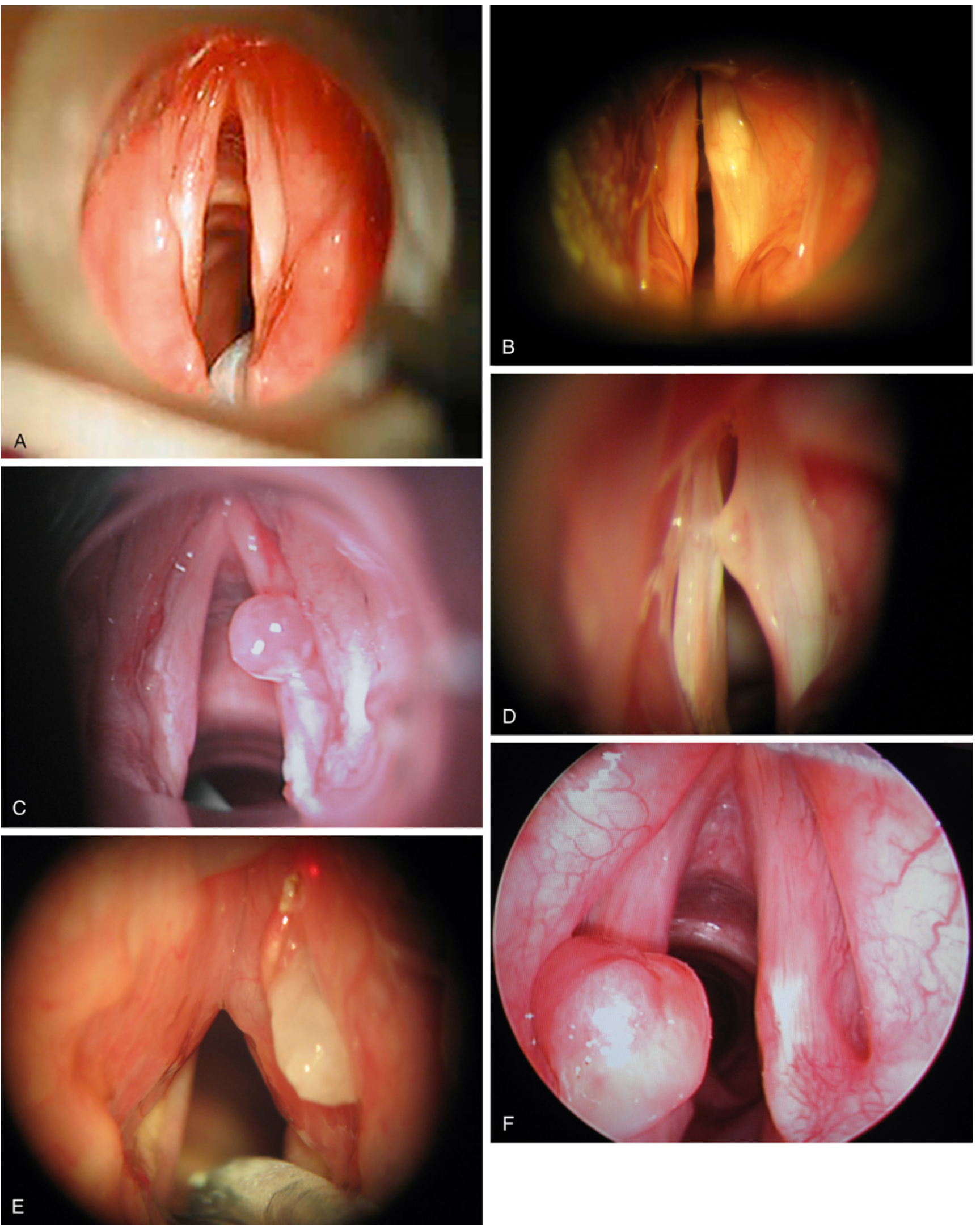
1. Vocal Cord Nodules (Singer's Nodes / Screamer's Nodules)
- Pathology: Smooth, hemispherical protrusions, usually <0.5 cm, on the true vocal cords. Composed of fibrous tissue covered by stratified squamous mucosa (may be ulcerated by contact with the opposite cord).
- Location: Bilateral, at the junction of the anterior 1/3 and posterior 2/3 of the vocal fold (the point of maximal vibratory impact).
- Cause: Vocal trauma / overuse (professional singers, teachers, children who scream).
- Treatment: Voice rest + speech therapy (first-line); surgical excision rarely needed.
- Robbins Basic Pathology, p. 1696; KJ Lee's Essential Otolaryngology, p. 1224
2. Vocal Cord Polyps
- Pathology: Smooth, unilateral, pedunculated or sessile lesion, edematous stroma with dilated blood vessels.
- Location: Between the anterior 1/3 and posterior 2/3 of the vocal fold.
- Cause: Vocal trauma + smoking.
- Distinction from nodules: Nodules are bilateral and firmer; polyps are typically unilateral and softer/more vascular.
- Treatment: Microlaryngoscopy (micro DL) with excision.
- KJ Lee's Essential Otolaryngology, p. 1225
3. Laryngeal Papilloma (Squamous Papilloma)
- Appearance: Soft, raspberry-like excrescence, rarely >1 cm, usually on the true vocal cords.
- Histology: Fingerlike projections with central fibrovascular cores covered by orderly stratified squamous epithelium. No cytologic atypia.
- Etiology: Human papillomavirus (HPV) types 6 and 11.
- Key facts:
- Adults: usually single - lower recurrence risk
- Children: often multiple - called Recurrent Respiratory Papillomatosis (RRP); onset usually 2-4 years of age
- RRP tends to recur after excision but often spontaneously regresses at puberty
- Vertical transmission from infected mother during delivery is the most likely cause in children
- Occult sites include the nasopharynx and the undersurface of the true vocal folds
- Malignant transformation is rare
- HPV vaccine (protecting against types 6 and 11) offers a prevention opportunity
- Treatment:
- Micro DL with stripping and/or CO2 laser / microdebrider
- Intralesional cidofovir (5 mg/mL) - note risk of malignant transformation
- Avoid jet ventilation - can seed lower respiratory airways
- Robbins Basic Pathology, p. 1698-1700; KJ Lee's Essential Otolaryngology
4. Vocal Cord Cysts
- Types:
- Ductal cysts (75%): from obstructed mucous duct - most common
- Saccular cysts (24%): mucous-filled dilations of the laryngeal saccule
- Anterior type: protrudes anteromedially between true and false vocal folds
- Lateral type: extends to the false cord, aryepiglottic fold, or vallecula
- Location: May involve any laryngeal structure except the free edge of the true vocal cords.
- Lining: Ciliated pseudostratified columnar, columnar, squamous epithelium, or a combination.
- KJ Lee's Essential Otolaryngology, p. 1224
5. Reinke's Edema (Polypoid Corditis)
- Pathology: Accumulation of fluid in the superficial layer of the lamina propria (Reinke's space).
- Appearance: Bilateral, diffuse, edematous swelling of the entire vocal fold - "waterbed-like" appearance on laryngoscopy.
- Cause: Strongly associated with smoking (most important risk factor).
- Treatment: Smoking cessation (mild cases); surgical drainage in severe cases.
- KJ Lee's Essential Otolaryngology, p. 1225
6. Granuloma
- Location: Posteriorly, over the vocal process of the arytenoid cartilage.
- Cause: Extrinsic trauma (endotracheal intubation, GERD-related hyperfunction).
- Treatment: Speech therapy, proton pump inhibitor (PPI), and removal of the causative trauma.
- KJ Lee's Essential Otolaryngology, p. 1225
7. Granular Cell Tumor
- Appearance: Yellow submucosal lesion on the posterior 1/3 of the vocal fold.
- Also found on tongue, skin, breast, subcutaneous tissues, and respiratory tract.
- Histology: Pseudoepitheliomatous hyperplasia (can mimic squamous cell carcinoma - important diagnostic pitfall).
- Malignant transformation: ~3% of cases.
- Treatment: Conservative excision.
- KJ Lee's Essential Otolaryngology, p. 1225
8. Amyloidosis (Laryngeal)
- The larynx is the most common site of airway amyloid deposition.
- Presents as a submucosal mass of the true or false vocal fold.
- Histology: Apple-green birefringence on Congo red staining (under polarized light).
- Treatment: Surgical excision.
- KJ Lee's Essential Otolaryngology, p. 1224
9. Chondroma
- Firm, smooth benign cartilaginous tumor.
- Usually involves the posterior cricoid cartilage (most common site for laryngeal chondroma).
- Treatment: Surgical excision.
- KJ Lee's Essential Otolaryngology, p. 1224
Comparative Summary Table
| Lesion | Location | Laterality | Key Feature | Treatment |
|---|---|---|---|---|
| Nodule | Junction ant 1/3 : post 2/3 | Bilateral | Vocal overuse/trauma | Speech therapy |
| Polyp | Junction ant 1/3 : post 2/3 | Unilateral | Smoking + trauma | Micro DL excision |
| Papilloma | True vocal cord (free edge) | Single (adult), multiple (child) | HPV 6 & 11 | CO2 laser / microdebrider |
| Reinke's edema | Entire vocal fold (Reinke's space) | Bilateral | Smoking | Smoking cessation; surgery |
| Cyst | Any laryngeal structure except TVC free edge | Usually unilateral | Obstructed duct / saccule | Excision |
| Granuloma | Vocal process of arytenoid (posterior) | Unilateral | Intubation / GERD | PPI + speech therapy |
| Granular cell tumor | Posterior 1/3 vocal fold | Unilateral | Yellow lesion; pseudo-SCC histology | Conservative excision |
| Amyloid | True or false vocal fold | Variable | Congo red (+), apple-green birefringence | Excision |
| Chondroma | Posterior cricoid cartilage | - | Firm, smooth mass | Excision |
Key Clinical Points
- Hoarseness (dysphonia) is the universal presenting symptom for all vocal cord lesions.
- Hoarseness persisting >4 weeks warrants otolaryngology referral and laryngoscopy.
- Nodules are the most common benign lesion and are managed conservatively with voice therapy.
- Papillomas are the most common benign neoplasm of the larynx; HPV 6/11 are causative.
- In RRP in children, avoid jet ventilation (risk of distal seeding).
- Malignancy must always be excluded, especially in smokers - squamous cell carcinoma accounts for >90% of laryngeal malignancies.
Neoplastic tumor
Of vocal cord
squamous cell carcinoma larynx vocal cord malignant tumor
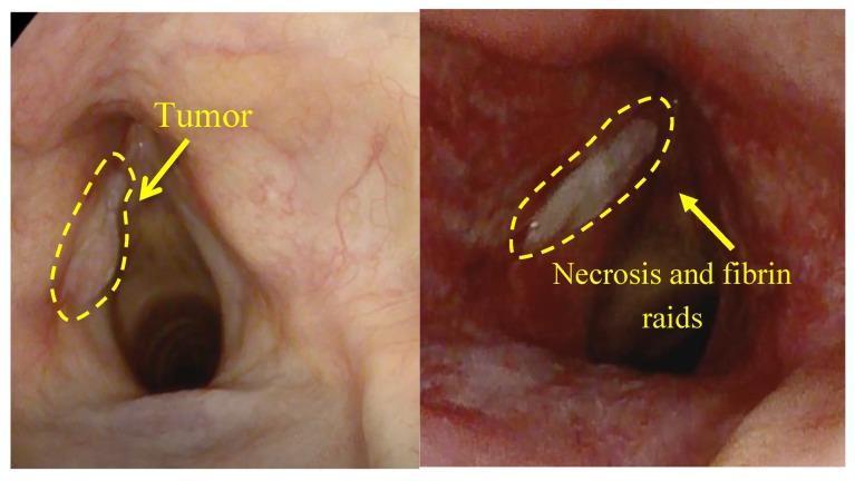
A side-by-side comparison chart featuring two endoscopic views of the larynx, specifically the vocal cords, demonstrating the effects of Photodynamic Therapy (PDT) on squamous cell carcinoma. The left image (pre-treatment) shows a protruding, off-white mass on the left vocal cord, demarcated by a yellow dashed oval and labeled 'Tumor'; the surrounding laryngeal mucosa appears pink with healthy vascularization. The right image (post-treatment, 7 days later) displays the same anatomical region showing significant inflammatory changes with deep red, hyperemic tissue. A yellow dashed oval highlights a localized area of 'Necrosis and fibrin raids,' appearing as a thick, whitish-gray plaque replacing the previous tumor site. This comparison illustrates the transition from a malignant neoplasm to therapeutic tissue destruction and necrosis. The images are relevant for oncology and otolaryngology, specifically for assessing treatment efficacy and the natural progression of tissue healing following PDT for laryngeal cancer.
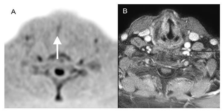
This diagnostic imaging comparison displays two axial MRI views of the larynx in an 80-year-old patient with squamous cell carcinoma of the right vocal cord (pT1a). Image A is a Diffusion-Weighted Whole-body Imaging with Background Body Signal Suppression (DWIBS) scan. It shows a focal area of high signal intensity (hyperintensity) on the right vocal cord, indicated by a white arrow, which represents restricted diffusion characteristic of malignant tissue. While the resolution in DWIBS is lower and anatomical boundaries are less distinct, the lesion is clearly identified. Image B is a contrast-enhanced transversal T1-weighted scan with fat saturation. This sequence provides superior anatomical resolution of laryngeal cartilages and surrounding soft tissues but fails to clearly distinguish the small T1a carcinoma from the normal vocal cord morphology or enhancement. The comparison illustrates the clinical utility of functional imaging (DWIBS) in detecting early-stage (pT1a) glottic cancer that may be occult on standard morphological MRI sequences.
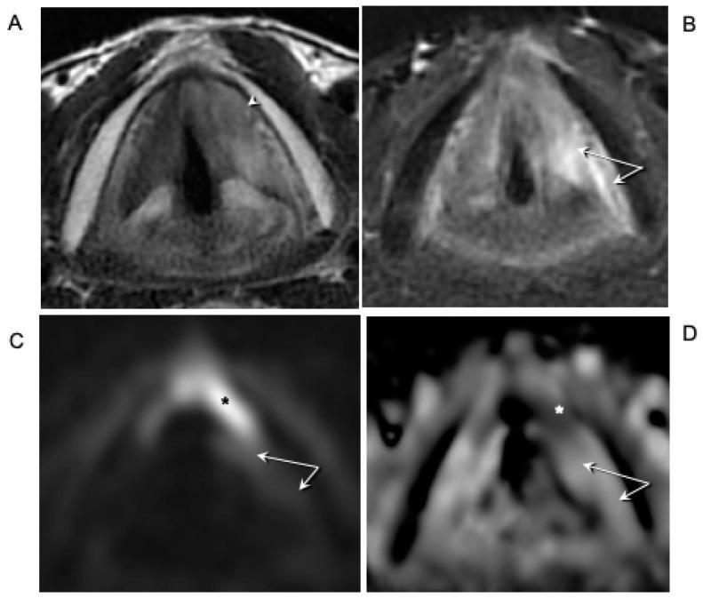
This composite diagnostic image displays axial MRI sequences of the larynx in a patient with recurrent glottic squamous cell carcinoma. Panel (A) is a T2-weighted axial image showing intermediate signal intensity neoplastic tissue in the anterior third of the left true vocal cord, extending across the anterior commissure and invading the left inferior paraglottic space (white arrowhead). Panel (B) utilizes T2-weighted imaging with fat suppression to highlight hyperintense signal alterations (white arrows) in the posterior aspect of the left vocal cord and posterior paraglottic space, indicative of peritumoral edema rather than tumor infiltration. Panels (C) and (D) show Diffusion-Weighted Imaging (DWI) and the corresponding Apparent Diffusion Coefficient (ADC) map, respectively. The tumor demonstrates restricted diffusion characterized by hyperintensity on DWI (black asterisk) and hypointensity on the ADC map (white asterisk). The absence of restricted diffusion in the posterior larynx (white arrows in C and D) confirms the edematous nature of those signal changes, supporting the clinical determination of a tumor-free posterior paraglottic space despite fixed vocal cord mobility.
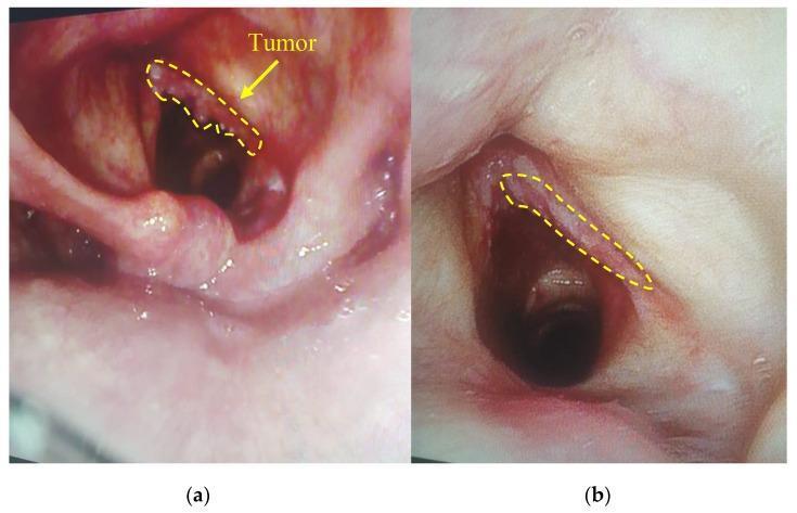
This comparison clinical photograph features two side-by-side endoscopic images of the laryngeal region, specifically focusing on the vocal cords before and after photodynamic therapy (PDT) for squamous cell carcinoma. Image (a) represents the pre-treatment state, showing an irregular, exophytic mass labeled as 'Tumor' on the right vocal cord. The lesion exhibits a rough, nodular surface with mottled pink and white coloration and signs of increased hypervascularity and tissue density. Image (b) shows the same anatomical site one week after PDT. In the post-treatment image, the previously highlighted region demonstrates significant tumor regression, characterized by a smoother surface texture, more uniform pink mucosal color, and a reduction in both tissue volume and vascular prominence. This comparative view illustrates the efficacy of PDT in inducing necrosis and flattening of malignant laryngeal neoplasms, highlighting its clinical utility in treating glottic squamous cell carcinoma.
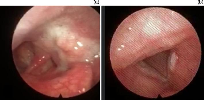
This clinical photograph displays two fiber-optic endoscopic views of the larynx and pharynx, illustrating primary malignancies. Panel (a) shows a nasendoscopic view of the left piriform fossa containing a fleshy, erythematous mass with an irregular, bumpy surface. The lesion demonstrates areas of white leukoplakia and prominent superficial vascularization, characteristic of piriform fossa squamous cell carcinoma. Panel (b) provides a view of the glottis showing the vocal cords in a narrow V-shaped configuration. A distinct, opaque, white exophytic mass is visible on the edge of the left vocal cord, representing a vocal cord squamous cell carcinoma. The images were captured using a smartphone-integrated flexible endoscope. These views are essential for otolaryngology training in identifying malignant upper aerodigestive tract lesions and distinguishing them from benign pathology through visual characteristics such as irregular margins, atypical color changes, and tissue distortion.
Neoplastic Tumors of the Vocal Cord (Larynx)
Endoscopic Views: Malignant Vocal Cord Lesions
A. Squamous Cell Carcinoma (SCC) of the Larynx
Epidemiology
- Represents only ~2% of all cancers
- Most common after age 40; M:F ratio = 7:1 (highest gender disparity of all H&N cancers)
- In the US, ~13,150 new cases/year; ~3,710 deaths/year
- Worldwide incidence is declining due to reduced smoking in developed countries
Risk Factors
| Risk Factor | Notes |
|---|---|
| Tobacco | Most important; risk is proportional to intensity/duration; takes ≥15 years after cessation to return toward baseline |
| Alcohol | Synergistic with tobacco |
| HPV 16 and 18 | Found in ~15% of tumors; these tend to have a better prognosis |
| GERD | Chronic irritation |
| Radiation exposure | Prior neck/head RT |
| Asbestos exposure | Occupational risk |
Anatomy: Sites of Origin
| Site | Frequency | Key Feature |
|---|---|---|
| Glottis (true vocal cords) | 60-75% | Most common; early hoarseness; sparse lymphatics |
| Supraglottis (above cords) | 25-40% | Rich lymphatics; late symptoms; high nodal spread |
| Subglottis (below cords) | <5% | Late presentation; worst prognosis |
Gross & Histological Appearance
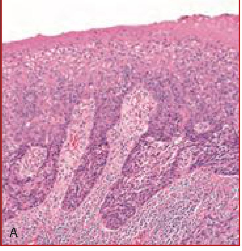
- Gross: Pearly gray, wrinkled plaque that ulcerates with tumor progression (can also appear ulcerative, exophytic, sessile, or polypoid on laryngoscopy)
- Histology: Keratinizing, well-to-moderately differentiated SCC most common in glottic tumors
- Adjacent mucosa often shows squamous hyperplasia, dysplasia, or carcinoma in situ (precursor lesions from chronic carcinogen exposure)
B. TNM Staging (AJCC) - Laryngeal SCC
Glottic SCC (Most Common)
| Stage | Description |
|---|---|
| Tis | Carcinoma in situ |
| T1a | Tumor limited to one vocal cord, normal mobility |
| T1b | Tumor involving both vocal cords, normal mobility |
| T2 | Tumor extends to supraglottis/subglottis, or impaired vocal cord mobility |
| T3 | Tumor limited to larynx with vocal cord fixation |
| T4a | Tumor invades thyroid cartilage and/or extends beyond larynx |
| T4b | Tumor invades prevertebral space, encases carotid, or invades mediastinum |
Supraglottic SCC
| Stage | Description |
|---|---|
| T1 | Tumor limited to one subsite of supraglottis, normal mobility |
| T2 | Invades mucosa of >1 subsite or extends to glottis/vallecula/medial pyriform wall |
| T3 | Vocal cord fixation OR invasion of post-cricoid area, pre-epiglottic space, paraglottic space, thyroid cartilage (minor erosion) |
| T4a/b | Invasion through thyroid cartilage or into adjacent structures |
C. Clinical Presentation
| Site | Cardinal Symptom | Why |
|---|---|---|
| Glottis | Early hoarseness (dysphonia) | Even small lesion alters cord vibration |
| Supraglottis | Dysphagia, odynophagia, referred otalgia, altered vocal resonance, neck mass | Tumors grow larger before obstructing; rich lymphatics allow early nodal spread |
| Subglottis | Stridor, dyspnea (late); often misdiagnosed as asthma | No early symptoms until advanced |
D. Lymph Node Drainage & Spread
- Glottis: Sparse lymphatics - low rate of nodal spread (favorable)
- Supraglottis: Rich lymphatics - drains to levels II, III, IV (bilateral)
- Subglottis: Drains to levels VI (paratracheal) and upper mediastinum
- Neck dissection: Bilateral selective neck dissection (levels II-IV) for clinically normal necks in supraglottic and advanced (T3-T4) laryngeal tumors
E. Treatment
Early Disease (Stage I & II)
- Surgery OR radiotherapy (XRT) - comparable survival rates
- Choice is guided by voice quality outcomes, cost, patient factors, and whether RT may be needed later for a second primary
| Procedure | Indication |
|---|---|
| Glottic CIS | Serial micro DL, vocal cord stripping, CO2 laser excision |
| Cordectomy | Removal of diseased true vocal cord via laryngofissure |
| Vertical hemilaryngectomy | Unilateral T1-T2 disease; can involve anterior commissure; <1 cm subglottic extension |
| Supraglottic laryngectomy | T1, T2, or T3 (pre-epiglottic space only) supraglottic lesions; preserves true cords, arytenoids, thyroid cartilage |
| Supracricoid laryngectomy | Anterior glottic tumors; preserves cricoid and ≥1 arytenoid; ~50% remain tracheostomy-dependent |
- T1 glottic SCC: 5-year local control 81-90%, laryngeal preservation 90-98%
- T2 with normal mobility: local control 64-87%, laryngeal preservation 75-87%
Advanced Disease (Stage III & IV)
- Concurrent chemoradiotherapy (chemoRT) - organ preservation protocol
- Total laryngectomy ± XRT - for T3/T4 with cartilaginous invasion, extralaryngeal or neck involvement
- After total laryngectomy: tracheoesophageal speech with prosthesis
F. Prognosis
| Site | Prognosis | Reason |
|---|---|---|
| Glottis | Best - ~90% confined to larynx at diagnosis | Early symptoms + sparse lymphatics |
| Supraglottis | Intermediate | Rich lymphatics, late symptoms |
| Subglottis | Worst | Late presentation, advanced stage |
- Overall: with surgery, RT, or combination - many patients can be cured
- ~1/3 of patients die of disease (usually from widespread metastasis)
- HPV-positive tumors have a better prognosis than tobacco/alcohol-related SCC
G. Other (Less Common) Malignant Tumors of the Larynx
| Tumor | Notes |
|---|---|
| Adenoid cystic carcinoma | Minor salivary gland origin; subglottis; perineural spread characteristic |
| Mucoepidermoid carcinoma | Minor salivary gland origin |
| Chondrosarcoma | Arises from posterior cricoid cartilage; treated with narrow excision - no postoperative radiation |
| Adenocarcinoma | Rare (<5% of laryngeal cancers) |
| Neuroendocrine carcinoma | Rare; supraglottis most common site |
H. Precursor / Premalignant Lesions
- Leukoplakia: white patch on vocal cord mucosa - requires biopsy
- Erythroplakia: red patch - higher malignant potential than leukoplakia
- Dysplasia (mild/moderate/severe): spectrum leading to CIS
- Carcinoma in situ (CIS): full-thickness epithelial dysplasia, no basement membrane violation - treated with serial CO2 laser excision / micro DL stripping
Summary: Vocal Cord Fixation = T3
Vocal cord fixation, regardless of tumor size, automatically upgrades the tumor to at least T3. Fixed cords result from invasion of the thyroarytenoid muscle.