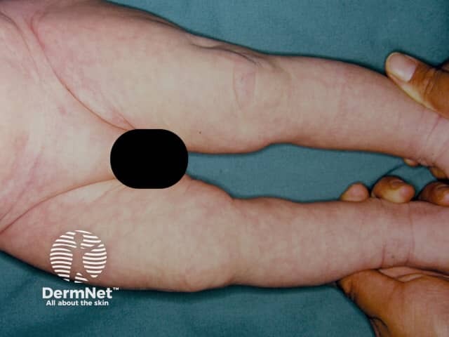
attachment
attachment

What are these red spots on my baby she's only 16 days old

analyse_media
Reading File
Reading File
Finding Sources
Searching the Web

newborn baby small red pinpoint spots skin 2 weeks old causes

Quick answer

Looking at your photo, I can see a couple of small, distinct, bright red pinpoint-to-pinhead sized spots on your baby's scalp/hairline area. In a 16-day-old, isolated small red spots like this are most commonly one of the following, all generally harmless:
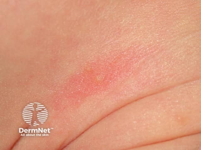
  1. Neonatal/newborn rash (erythema toxicum neonatorum) - common in the first couple weeks, appears as small red spots, often with a tiny pale bump in the center, comes and goes on different parts of the body.
  2. Insect bites (like mosquito bites) - small, round, isolated red spots, sometimes with a tiny central punctum, which matches what's visible here.
  3. Minor skin irritation or scratch marks - newborns often scratch themselves lightly with their own fingernails, causing small red marks.
  4. Petechiae (tiny red/purple spots that do NOT fade when you press on them) - less likely here since these look like ordinary red spots, but this is the one thing you want to specifically rule out.
A simple check you can do at home: gently press a finger or a clear glass against the spot.
  • If it fades/blanches and comes back when you release pressure - reassuring, likely one of the benign causes above.
  • If it does NOT fade with pressure - this is petechiae, which in a newborn can (rarely) signal something more serious (infection, low platelets, or a clotting problem) and needs same-day medical evaluation.
See a doctor promptly (same day) if:
  • The spots don't blanch with pressure
  • New spots are appearing or spreading
  • Baby has fever, is unusually sleepy/floppy, feeding poorly, or irritable
  • The spots are bruising, bleeding, or look like tiny blood blisters
  • There's any swelling, pus, or the area feels warm
Given that this is a newborn only 16 days old, if you have any doubt at all, it's reasonable to have your pediatrician or a nurse take a quick look, either at a scheduled visit or a same-day call, just for peace of mind. Newborns should generally have low threshold for a check given how quickly things can change at this age.
I'm not able to make a definitive diagnosis from a photo alone. This is general guidance, not a substitute for an in-person exam by your pediatrician.
Finding Sources
Looking at the photo, I can see 2 small, discrete, flat, pinpoint red/pink dots on the scalp/hairline area - they're isolated (not clustered), don't have a white or yellow bump in the center, and aren't spread widely over the body.
Most likely, benign explanations for isolated spots like this in a newborn:
  • Scratch marks from her own fingernails - very common at this age since babies can't control their hand movements and newborn nails are sharp.
  • Minor pressure/friction marks - from a hair tie, seam, or her own position against bedding (I noticed a hair accessory in the photo near her head).
  • Petechiae from minor trauma - tiny broken capillaries from crying, coughing, or pressure during birth/positioning. These are pinpoint, flat, and do not fade (blanch) when you press on them.
  • Nevus simplex ("stork bite"/"angel kiss") - a common pink birthmark, usually on the forehead, eyelids, or nape of neck. These do blanch (turn pale) when pressed and are harmless.
  • Insect bite - possible if she's had any exposure to mosquitoes or other bugs.
What this does not look like from the photo is erythema toxicum (which shows larger red blotches with a white/yellow bump in the center) or milia (which are white, not red).
Do this simple check now: Gently press a fingertip on one of the spots for a second and release.
  • If it turns pale/white and then the red color returns - it's a blood vessel (blanches), like a birthmark or dilated capillary - not concerning.
  • If it stays exactly the same red/purple color and does not fade with pressure - that's petechiae (tiny bleeding under the skin).
Why this matters more at 16 days old: In infants under 28 days, isolated petechiae are usually harmless (from minor trauma or crying), but this age group has a low threshold for concern because petechiae can occasionally be an early sign of serious infection, especially when they show up alongside other signs of illness. Pediatric emergency medicine references note that in neonates, petechiae combined with fever, lethargy, poor feeding, mottled skin, a bulging soft spot, or fast breathing warrant urgent evaluation (Rosen's Emergency Medicine, "Infants 0 to 28 Days Old").
See a doctor today/urgently if you notice any of:
  • Fever (rectal temp ≥ 100.4°F/38°C) - always urgent at this age
  • More spots appearing or spreading over the body
  • Baby seems unusually sleepy, floppy, hard to wake, or not feeding well
  • Spots don't blanch with pressure
  • Bruising elsewhere, or bleeding from gums/nose
  • Baby seems irritable/inconsolable, or the soft spot on her head looks bulging
If she otherwise looks well (feeding normally, good color, active, no fever, alert), and the spots blanch with pressure or look like simple scratch/pressure marks, it's reasonable to just keep an eye on it and mention it at her next pediatric visit - but given she's only 16 days old, I'd lean toward having a pediatrician or nurse look at it in person (or a quick photo sent to your pediatrician's nurse line) just to be safe, since visual assessment in person is more reliable than a photo.
I can't give you a definitive diagnosis from a photo alone - if anything changes or you're unsure, it's always reasonable to have a newborn checked promptly.

Find clinical images of common newborn skin conditions

Searching Images

erythema toxicum neonatorum newborn rash

Imaging modality: Clinical photography; Technique: close-up, color standard photograph of neonatal skin under balanced lighting. Anatomical location: neonatal trunk and proximal limbs; Cutaneous findings: scattered erythematous macules and papules with occasional small pustules on an erythematous base, most prominent on the torso and arms. Pathology and description: erythema toxicum neonatorum; eosinophilic pustules within inflamed epidermis without systemic symptoms; benign inflammatory eruption that often appears within the first 24-48 hours of life and resolves spontaneously within 1-2 weeks. Diagnostic significance: common, self-limited neonatal rash requiring only observation; differentiation from infection (neonatal herpes simplex, candidiasis) or other pustular conditions is essential. Clinical correlation: no fever or irritability typically; may be associated with transient eosinophilia; management is reassurance. Context: used in dermatology education and neonatal care to illustrate benign neonatal dermatoses; useful for clinicians assessing newborn rashes and teaching about differential diagnosis, natural history, and parental counseling. Imaging features: non‑bullous pustules on erythematous base, variable distribution across trunk and proximal extremities; resolution aligns with maturation of skin. This image demonstrates classic erythema toxicum features without crusting or vesicular breakdown. Clinical notes emphasize observation, supportive care, and parental reassurance; no antibiotics are routinely required unless signs of secondary infection or systemic illness emerge. Uncommonly.

Imaging modality: Clinical photography; Technique: close-up, color standard photograph of neonatal skin under balanced lighting. Anatomical location: neonatal trunk and proximal limbs; Cutaneous findings: scattered erythematous macules and papules with occasional small pustules on an erythematous base, most prominent on the torso and arms. Pathology and description: erythema toxicum neonatorum; eosinophilic pustules within inflamed epidermis without systemic symptoms; benign inflammatory eruption that often appears within the first 24-48 hours of life and resolves spontaneously within 1-2 weeks. Diagnostic significance: common, self-limited neonatal rash requiring only observation; differentiation from infection (neonatal herpes simplex, candidiasis) or other pustular conditions is essential. Clinical correlation: no fever or irritability typically; may be associated with transient eosinophilia; management is reassurance. Context: used in dermatology education and neonatal care to illustrate benign neonatal dermatoses; useful for clinicians assessing newborn rashes and teaching about differential diagnosis, natural history, and parental counseling. Imaging features: non‑bullous pustules on erythematous base, variable distribution across trunk and proximal extremities; resolution aligns with maturation of skin. This image demonstrates classic erythema toxicum features without crusting or vesicular breakdown. Clinical notes emphasize observation, supportive care, and parental reassurance; no antibiotics are routinely required unless signs of secondary infection or systemic illness emerge. Uncommonly.

Searching Images

milia newborn face tiny white bumps

Searching Images

nevus simplex salmon patch stork bite newborn

Searching Images

Mongolian spot newborn bluish birthmark

Searching Images

neonatal acne baby face red pimples

Searching Images

cutis marmorata mottled skin infant

Searching Images

infantile hemangioma strawberry birthmark

Searching Images

neonatal pustular melanosis

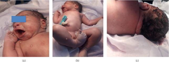
Clinical photographs of a term newborn presenting with a generalized vesiculopustular eruption. The images are divided into three panels: (a) a facial and upper chest view showing multiple erythematous macules and distinct white-to-yellow pustules on the cheeks and torso; (b) a full-body view demonstrating extensive distribution of pustular and bullous lesions across the trunk, abdomen, and lower extremities; (c) a posterior view of the neck and scalp showing isolated pustules. The lesions vary in size from small, tense pustules with turbid contents to larger, fluid-filled bullae. Surrounding the lesions is mild, patchy erythema. The clinical presentation is consistent with neonatal pustular dermatosis, requiring differentiation between infectious etiologies such as bullous impetigo or Congenital Cutaneous Candidiasis, and non-infectious conditions like Erythema Toxicum Neonatorum or Transient Neonatal Pustular Melanosis. This visual serves as an educational reference for neonatal dermatology and the diagnostic evaluation of skin eruptions at birth.
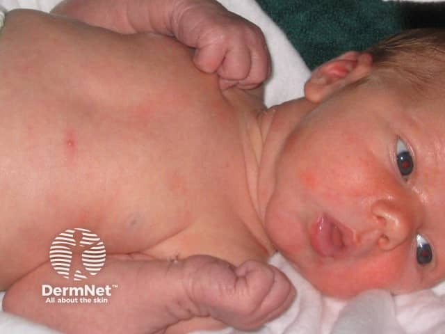
Clinical photographs of a term newborn presenting with a generalized vesiculopustular eruption. The images are divided into three panels: (a) a facial and upper chest view showing multiple erythematous macules and distinct white-to-yellow pustules on the cheeks and torso; (b) a full-body view demonstrating extensive distribution of pustular and bullous lesions across the trunk, abdomen, and lower extremities; (c) a posterior view of the neck and scalp showing isolated pustules. The lesions vary in size from small, tense pustules with turbid contents to larger, fluid-filled bullae. Surrounding the lesions is mild, patchy erythema. The clinical presentation is consistent with neonatal pustular dermatosis, requiring differentiation between infectious etiologies such as bullous impetigo or Congenital Cutaneous Candidiasis, and non-infectious conditions like Erythema Toxicum Neonatorum or Transient Neonatal Pustular Melanosis. This visual serves as an educational reference for neonatal dermatology and the diagnostic evaluation of skin eruptions at birth.

Clinical photography of a term neonate demonstrating erythematous papules and vesicles distributed primarily on the trunk and face. Imaging modality: Clinical photography; technique: high-resolution color image captured in natural or balanced lighting, frontal/anterior view with the infant supine. Anatomical localization: integumentary system; neonatal skin (epidermis and superficial dermis). Visual features include numerous small 1–3 mm erythematous papules and vesicles on an erythematous base, some showing a pale or yellowish vesicular center; lesions are non-follicular and non-desquamative, arranged in scattered coalescent patches but with overall benign appearance. There is no edema, crusting, or purpura; mucous membranes spared; no systemic signs of illness. Clinically diagnostic for erythema toxicum neonatorum (ETN), a common benign neonatal eruption typically appearing within 24–72 hours of birth and resolving spontaneously within 1–2 weeks. Haematologic or infectious etiologies are not supported by the image alone; no systemic symptoms; differential considerations include milia, miliaria rubra, transient neonatal pustular melanosis, neonatal pustular psoriasis, and less commonly neonatal herpes. Pathophysiology involves eosinophilic inflammatory response; management is reassurance and observation; no antimicrobial therapy required unless accompanied by fever or sepsis concern. The image is relevant for educational purposes in dermatology, pediatrics, and neonatology to illustrate ETN morphology, distribution, natural history, and differentiation from other neonatal pustular conditions.

Clinical photography of a term neonate demonstrating erythematous papules and vesicles distributed primarily on the trunk and face. Imaging modality: Clinical photography; technique: high-resolution color image captured in natural or balanced lighting, frontal/anterior view with the infant supine. Anatomical localization: integumentary system; neonatal skin (epidermis and superficial dermis). Visual features include numerous small 1–3 mm erythematous papules and vesicles on an erythematous base, some showing a pale or yellowish vesicular center; lesions are non-follicular and non-desquamative, arranged in scattered coalescent patches but with overall benign appearance. There is no edema, crusting, or purpura; mucous membranes spared; no systemic signs of illness. Clinically diagnostic for erythema toxicum neonatorum (ETN), a common benign neonatal eruption typically appearing within 24–72 hours of birth and resolving spontaneously within 1–2 weeks. Haematologic or infectious etiologies are not supported by the image alone; no systemic symptoms; differential considerations include milia, miliaria rubra, transient neonatal pustular melanosis, neonatal pustular psoriasis, and less commonly neonatal herpes. Pathophysiology involves eosinophilic inflammatory response; management is reassurance and observation; no antimicrobial therapy required unless accompanied by fever or sepsis concern. The image is relevant for educational purposes in dermatology, pediatrics, and neonatology to illustrate ETN morphology, distribution, natural history, and differentiation from other neonatal pustular conditions.

query_images_db
query_images_db
query_images_db
query_images_db
query_images_db
query_images_db
query_images_db
query_images_db
Here's a visual guide to common newborn skin findings, pulled from clinical dermatology references. All of these are typically benign and self-limited, but I've noted quick differentiators for each.

1. Erythema Toxicum Neonatorum

The most common newborn rash (up to 50% of babies), appearing in the first 2-3 days of life. Red blotchy macules/papules, often with a tiny white or yellow pustule in the center, scattered on the trunk and limbs. Resolves on its own within 1-2 weeks.
Erythema toxicum neonatorum

2. Milia

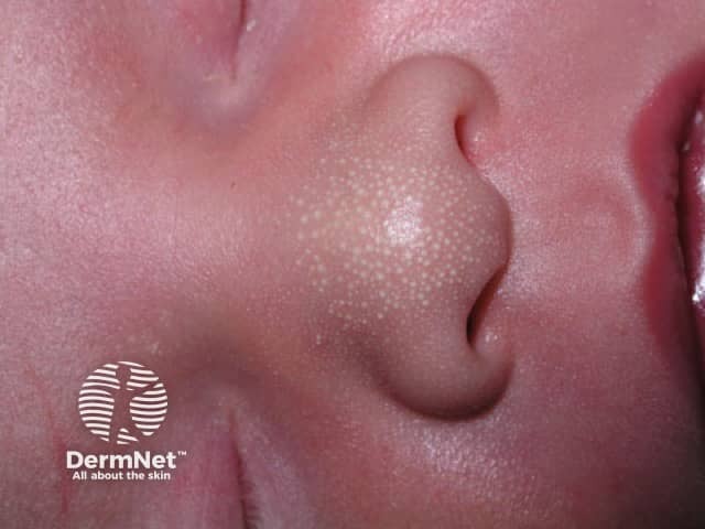
Tiny (1-2mm) pearly white bumps, usually clustered on the nose and cheeks. These are keratin-filled cysts, not infected, and clear up within weeks to months without treatment.
Milia on infant face

3. Neonatal (Baby) Acne

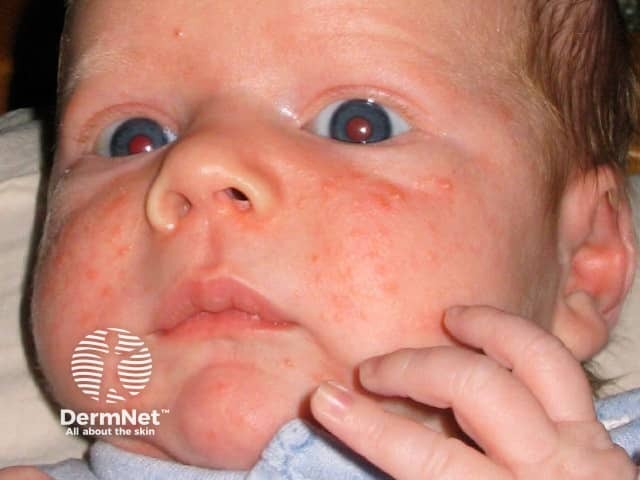
Small red papules and pustules on the cheeks, nose, and forehead, usually starting at 2-4 weeks of age from residual maternal hormone exposure. Resolves in weeks to a few months; avoid oils/lotions on the area.
Neonatal acne on infant face

4. Salmon Patch / Nevus Simplex ("Angel Kiss" or "Stork Bite")
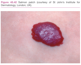
A flat, well-demarcated pink-red patch, most common on the eyelids, forehead, or nape of the neck. It blanches with pressure and usually fades over the first year or two.
Salmon patch birthmark

5. Port-Wine Stain (Capillary Malformation)
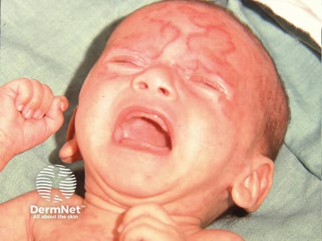
Unlike the salmon patch, this is a congenital vascular birthmark that is darker (red to violaceous), persists and can darken with age rather than fading. Facial lesions in the eye/forehead area occasionally need evaluation for Sturge-Weber syndrome.
Port-wine stain on scalp and forehead

6. Mongolian Spot (Dermal Melanocytosis)
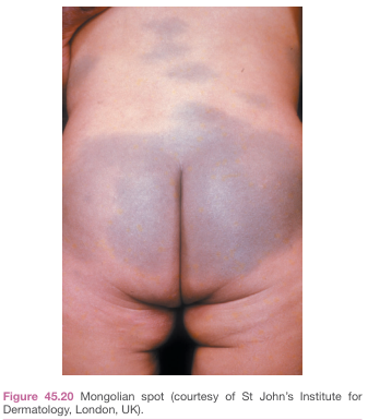
Flat, bluish-grey patch(es), most often over the lower back/buttocks, very common in babies with darker skin tones. Benign, congenital, and usually fades over several years - important not to mistake for bruising.
Mongolian spot on lower back and buttocks

7. Cutis Marmorata

A lacy, mottled, red-purple/blue pattern on the skin (often limbs) triggered by cold exposure. It's a normal, temporary vasomotor response in newborns and resolves with warming.
Cutis marmorata mottled skin pattern on infant hands

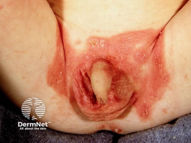
8. Diaper (Napkin) Dermatitis

Erythema and irritation confined to the diaper area from moisture/friction; can be complicated by Candida (look for satellite red papules beyond the main rash border).
Diaper dermatitis in infant

9. Caput Succedaneum / Cephalohematoma (birth-related scalp swelling)
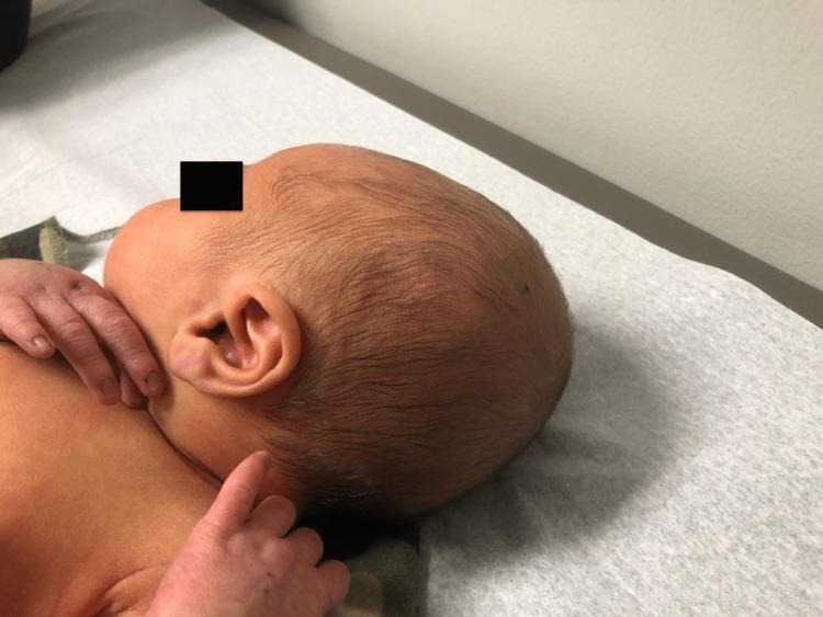
Soft tissue swelling of the scalp from birth trauma - caput succedaneum crosses suture lines and resolves in days; cephalohematoma is confined to one bone and takes longer to resolve.
Neonate with resolving scalp swelling after birth

Quick rule of thumb for any newborn rash: if it blanches with gentle pressure, is flat or mildly raised without pus, and the baby is feeding well/acting normally, it's usually benign. Seek same-day medical review for fever, lethargy, poor feeding, spreading pustules/blisters, or spots that don't blanch (possible petechiae), especially in a baby under 4 weeks old.
This is a shared conversation. Sign in to Orris to start your own chat.