
Thyroid masses pathology and diagnostic
Reading File
Now I'll run parallel searches across the medical library for thyroid masses pathology and diagnostics.
Finding Sources
Finding Sources
Searching PubMed
"thyroid nodule" AND diagnosis
Excellent finds. Now I'll read the most comprehensive sources in parallel - Robbins pathology for pathology, Sabiston for surgical/clinical, and Scott-Brown for ENT perspective.
Reading File
Reading File
Reading File
Excellent data from Robbins. Now let me get more Robbins pathology content (follicular, medullary, anaplastic carcinoma) and the diagnostic sections in parallel.
Reading File
Finding Sources
Finding Sources
Good. Now let me get medullary + anaplastic carcinoma from Robbins, the FNA/evaluation section from Cummings, and fetch the key pathology images in parallel.
Reading File
Reading File
Loading Image
Loading Image
Excellent content from Robbins. Let me now get the medullary carcinoma images, more Robbins content on Bethesda system/FNA, and Cummings on the rest of the diagnostic workup.
Reading File
Reading File
Finding Sources
Now let me get the Robbins key concepts/molecular section, the Cummings diagnostic studies (FNAC, ultrasound), and fetch medullary carcinoma images.
Reading File
Reading File
Loading Image
I now have excellent, comprehensive content. Let me compile the full, detailed answer.
Thyroid Masses: Pathology and Diagnostics
Overview
Thyroid nodules are common - with a lifetime incidence of 4-7% and an annual incidence of approximately 0.1% in the United States (~300,000 new nodules/year). The majority are benign (colloid nodules, adenomas, cysts, focal thyroiditis). Only approximately 5% harbor carcinoma, though approximately 1 in 200 nodules is lethal. The diagnostic challenge is separating benign from malignant disease while balancing the morbidity of treatment. - Cummings Otolaryngology, p. 2375
Classification of Thyroid Masses
Benign Lesions
| Type | Key Features |
|---|---|
| Colloid (adenomatoid) nodule | Most common; degenerative change in multinodular goiter |
| Follicular adenoma | Solitary, encapsulated, no capsular/vascular invasion |
| Thyroid cyst | Simple or complex; may result from degeneration |
| Hashimoto thyroiditis | Autoimmune; can form a mass; associated with lymphoma risk |
| Thyroglossal duct cyst | Congenital; midline, moves with swallowing; lined by squamous or respiratory epithelium |
| Subacute (de Quervain) thyroiditis | Painful; granulomatous inflammation |
Malignant Lesions (overview)
| Type | Frequency | Cell of Origin |
|---|---|---|
| Papillary carcinoma | ~85% | Follicular epithelium |
| Follicular carcinoma | 5-15% | Follicular epithelium |
| Oncocytic (Hürthle cell) carcinoma | Subset of follicular | Follicular epithelium |
| Medullary carcinoma | ~5% | Parafollicular C cells |
| Anaplastic carcinoma | <5% | Dedifferentiated |
| Thyroid lymphoma | Rare | B lymphocytes (often on Hashimoto background) |
Malignant Tumors: Detailed Pathology
1. Papillary Thyroid Carcinoma (PTC)
Epidemiology: Most common thyroid cancer (~85%). Peak age 25-50 years. Strongly associated with prior ionizing radiation exposure. More common in women.
Macroscopy: May be solitary or multifocal. Some well-circumscribed, others infiltrative with ill-defined margins. Often cystic; may contain fibrosis and calcification.
Histopathology (Fig. 24.19):
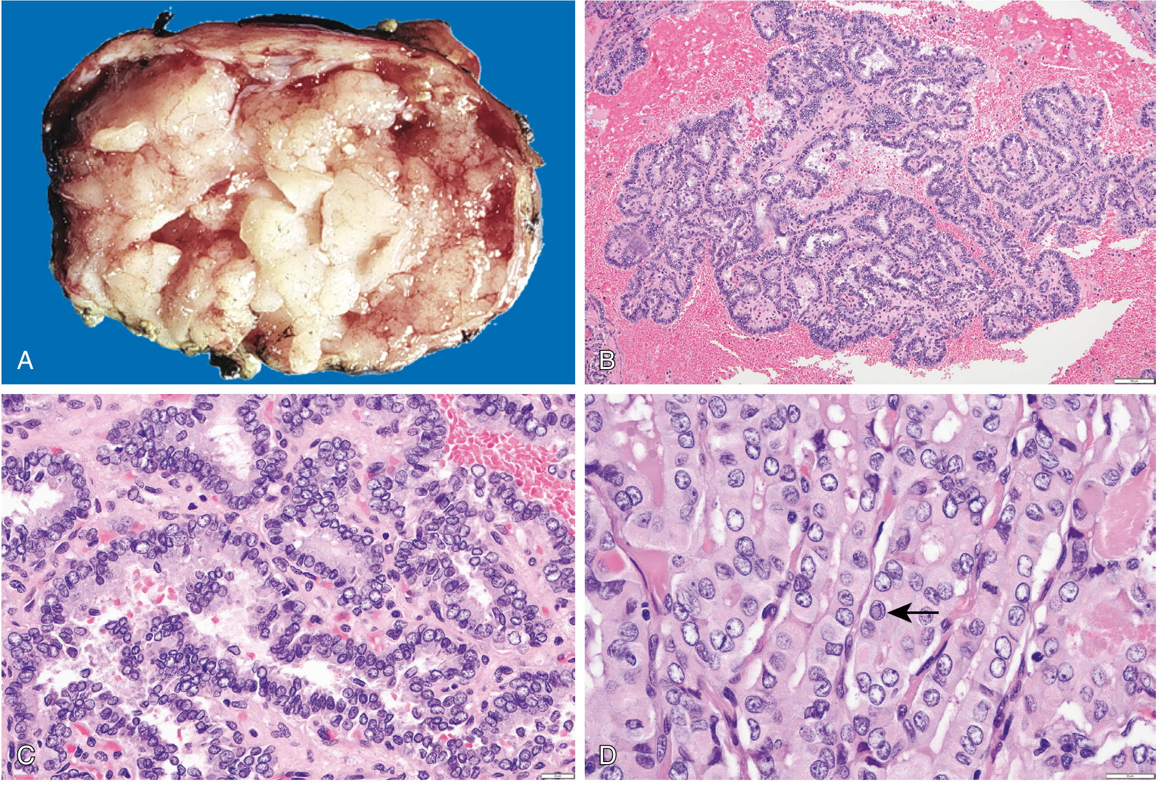
Fig. 24.19 - Papillary carcinoma of the thyroid. (A) Gross - papillary structures visible on cut surface. (B) Branching papillae with fibrovascular cores. (C) Crowded cells with characteristic nuclear features. (D) "Orphan Annie eye" nuclei with nuclear pseudo-inclusions (arrow). (Robbins & Cotran Pathologic Basis of Disease)
Key microscopic hallmarks:
- Branching papillae with fibrovascular stalks covered by cuboidal epithelial cells
- "Orphan Annie eye" (ground-glass) nuclei - finely dispersed chromatin, optically clear appearance
- Nuclear grooves (coffee-bean shape) and nuclear pseudoinclusions (cytoplasmic invaginations)
- Psammoma bodies - concentrically calcified structures in papillary cores/stroma; virtually pathognomonic (absent in follicular and medullary carcinoma)
- Lymphatic invasion common; blood vessel invasion uncommon in smaller lesions
Spread: Lymph node metastases to cervical nodes in up to 50% of cases. Hematogenous metastasis (to lung) less common.
Molecular drivers: BRAF V600E point mutation (most common) or fusion genes (RET/PTC, NTRK, BRAF fusions).
Prognosis: Excellent - 10-year survival >95%. 5-20% local/regional recurrence; 10-15% distant metastases. Prognosis worsens with age >55, extrathyroidal extension, distant metastases.
2. Follicular Carcinoma
Epidemiology: 5-15% of thyroid cancers (up to 25-40% in iodine-deficient regions). Peak age 40-60 years.
Macroscopy:
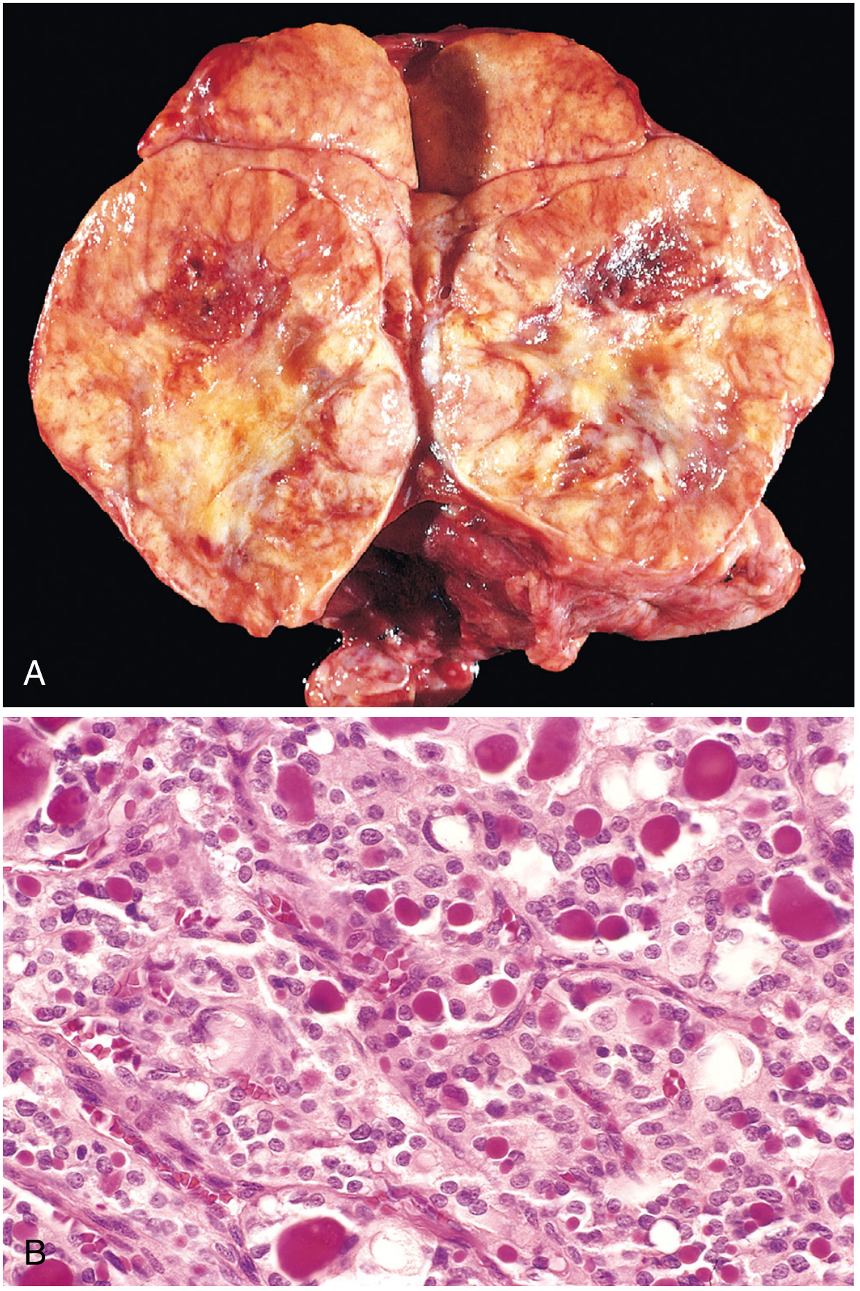
Fig. 24.20 - Follicular carcinoma. (A) Cut surface showing tan appearance with foci of hemorrhage. (B) Glandular lumens containing recognizable colloid. (Robbins & Cotran)
Solitary nodule; may be circumscribed or widely infiltrative. Gray-tan-pink on cut surface; may be translucent (large colloid-filled follicles). Larger lesions may penetrate the capsule.
Histopathology: Uniform cells forming small follicles with colloid - resembles normal thyroid. No ground-glass nuclei, no psammoma bodies. A subset shows oncocytic (Hürthle cell) features with abundant granular eosinophilic cytoplasm.
Critical distinction from adenoma: Requires demonstration of capsular invasion and/or vascular invasion on histology. FNA alone cannot distinguish follicular adenoma from minimally invasive follicular carcinoma - extensive capsular sampling is mandatory.
- Vascular invasion involves venous vessels within and just beyond the capsule
- Widely invasive follicular carcinoma: obvious infiltration of thyroid parenchyma and extrathyroidal soft tissues
Spread: Unlike PTC, hematogenous spread predominates (lung, bone). Lymphatic spread is uncommon.
Molecular drivers: Oncogenic RAS mutations and PAX8:PPARG fusions.
3. NIFTP (Noninvasive Follicular Thyroid Neoplasm with Papillary-Like Nuclear Features)
A reclassified entity - formerly "encapsulated follicular variant of PTC." Encapsulated, no invasion. Has PTC-like nuclear features but behaves as a benign/borderline lesion with excellent prognosis after surgical resection alone. This reclassification was made to avoid overtreatment.
4. Medullary Thyroid Carcinoma (MTC)
Epidemiology: ~5% of thyroid neoplasms. Derived from parafollicular C cells (neuroendocrine). 70% sporadic; 30% familial (MEN2A, MEN2B, familial MTC) - all caused by germline RET proto-oncogene mutations.
Macroscopy: Sporadic = solitary nodule. Syndromic = bilateral and multicentric. Firm, pale gray to tan, infiltrative. Areas of necrosis and hemorrhage in larger lesions.
Histopathology:
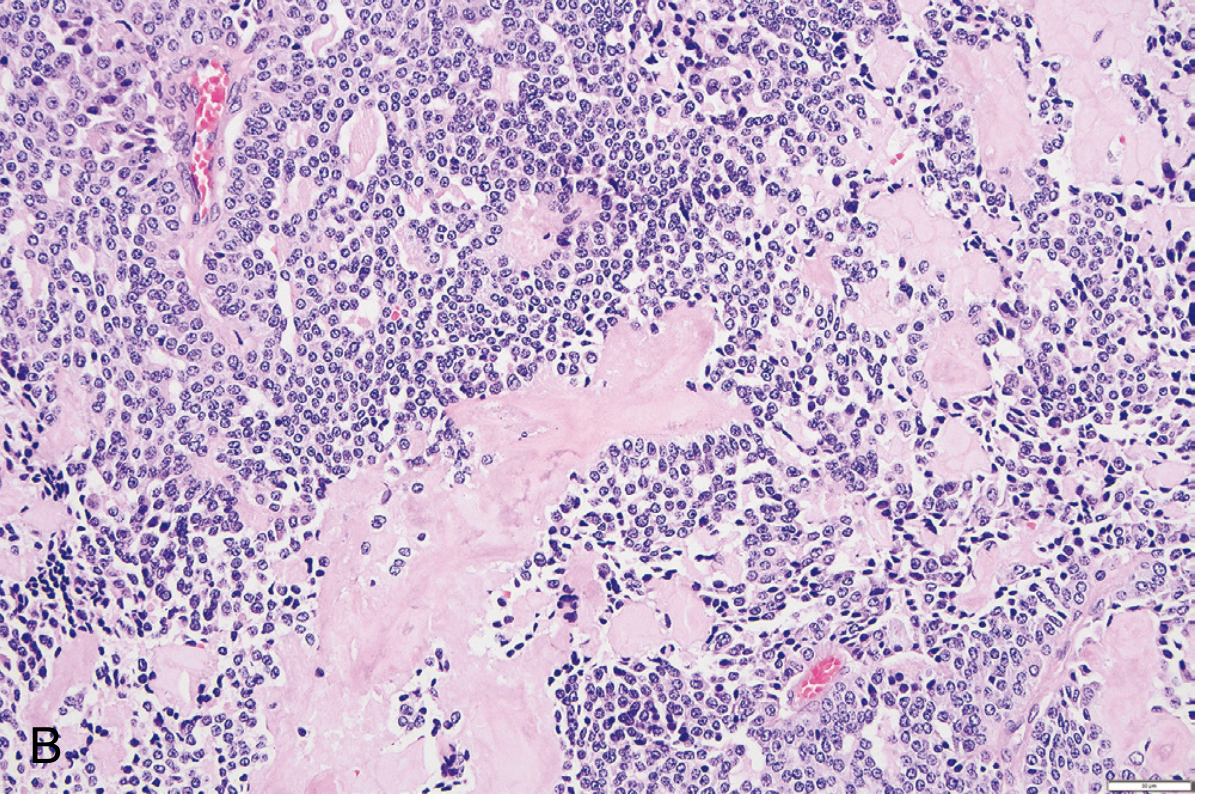
Fig. 24.22B - Medullary carcinoma. Nested architecture with round nuclei, stippled (neuroendocrine) chromatin, and intercellular amyloid (pink material) derived from calcitonin peptides. (Robbins & Cotran)
- Polygonal to spindle-shaped cells in nests, trabeculae, or follicles
- Amyloid deposits in stroma (calcitonin-derived) - Congo red positive
- Immunohistochemistry positive for calcitonin, CEA, synaptophysin, chromogranin
- Electron microscopy: membrane-bound electron-dense secretory granules
- Syndromic cases: C-cell hyperplasia in surrounding thyroid (precursor lesion) - often in contralateral lobe
Biomarkers:
- Serum calcitonin - key diagnostic and postoperative follow-up marker
- Serum CEA - useful especially in calcitonin-negative tumors
- Also may secrete: serotonin, corticotropin (ACTH), VIP
Clinical presentations: Neck mass; may have diarrhea (VIP), Cushing syndrome (ACTH). Hypocalcemia is NOT prominent despite elevated calcitonin.
Genetics: All MEN2 patients carry germline RET mutations. Prophylactic thyroidectomy offered to asymptomatic carriers. Treatment of advanced disease: RET tyrosine kinase inhibitors.
5. Poorly Differentiated and Anaplastic Thyroid Carcinoma
Anaplastic carcinoma is one of the most aggressive cancers known. It typically arises from dedifferentiation of pre-existing well-differentiated carcinoma. Associated with TP53 and TERT promoter mutations (in addition to RAS/BRAF as initiating events).
Macroscopy/Clinical: Rapidly enlarging bulky neck mass. At presentation, disease has usually spread beyond the thyroid or metastasized to the lungs. Symptoms of compression/invasion (dyspnea, dysphagia, hoarseness, cough) are common. Death occurs within 1 year in most cases.
Histopathology: Three main patterns - spindle cell, pleomorphic giant cell, and squamoid. Extensive necrosis. Marked nuclear pleomorphism and mitoses.
Diagnostic Approach
Step 1 - Clinical Assessment
High-risk history features:
- Age <20 years (20-50% malignancy rate for solitary nodule)
- Men >40 years, women >50 years with rapid growth
- Prior head/neck radiation exposure
- Family history of thyroid cancer, MEN2, pheochromocytoma, hyperparathyroidism
- Associated syndromes: Gardner syndrome, Cowden disease
High-risk physical exam features (from Cummings):
- Hard, fixed lesion
- Rapid growth
- Associated lymphadenopathy
- Vocal cord paralysis (perform laryngoscopy in all cases)
- Aerodigestive compromise (dysphagia, stridor)
- Pemberton sign positive (arms raised - facial suffusion/venous engorgement = substernal involvement)
Step 2 - Laboratory Studies
| Test | Indication/Role |
|---|---|
| TSH | First-line screening; hypothyroid/hyperthyroid shifts workup away from cancer |
| Serum calcitonin | If family history MTC, MEN2, or FNA suspicious for MTC |
| CEA | Useful in MTC monitoring |
| Thyroglobulin (Tg) | NOT useful at initial presentation; used postoperatively after total thyroidectomy for differentiated cancer |
| Calcium | Exclude hyperparathyroidism if MEN2 suspected |
| 24h urine metanephrines/catecholamines + abdominal MRI | If RET mutation present (exclude pheochromocytoma before surgery) |
Step 3 - Imaging
Ultrasound (US) - First-line imaging modality for all thyroid nodules.
Suspicious sonographic features:
- Hypoechoic solid nodule
- Irregular/lobulated margins
- Microcalcifications (correlate with psammoma bodies in PTC)
- Taller-than-wide shape
- Absent halo
- Increased intranodular vascularity
- Associated suspicious lymphadenopathy
The ACR TI-RADS (Thyroid Imaging Reporting and Data System) provides a standardized risk stratification to guide FNA decisions.
Radionuclide Scintigraphy (¹²³I or ⁹⁹ᵐTc)
- "Cold" (non-functioning) nodule: higher malignancy risk - warrants FNA
- "Hot" (hyperfunctioning) nodule: almost never malignant; warrants investigation for thyrotoxicosis
- FDG PET-CT is of limited value for pre-operative assessment (both benign and malignant lesions may be FDG-avid). However, it has an important role in post-treatment surveillance of differentiated thyroid cancer, particularly in patients with elevated thyroglobulin but negative ¹³¹I scan (pooled sensitivity 95%, specificity 81%). - Scott-Brown's Otorhinolaryngology, p. 597
CT/MRI - Used for:
- Substernal goiter assessment
- Evaluation of tracheal displacement/compression
- Staging of known malignancy and lymph node mapping
- Suspected extrathyroidal extension
Step 4 - Fine-Needle Aspiration Cytology (FNAC) - Cornerstone of Diagnosis
FNAC is the procedure of choice for evaluating thyroid nodules. It has dramatically reduced unnecessary surgeries by 35-75% and nearly tripled the malignancy yield in operated patients. Accuracy of PTC diagnosis by FNAC: 99% with false-positive rate <1%. - Cummings, p. 2375
Indications (general thresholds):
- Nodules ≥1 cm with any suspicious sonographic features
- All nodules ≥2 cm (due to higher false-negative rate for larger lesions)
- Nondiagnostic aspirates: repeat with ultrasound guidance; persistent nondiagnostic = surgical excision for solid nodules
The Bethesda System for Reporting Thyroid Cytopathology (TBSRTC):
| Bethesda Category | Diagnosis | Risk of Malignancy | Management |
|---|---|---|---|
| I | Nondiagnostic | 5-10% | Repeat US-guided FNA |
| II | Benign | 0-3% | Clinical follow-up |
| III | Atypia of undetermined significance (AUS/FLUS) | 10-30% | Repeat FNA, molecular testing |
| IV | Follicular neoplasm / Suspicious for follicular neoplasm | 25-40% | Surgical lobectomy |
| V | Suspicious for malignancy | 50-75% | Near-total thyroidectomy or lobectomy |
| VI | Malignant | 97-99% | Total thyroidectomy |
Limitation: FNAC cannot distinguish follicular adenoma from follicular carcinoma - this requires surgical resection and histologic demonstration of capsular/vascular invasion.
Step 5 - Molecular Testing
When FNA cytology is indeterminate (Bethesda III-IV), molecular testing significantly improves diagnostic accuracy:
- BRAF V600E mutation - highly specific for PTC malignancy (>99%)
- RET/PTC rearrangements, NTRK fusions - indicate papillary carcinoma
- RAS mutations - associated with follicular neoplasms (adenoma or carcinoma)
- PAX8:PPARG fusion - associated with follicular carcinoma
- TERT promoter mutations - marker of aggressive behavior/dedifferentiation
- TP53 mutations - associated with anaplastic transformation
- Germline RET mutation testing - all patients diagnosed with MTC and their first-degree relatives
Commercial panels (e.g., ThyroSeq, Afirma) combine multiple molecular markers for risk stratification of indeterminate nodules.
Summary Table: Key Distinguishing Features
| Feature | Papillary | Follicular | Medullary | Anaplastic |
|---|---|---|---|---|
| Frequency | 85% | 5-15% | ~5% | <5% |
| Origin | Follicular cells | Follicular cells | C cells | Dedifferentiated |
| Nuclear features | Ground-glass, grooves, pseudoinclusions | Normal/bland | Neuroendocrine (stippled) | Pleomorphic |
| Psammoma bodies | Yes | No | No | No |
| Amyloid in stroma | No | No | Yes | No |
| Spread | Lymphatic | Hematogenous | Both | Rapid local invasion |
| Key biomarker | Thyroglobulin (post-op) | Thyroglobulin (post-op) | Calcitonin + CEA | None reliable |
| Molecular marker | BRAF V600E | RAS, PAX8:PPARG | RET (germline/somatic) | TP53, TERT |
| FNA diagnosis | Reliable | Cannot distinguish from adenoma | Reliable | Reliable |
| Prognosis | Excellent (>95% 10-yr) | Good (capsular) / Guarded (widely invasive) | Intermediate | Very poor (<1 yr median) |
Sources: Robbins, Cotran & Kumar Pathologic Basis of Disease (9780443264528); Cummings Otolaryngology Head and Neck Surgery (9780323612173); Scott-Brown's Otorhinolaryngology Head & Neck Surgery Vol 1 (9781138094611)
Explain
Your credits are exhausted. You can purchase a top-up or subscribe to a plan to continue using Orris. If you're already subscribed, your daily credits will reset tomorrow.
This is a shared conversation. Sign in to Orris to start your own chat.