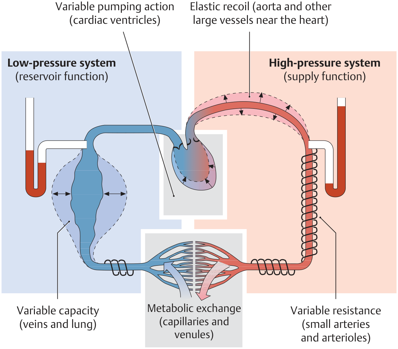
Is vena leva included in pulmonary nd systemic both circuits?
- Vena cava (superior/inferior) - these are the large veins returning deoxygenated blood to the right atrium
- Left side of the heart in general
pulmonary and systemic circulation diagram heart vena cava
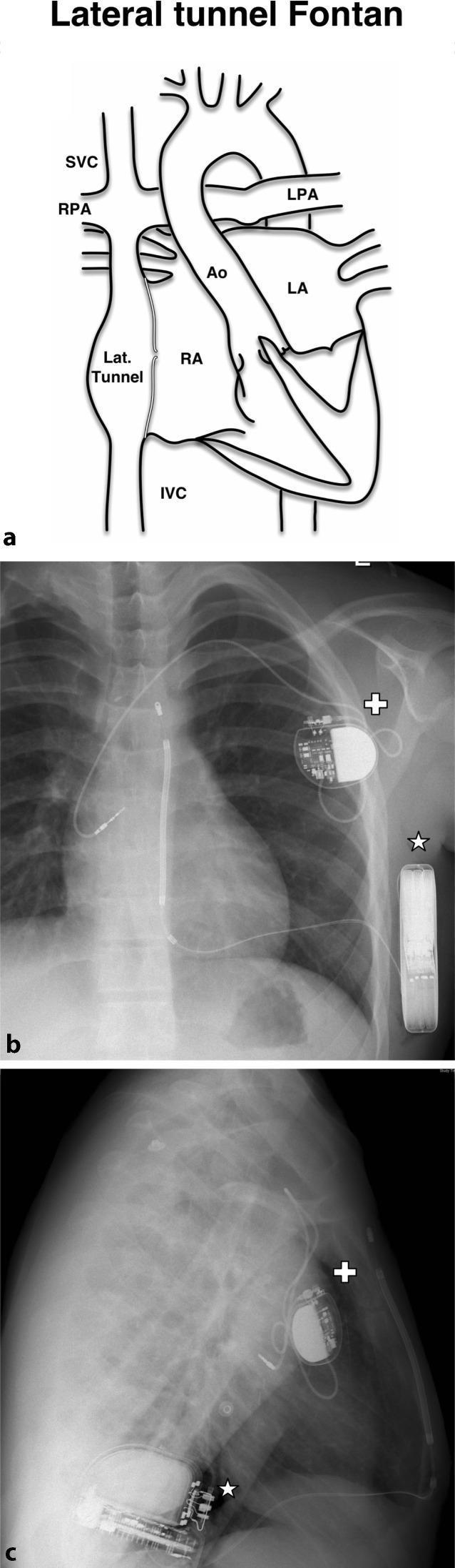
This composite educational resource illustrates a hybrid cardiac device management strategy in a patient with a lateral tunnel Fontan procedure for congenital heart disease. Panel (a) is an anatomical diagram showing the Fontan circulation, highlighting the lateral tunnel connecting the inferior vena cava (IVC) to the pulmonary arteries, bypassing the right heart. Panels (b) and (c) are posteroanterior and lateral chest radiographs demonstrating the dual-device configuration. A single-chamber transvenous atrial pacemaker (marked with a white cross) is situated in the upper right chest. Its radiopaque lead is seen extending into the systemic venous portion of the right atrium to provide bradycardia support. Simultaneously, a subcutaneous implantable cardioverter-defibrillator (S-ICD, marked with a white star) is positioned laterally along the ribcage in the PA view and anteriorly in the lateral view. The S-ICD provides defibrillation capabilities without requiring intracardiac leads, which is advantageous in complex post-surgical congenital heart anatomy where venous access to the ventricles may be limited or contraindicated. This case illustrates the integration of transvenous pacing and subcutaneous defibrillation technologies.
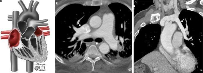
This composite educational material demonstrates Partial Anomalous Pulmonary Venous Return (PAPVR) through medical illustration and diagnostic imaging. Panel A is a conceptual anatomical diagram of the heart and great vessels showing pulmonary veins draining aberrantly into the right atrium and superior vena cava (SVC), rather than the left atrium, creating a left-to-right shunt. Panel B shows an axial contrast-enhanced CT image of an adult. A white arrow indicates an anomalous right upper lobe pulmonary vein draining directly into the SVC. Panel C displays a double oblique contrast-enhanced CT reconstruction. The white arrow highlights an anomalous left upper lobe pulmonary vein (vertical vein) that drains superiorly into the left brachiocephalic vein. The collection illustrates common variants of PAPVR, a congenital cardiovascular condition where some pulmonary veins drain into systemic venous circulation. These images are clinically significant for diagnosing incidental left-to-right shunts in adults, which may be asymptomatic or lead to right heart volume overload depending on the degree of anomalous flow.
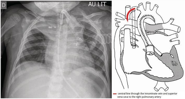
Anteroposterior (AP) chest radiograph and corresponding anatomical diagram demonstrating the trajectory of a peripherally inserted central catheter (PICC) in a pediatric patient with complex congenital heart disease. The radiograph shows a tracheostomy tube and several radiopaque surgical clips in the upper mediastinum. A radiopaque catheter enters from the left peripheral vasculature, coursing medially through the left innominate vein into the superior vena cava (SVC). Uniquely, due to a Fontan anastomosis (depicted in the adjacent diagram), the catheter passes directly from the SVC into the right pulmonary artery (RPA) rather than the right atrium. This specific positioning allows for direct monitoring of pulmonary arterial pressures. The anatomical diagram clarifies the post-surgical cardiovascular geometry, with a red arrow highlighting the path from the systemic venous return to the pulmonary circulation, bypassing the ventricle. The lungs, ribs, and clavicles serve as anatomical landmarks for verifying the catheter's mediastinal path and distal tip placement within the pulmonary vasculature.
Is the Vena Cava Part of Both Pulmonary and Systemic Circuits?
The Answer: NO - Vena Cava is Part of the Systemic Circuit ONLY
- Deoxygenated blood from the body returns via the superior and inferior vena cava to the right atrium
- Right atrium → right ventricle → pulmonary arteries → lungs (gas exchange)
- Oxygenated blood returns via pulmonary veins → left atrium
- Oxygenated blood leaves the left ventricle via the aorta
- Distributes to all organs and tissues
- Deoxygenated blood collects into the superior vena cava (from upper body) and inferior vena cava (from lower body)
- Returns to the right atrium - completing the systemic circuit
Summary Table
| Structure | Pulmonary Circuit | Systemic Circuit |
|---|---|---|
| Superior vena cava | ✗ No | ✓ Yes (returns blood TO right atrium) |
| Inferior vena cava | ✗ No | ✓ Yes (returns blood TO right atrium) |
| Pulmonary arteries | ✓ Yes | ✗ No |
| Pulmonary veins | ✓ Yes | ✗ No |
| Aorta | ✗ No | ✓ Yes |
Why the Confusion?