

Lable the image
A labeled histology diagram of H&E stained bone tissue section showing active bone remodeling. The image should clearly label the following structures with arrows pointing to their locations: 1) Osteoclast - large multinucleated giant cell in a resorption bay at the top center; 2) Howship's Lacuna (Resorption Bay) - the cavity where the osteoclast sits; 3) Osteoblasts - single layer of cuboidal cells lining the bone trabecula margin at the lower right; 4) Osteocytes - mature bone cells embedded within the pink bone matrix in lacunae; 5) Osteocyte Lacunae - small clear spaces within the bone matrix; 6) Bone Matrix (Trabecular Bone) - dense eosinophilic pink extracellular matrix; 7) Bone Marrow Space / Fibrovascular Stroma - cellular loose connective tissue in the lower left; 8) Stromal/Osteoprogenitor Cells - spindle-shaped cells in the marrow space. Style: clean medical/anatomical illustration with pink and purple H&E color scheme, white label text with arrows, dark background for labels.
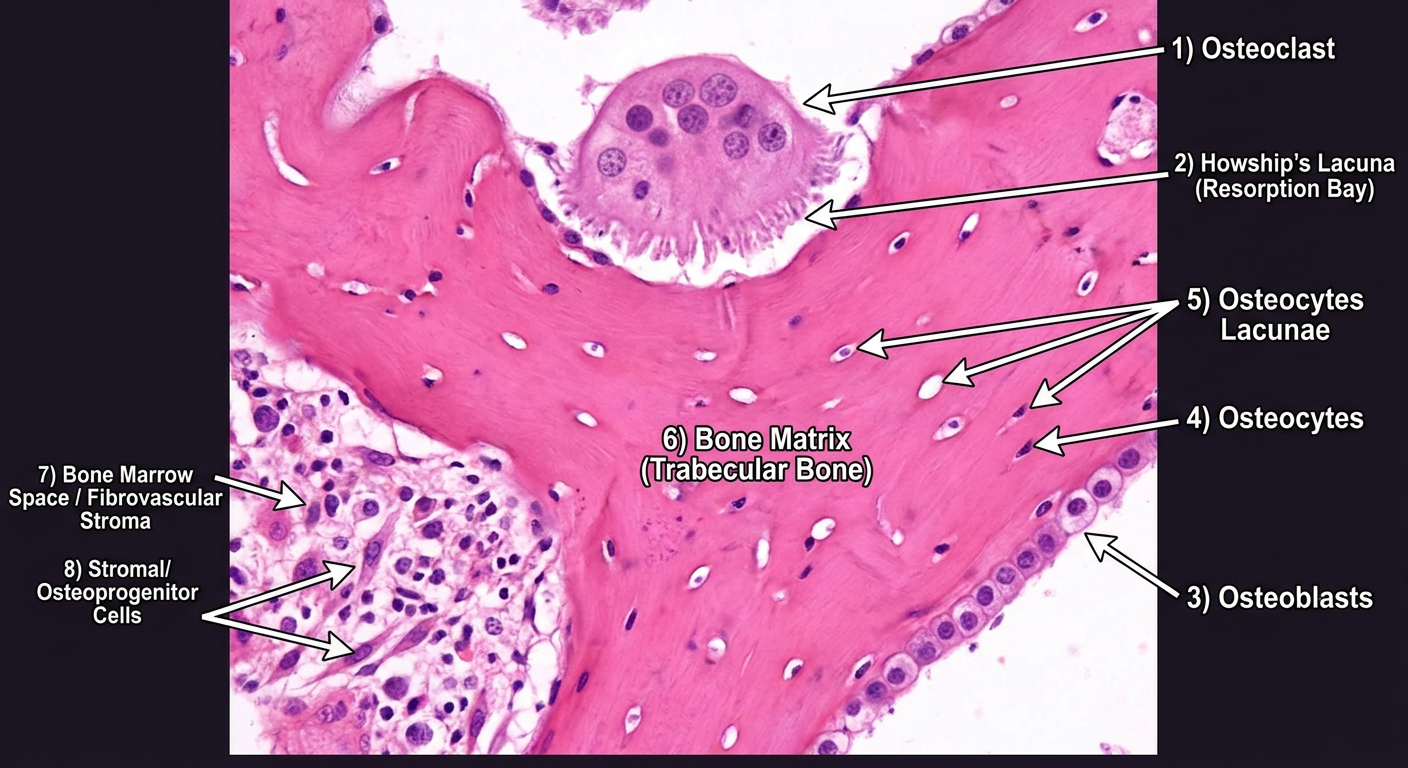
Labeled H&E Section - Active Bone Remodeling
Cell Types
| Label | Structure | Description |
|---|---|---|
| 1 | Osteoclast | Large multinucleated giant cell (upper-center). Has granular eosinophilic (pink) cytoplasm with multiple distinct nuclei. Responsible for bone resorption. |
| 2 | Osteoblasts | Single layer of cuboidal, mononuclear cells lining the lower-right margin of the trabecula. They actively synthesize new bone matrix (osteoid). |
| 3 | Osteocytes | Mature bone cells embedded individually within the pink bone matrix, housed inside lacunae. |
| 4 | Stromal / Osteoprogenitor Cells | Spindle-shaped cells in the loose connective tissue of the marrow space (lower-left). |
Structural Features
| Label | Structure | Description |
|---|---|---|
| 5 | Howship's Lacuna (Resorption Bay) | The shallow erosion cavity in the bone surface where the osteoclast is actively resorbing bone. |
| 6 | Osteocyte Lacunae | Small, clear oval spaces within the bone matrix that house the osteocytes. |
| 7 | Bone Matrix (Trabecular Bone) | Dense, bright eosinophilic (pink) extracellular matrix forming the bony spicule/trabecula. |
| 8 | Bone Marrow Space / Fibrovascular Stroma | The cellular loose connective tissue adjacent to the trabecula (lower-left), containing blood vessels, collagen, and progenitor cells. |
Key Teaching Point
Show in that same image
ls /tmp/workspace/ 2>/dev/null || mkdir -p /tmp/workspace/bone-histology
mkdir -p /tmp/workspace/bone-histology && curl -s -o /tmp/workspace/bone-histology/original.jpg "https://d361swluvexzqs.cloudfront.net/chats-attachments/IMG-20260806-WA0038-1785989196345-j4bzkj.jpg" && echo "Downloaded OK" && python3 -c "from PIL import Image; img = Image.open('/tmp/workspace/bone-histology/original.jpg'); print(img.size)"
~/bone-histology/label.py
from PIL import Image, ImageDraw, ImageFont
import os

img = Image.open("/tmp/workspace/bone-histology/original.jpg").convert("RGBA")
W, H = img.size  # 444 x 337

# Create overlay layer for arrows and labels
overlay = Image.new("RGBA", img.size, (0, 0, 0, 0))
draw = ImageDraw.Draw(overlay)

# Try to get a font; fall back to default
try:
    font = ImageFont.truetype("/usr/share/fonts/truetype/dejavu/DejaVuSans-Bold.ttf", 13)
    small_font = ImageFont.truetype("/usr/share/fonts/truetype/dejavu/DejaVuSans.ttf", 11)
except:
    font = ImageFont.load_default()
    small_font = font

# -------------------------------------------------------
# Label definitions: (label_text, arrow_start_xy, arrow_end_xy, text_anchor)
# arrow_start = tip of arrow (points AT the structure)
# arrow_end   = where the label text box is
# -------------------------------------------------------
labels = [
    # (text, tip_xy, label_xy, color)
    ("Osteoclast",           (215, 72),  (270, 28),   (255, 80, 80)),      # top center - large purple cell
    ("Howship's\nLacuna",    (205, 95),  (55,  30),   (255, 160, 50)),     # resorption bay under osteoclast
    ("Osteoblasts",          (310, 175), (355, 125),  (80, 200, 80)),      # cuboidal cells lower right
    ("Osteocytes",           (270, 155), (310, 220),  (100, 180, 255)),    # cells in matrix
    ("Osteocyte\nLacunae",   (240, 130), (130, 180),  (180, 130, 255)),    # clear spaces in matrix
    ("Bone Matrix\n(Trabecula)", (290, 105),(355, 60),(255, 220, 80)),     # pink dense bone
    ("Bone Marrow\nSpace",   (115, 220), (20,  250),  (200, 200, 200)),    # loose stroma lower left
    ("Stromal /\nProgenitor\nCells", (145, 240),(20, 295),(255, 200, 150)),# spindle cells
]

def draw_arrow(draw, tip, base, color, width=2):
    """Draw a line from base to tip (tip has arrowhead)."""
    draw.line([base, tip], fill=color + (230,), width=width)
    # Simple arrowhead: small filled circle at tip
    r = 4
    draw.ellipse([tip[0]-r, tip[1]-r, tip[0]+r, tip[1]+r], fill=color + (255,))

def draw_label_box(draw, text, pos, color, font):
    """Draw a semi-transparent box with text."""
    lines = text.split("\n")
    # Measure text size
    max_w = max(draw.textlength(line, font=font) for line in lines)
    line_h = 15
    box_w = int(max_w) + 8
    box_h = len(lines) * line_h + 6
    x, y = pos
    # Clamp to image bounds
    x = max(2, min(x, W - box_w - 2))
    y = max(2, min(y, H - box_h - 2))
    # Background box
    draw.rectangle([x-2, y-2, x+box_w, y+box_h], fill=(0, 0, 0, 160))
    draw.rectangle([x-2, y-2, x+box_w, y+box_h], outline=color+(220,), width=1)
    # Text
    for i, line in enumerate(lines):
        draw.text((x+2, y + i*line_h + 2), line, fill=color+(255,), font=font)
    # Return center of box for arrow base
    return (x + box_w//2, y + box_h//2)

for (text, tip, label_pos, color) in labels:
    base = draw_label_box(draw, text, label_pos, color, font)
    draw_arrow(draw, tip, base, color, width=2)

# Composite
result = Image.alpha_composite(img, overlay).convert("RGB")
result.save("/tmp/workspace/bone-histology/labeled.jpg", quality=95)
print("Saved labeled.jpg, size:", result.size)
python3 /tmp/workspace/bone-histology/label.py
labeled
Image · JPG
- 🔴 Osteoclast - large multinucleated giant cell (top center)
- 🟠 Howship's Lacuna - resorption bay beneath the osteoclast
- 🟢 Osteoblasts - cuboidal cells lining the trabecula (lower right)
- 🔵 Osteocytes - cells embedded in the bone matrix
- 🟣 Osteocyte Lacunae - clear spaces housing osteocytes
- 🟡 Bone Matrix / Trabecula - dense pink eosinophilic structure
- ⚪ Bone Marrow Space - loose stroma (lower left)
- 🟤 Stromal/Progenitor Cells - spindle-shaped cells in the marrow
Is that pathology
However, it could represent pathology depending on quantity and clinical context:
| Finding | Normal | Pathological |
|---|---|---|
| Osteoclasts present | Yes - physiological | Excess = hyperparathyroidism, Paget's disease, giant cell tumor, brown tumor |
| Howship's lacuna | Yes - normal remodeling | Excessive resorption = osteoporosis, osteitis fibrosa cystica |
| Osteoblasts lining bone | Yes - normal formation | Reactive excess = Paget's, osteosarcoma |
| Spindle stromal cells | Yes - marrow stroma | Atypical spindle cells = fibrosarcoma, fibrous dysplasia |
Top differentials if this IS pathological:
- Hyperparathyroidism (Osteitis Fibrosa Cystica) - prominent osteoclastic resorption with fibrovascular replacement of marrow
- Paget's Disease of Bone - chaotic remodeling with both osteoclasts and osteoblasts unusually active
- Giant Cell Tumor - osteoclast-like giant cells in a stromal background
- Brown Tumor - collections of osteoclasts in a fibrous stroma
Bottom line:
You are not showing correct labeling in image
~/bone-histology/label_v2.py
from PIL import Image, ImageDraw, ImageFont
import math

img = Image.open("/tmp/workspace/bone-histology/original.jpg").convert("RGBA")
W, H = img.size  # 444 x 337

overlay = Image.new("RGBA", img.size, (0, 0, 0, 0))
draw = ImageDraw.Draw(overlay)

try:
    font = ImageFont.truetype("/usr/share/fonts/truetype/dejavu/DejaVuSans-Bold.ttf", 12)
except:
    font = ImageFont.load_default()

# -------------------------------------------------------
# Corrected coordinates from vision AI analysis:
# Osteoclast: centered ~(260, 150), spans x:210-340, y:120-220
# Howship's Lacuna: below osteoclast, ~(250, 210)
# Osteoblasts: lower right margin ~(330, 230)
# Osteocytes: in bone matrix, scattered ~(190,130), (300,105)
# Bone Matrix: central dense pink area ~(250, 130)
# Bone Marrow Space: lower left ~(110, 240)
# Stromal cells: lower left ~(130, 260)
# -------------------------------------------------------

labels = [
    # (text, tip_xy, label_xy, RGB_color)
    ("Osteoclast",            (265, 155), (330, 35),   (255, 80,  80 )),
    ("Howship's Lacuna",      (250, 215), (100, 25),   (255, 165, 0  )),
    ("Osteoblasts",           (355, 235), (360, 290),  (80,  200, 80 )),
    ("Osteocyte",             (190, 130), (50,  130),  (100, 180, 255)),
    ("Osteocyte Lacuna",      (300, 108), (330, 165),  (180, 130, 255)),
    ("Bone Matrix",           (250, 100), (220, 40),   (255, 220, 60 )),
    ("Bone Marrow Space",     (100, 255), (10,  255),  (200, 200, 200)),
    ("Stromal Cells",         (145, 285), (15,  295),  (255, 180, 120)),
]

def draw_label_with_arrow(draw, text, tip, label_pos, color, font, W, H):
    lines = text.split("\n")
    padding = 5
    line_heights = []
    line_widths  = []
    for line in lines:
        bbox = font.getbbox(line)
        lw = bbox[2] - bbox[0]
        lh = bbox[3] - bbox[1]
        line_widths.append(lw)
        line_heights.append(lh)
    box_w = max(line_widths) + padding * 2
    box_h = sum(line_heights) + padding * 2 + (len(lines) - 1) * 2

    # Clamp label box
    lx = max(2, min(label_pos[0], W - box_w - 2))
    ly = max(2, min(label_pos[1], H - box_h - 2))

    # Draw background box
    draw.rectangle([lx, ly, lx + box_w, ly + box_h],
                   fill=(0, 0, 0, 180), outline=color + (230,), width=1)

    # Draw text
    cy = ly + padding
    for i, line in enumerate(lines):
        draw.text((lx + padding, cy), line, fill=color + (255,), font=font)
        cy += line_heights[i] + 2

    # Arrow: from center of box to tip
    box_cx = lx + box_w // 2
    box_cy = ly + box_h // 2

    # Find closest edge of box to tip, and draw arrow from that edge
    # Simple: just draw line from box center to tip
    draw.line([(box_cx, box_cy), tip], fill=color + (220,), width=2)

    # Arrowhead (small filled triangle toward tip)
    dx = tip[0] - box_cx
    dy = tip[1] - box_cy
    dist = math.hypot(dx, dy)
    if dist > 0:
        ux, uy = dx / dist, dy / dist
        # Arrowhead tip is `tip`, base is 8px back
        base_x = tip[0] - ux * 8
        base_y = tip[1] - uy * 8
        perp_x, perp_y = -uy * 4, ux * 4
        triangle = [
            tip,
            (base_x + perp_x, base_y + perp_y),
            (base_x - perp_x, base_y - perp_y),
        ]
        draw.polygon(triangle, fill=color + (255,))

for (text, tip, label_pos, color) in labels:
    draw_label_with_arrow(draw, text, tip, label_pos, color, font, W, H)

result = Image.alpha_composite(img, overlay).convert("RGB")
result.save("/tmp/workspace/bone-histology/labeled_v2.jpg", quality=95)
print("Done. Size:", result.size)
python3 /tmp/workspace/bone-histology/label_v2.py
labeled v2
Image · JPG
| Color | Label | Location in image |
|---|---|---|
| 🔴 Red | Osteoclast | Large multinucleated cell, upper-center (x:210-340, y:120-220) |
| 🟠 Orange | Howship's Lacuna | Resorption bay just below the osteoclast |
| 🟢 Green | Osteoblasts | Cuboidal cells along lower-right bone margin |
| 🔵 Blue | Osteocyte | Cell embedded in bone matrix, left-center |
| 🟣 Purple | Osteocyte Lacuna | Clear space housing osteocyte, right-center |
| 🟡 Yellow | Bone Matrix | Dense pink trabecula, upper-center |
| ⚪ White | Bone Marrow Space | Loose tissue, lower-left |
| 🟤 Tan | Stromal Cells | Spindle cells in marrow, lower-left |