
Immunopathology and AIDS 1.Hypersensitivity Reactions-define, Classify,and discuss 2.Type 1 Hypersensitivity Reaction Discuss and examples** 3.Graft rejection-mechanism and role of HLA antigens 4.Autoimmunity-define,pathogenesis. Autoimmune disorders-mechanisms* 5.SLE- pathogenesis,morphology And diagnosis* 6.HIV- life cycle,etiopathogenesis,lab Diagnosis,clinical stages of AIDS, Neoplasms found in AIDS patients** 2 mbbs pathology refer robbin add easy to draw diagram coloured , answer all question important topics frequently ask topic in muhs exam systemically aswer all
Reading File
Now let me run parallel searches for all topics simultaneously:
Finding Sources
Finding Sources
Finding Sources
Finding Sources
Finding Sources
Finding Sources
Finding Sources
Finding Sources
Now let me read the key Robbins sections in parallel:
Reading File
Reading File
Reading File
Excellent data found. Let me now read SLE, autoimmunity and graft rejection from Robbins in parallel:
Reading File
Reading File
Reading File
I have a wealth of content. Let me now fetch the key diagrams and read a few more critical sections (SLE morphology, graft rejection, HIV life cycle diagrams):
Loading Image
Reading File
Reading File
Loading Image
Reading File
I now have comprehensive content from Robbins. Let me update the todos and compile the full answer:Here is your complete, exam-ready 2nd MBBS Pathology notes on Immunopathology and AIDS, based on Robbins & Kumar (Basic Pathology + Pathologic Basis of Disease), organized systematically for MUHS exams.
IMMUNOPATHOLOGY AND AIDS
2nd MBBS Pathology - MUHS Exam Notes
(Reference: Robbins & Kumar - Basic Pathology / Pathologic Basis of Disease)
QUESTION 1: HYPERSENSITIVITY REACTIONS
Definition
Hypersensitivity refers to injurious immune reactions that cause tissue damage. These reactions arise when the immune response is:
- Inadequately controlled
- Directed against normally harmless antigens (environmental allergens)
- Inappropriately targeted to host tissues (autoimmunity)
The term implies an excessive or harmful reaction to an antigen in a previously sensitized individual.
Causes of Hypersensitivity
| Cause | Example |
|---|---|
| Autoimmunity (self antigens) | SLE, rheumatoid arthritis |
| Reactions against microbes | Post-streptococcal GN, TB |
| Environmental antigens | Pollen, dust mites, drugs |
Classification (Gell and Coombs - 4 Types)
┌─────────────────────────────────────────────────────────────────────┐
│           CLASSIFICATION OF HYPERSENSITIVITY REACTIONS              │
├────────┬───────────────┬───────────────┬────────────────────────────┤
│ TYPE   │ MECHANISM     │ MEDIATORS     │ EXAMPLES                   │
├────────┼───────────────┼───────────────┼────────────────────────────┤
│ I      │ IgE-mediated  │ IgE, Mast     │ Anaphylaxis, Asthma,       │
│ (Imm.) │ (Immediate)   │ cells,        │ Allergic rhinitis,         │
│        │               │ Eosinophils   │ Urticaria, Food allergy    │
├────────┼───────────────┼───────────────┼────────────────────────────┤
│ II     │ Antibody vs.  │ IgG, IgM,     │ Autoimmune hemolytic       │
│ (Cyto.)│ cell/tissue   │ Complement,   │ anemia, Goodpasture,       │
│        │ antigens      │ Phagocytes    │ Graves disease, MG         │
├────────┼───────────────┼───────────────┼────────────────────────────┤
│ III    │ Immune complex │ IgG, Comple-  │ SLE, Post-strep GN,        │
│(IC-med)│ deposition    │ ment, Neutro- │ Serum sickness,            │
│        │               │ phils         │ Arthus reaction            │
├────────┼───────────────┼───────────────┼────────────────────────────┤
│ IV     │ T cell-       │ CD4+ (DTH),   │ Contact dermatitis, TB     │
│ (DTH)  │ mediated      │ CD8+ (CTL),   │ granuloma, Type 1 DM,      │
│        │               │ Macrophages   │ Multiple sclerosis         │
└────────┴───────────────┴───────────────┴────────────────────────────┘
- Types I, II, III = Antibody-mediated
- Type IV = Cell-mediated (Delayed-type Hypersensitivity)
QUESTION 2: TYPE I HYPERSENSITIVITY (IMMEDIATE / IgE-MEDIATED) ⭐⭐
Definition
Type I hypersensitivity is an immediate (within minutes) immunologic reaction triggered by IgE antibodies bound to mast cells, upon re-exposure to an allergen. Also called Allergy or Atopy.
Mechanism (Step by Step)
┌─────────────────────────────────────────────────────────────────────┐
│              TYPE I HYPERSENSITIVITY - MECHANISM                    │
└─────────────────────────────────────────────────────────────────────┘

SENSITIZATION PHASE (First exposure):
   Allergen → APC presentation → Th2 cell activation
       ↓
   Th2 cells secrete IL-4 and IL-13
       ↓
   B cell class switching → IgE production
       ↓
   IgE binds to FcεRI receptors on MAST CELLS and BASOPHILS
   (Mast cells become "armed/sensitized")

ELICITATION PHASE (Re-exposure):
   Same allergen re-enters → CROSS-LINKS IgE on mast cells
       ↓
   Mast cell DEGRANULATION (within seconds-minutes)
       ↓
   Release of PRIMARY MEDIATORS from granules:
     • Histamine (vasodilation, increased permeability, smooth muscle spasm)
     • ECF-A (eosinophil chemotaxis)
     • NCF-A (neutrophil chemotaxis)
     • Proteases (tryptase, chymase)
       ↓
   Release of SECONDARY MEDIATORS (newly synthesized):
     • Leukotrienes (LTC4, LTD4, LTE4) - sustained bronchospasm
     • Prostaglandin D2 - bronchoconstriction
     • PAF - aggregates platelets
     • Cytokines (IL-4, IL-5, TNF) - late phase
Two Phases of Type I Response
Immediate Phase (0-30 minutes):
- Vasodilation, increased vascular permeability
- Smooth muscle spasm
- Hypersecretion of mucus
- Due to: Histamine, prostaglandins
Late Phase (2-24 hours):
- Infiltration by eosinophils, basophils, neutrophils, T cells
- Tissue damage, mucosal edema
- Due to: Leukotrienes, cytokines, eosinophil products (MBP)
Mediators Summary Table
| Mediator | Source | Effect |
|---|---|---|
| Histamine | Granules | Vasodilation, bronchoconstriction, itch |
| LTC4, LTD4 | Newly formed | Sustained bronchospasm (SRS-A) |
| PGD2 | Membrane | Bronchoconstriction |
| IL-5 | Cytokines | Eosinophil activation |
| TNF-α | Cytokines | Inflammation |
| Tryptase | Granules | Marker of mast cell activation |
Clinical Examples
| Condition | Features |
|---|---|
| Anaphylaxis | Systemic - shock, laryngospasm, urticaria, can be fatal |
| Bronchial Asthma | Episodic wheeze, breathlessness, bronchospasm |
| Allergic Rhinitis (Hay Fever) | Sneezing, nasal congestion, watery discharge |
| Urticaria/Angioedema | Skin wheals, dermal edema |
| Food Allergy | Peanuts, shellfish - GI and systemic |
| Drug Allergy | Penicillin - urticaria to anaphylaxis |
| Atopic Dermatitis (Eczema) | Chronic pruritic skin inflammation |
MUHS Exam Tip: Anaphylaxis = most severe systemic Type I reaction. SRS-A (Slow-Reacting Substance of Anaphylaxis) = LTC4 + LTD4 + LTE4.
QUESTION 3: GRAFT REJECTION - MECHANISM AND ROLE OF HLA ANTIGENS ⭐
Types of Graft Rejection
┌────────────────────────────────────────────────────────────┐
│                    GRAFT REJECTION                         │
├──────────────┬───────────┬─────────────┬───────────────────┤
│ TYPE         │ TIMING    │ MECHANISM   │ MORPHOLOGY        │
├──────────────┼───────────┼─────────────┼───────────────────┤
│ Hyperacute   │ Minutes-  │ Preformed   │ Thrombosis,       │
│              │ hours     │ antibodies  │ ischemic necrosis │
│              │           │ against HLA │                   │
├──────────────┼───────────┼─────────────┼───────────────────┤
│ Acute        │ Days-     │ T cells     │ Tubulointerstitial │
│ Cellular     │ weeks     │ (CD4+, CD8+)│ nephritis,        │
│              │           │ react to    │ vasculitis        │
│              │           │ donor HLA   │                   │
├──────────────┼───────────┼─────────────┼───────────────────┤
│ Acute        │ Weeks-    │ Antibodies  │ Necrotizing        │
│ Humoral      │ months    │ to HLA      │ vasculitis         │
│              │           │ (de novo)   │                   │
├──────────────┼───────────┼─────────────┼───────────────────┤
│ Chronic      │ Months-   │ T cells +   │ Vascular           │
│              │ years     │ Antibodies  │ intimal thicken-   │
│              │           │             │ ing, fibrosis,     │
│              │           │             │ "graft vasculo-    │
│              │           │             │ pathy"             │
└──────────────┴───────────┴─────────────┴───────────────────┘
Mechanisms of Acute T-Cell Mediated Rejection
Two pathways by which T cells recognize alloantigens:
-
Direct pathway:
- Recipient T cells recognize intact donor MHC on donor APCs
- Responsible for early, acute rejection
-
Indirect pathway:
- Recipient APCs process donor antigens
- Present donor peptides (including HLA peptides) to recipient T cells
- More important in chronic rejection
Effector mechanisms:
- CD4+ Th1 cells → secrete IFN-γ → macrophage activation → DTH-type inflammation
- CD8+ CTLs → directly kill graft parenchymal cells (via perforins/granzymes)
- CD4+ Th17 cells → recruit neutrophils
Role of HLA (MHC) Antigens in Graft Rejection
HLA = Human Leukocyte Antigens (encoded by MHC genes on chromosome 6p)
┌──────────────────────────────────────────────────────┐
│              HLA ANTIGENS IN TRANSPLANTATION         │
├──────────────┬───────────────────────────────────────┤
│ CLASS I      │ HLA-A, HLA-B, HLA-C                  │
│ (HLA Class I)│ Present on ALL nucleated cells        │
│              │ Recognized by CD8+ T cells            │
├──────────────┼───────────────────────────────────────┤
│ CLASS II     │ HLA-DR, HLA-DQ, HLA-DP               │
│ (HLA Class II│ Present on APCs (DC, macrophage, B)   │
│              │ Recognized by CD4+ T cells            │
└──────────────┴───────────────────────────────────────┘
Why HLA antigens cause rejection:
- Each person has a unique set of HLA molecules (except identical twins)
- Donor HLA molecules on graft cells = foreign antigens to recipient
- Recipient immune system mounts both cellular and humoral attack
- Greater the HLA mismatch → greater the rejection risk
Clinical significance:
- HLA typing done before transplantation (crossmatching)
- Best match = HLA-identical sibling (lowest rejection)
- Immunosuppressants (cyclosporine, tacrolimus) block T cell activation
Hyperacute rejection:
- Caused by preformed anti-HLA antibodies (from prior transfusion, pregnancy, or transplant)
- Complement activation → thrombosis → immediate graft loss
QUESTION 4: AUTOIMMUNITY ⭐
Definition
Autoimmunity is the failure of self-tolerance resulting in immune reactions directed against one's own (self) tissues, leading to tissue injury and autoimmune diseases.
Self-tolerance: The normal state in which the immune system does not react against its own antigens.
Mechanisms of Self-Tolerance (Normal)
-
Central tolerance:
- In thymus (T cells) and bone marrow (B cells)
- Clonal deletion of self-reactive lymphocytes (negative selection)
- AIRE gene controls expression of tissue antigens in thymus
-
Peripheral tolerance:
- Clonal anergy: Self-reactive T cells encounter antigen without co-stimulation → become unresponsive
- Regulatory T cells (Tregs): CD4+CD25+FoxP3+ cells suppress self-reactive T cells
- Activation-induced cell death (AICD): Repeated antigenic stimulation → apoptosis via Fas-FasL
Pathogenesis of Autoimmunity
┌──────────────────────────────────────────────────────────────────┐
│              PATHOGENESIS OF AUTOIMMUNITY                        │
└──────────────────────────────────────────────────────────────────┘

    GENETIC FACTORS               ENVIRONMENTAL FACTORS
    • HLA genes (HLA-DR2,         • Infections (molecular mimicry)
      HLA-DR3, HLA-B27)           • Polyclonal B cell activation
    • Non-HLA genes               • Tissue injury releasing
    • PTPN22, CTLA4, FoxP3          sequestered antigens
    • AIRE mutations              • UV light (SLE)
              ↓                              ↓
         FAILURE OF SELF-TOLERANCE
         • Escape of autoreactive T cells from thymus
         • Loss of peripheral anergy
         • Deficient Treg function
         • Molecular mimicry
         • Bystander activation
              ↓
     AUTOIMMUNE DISEASE
     (organ-specific or systemic)
Mechanisms in Autoimmune Disorders
| Mechanism | Disease | How It Works |
|---|---|---|
| Antibodies vs cell antigens (Type II) | Autoimmune hemolytic anemia | Anti-RBC IgG → complement lysis |
| Antibodies vs receptor (Type II) | Graves disease | Anti-TSH receptor → stimulation |
| Antibodies blocking receptor (Type II) | Myasthenia Gravis | Anti-AChR → impaired NMJ |
| Immune complexes (Type III) | SLE | Anti-dsDNA complexes → glomerulonephritis |
| T-cell mediated (Type IV) | Type 1 Diabetes | CD8+ T cells destroy beta cells |
| T cell + antibody | Rheumatoid Arthritis | Anti-IgG (RF), T cells in synovium |
Key concept: Molecular mimicry - Streptococcal M protein mimics cardiac myosin → rheumatic fever. EBV, CMV, and HIV cause polyclonal B cell activation → autoantibodies.
QUESTION 5: SYSTEMIC LUPUS ERYTHEMATOSUS (SLE) ⭐⭐
Definition
SLE is a chronic, multisystem autoimmune disease characterized by widespread autoantibodies against nuclear and cytoplasmic antigens, leading to tissue injury via immune complex deposition.
Pathogenesis of SLE
┌─────────────────────────────────────────────────────────────────┐
│                    PATHOGENESIS OF SLE                          │
└─────────────────────────────────────────────────────────────────┘

GENETIC PREDISPOSITION:
HLA-DR2, HLA-DR3 → Susceptibility
C1q, C2, C4 deficiencies → Impaired clearance of apoptotic material

TRIGGER:
UV light, Estrogen (F:M = 9:1), Drugs (hydralazine, procainamide)

DEFECTIVE CLEARANCE OF APOPTOTIC CELLS:
↓ Apoptotic bodies release nuclear fragments (dsDNA, histones, RNA)
↓ Normally cleared; in SLE → persist and stimulate TLRs
↓ TLR activation in pDCs → excessive type I IFN production

LOSS OF SELF-TOLERANCE:
↓ Autoreactive B cells and T cells escape elimination
↓ CD4+ T cell help → B cell hyperactivation
↓ Production of ANA (Antinuclear Antibodies)

KEY AUTOANTIBODIES IN SLE:
• Anti-dsDNA (highly specific for SLE, correlates with disease activity)
• Anti-Sm (Smith antigen - highly specific)
• Anti-histone (drug-induced lupus)
• Anti-Ro/SSA, Anti-La/SSB (neonatal lupus, Sjogren's)
• Antiphospholipid antibodies (thrombosis, recurrent miscarriage)
• ANA (screening test - sensitive but not specific)

TISSUE INJURY:
• Immune complexes (IC) deposit in basement membranes
  → Complement activation → Inflammation → Organ damage
• Direct antibody attack on cells (anemia, thrombocytopenia)
• T cell-mediated tissue injury
Morphology of SLE
1. Skin:
- Butterfly (malar) rash - erythema over cheeks and nose
- Hematoxylin bodies (LE bodies) - characteristic nuclear debris in tissue
- Vasculitis, fibrinoid necrosis
2. Kidneys (Lupus Nephritis - most important):
| WHO/ISN Class | Morphology |
|---|---|
| I | Normal |
| II | Mesangial disease |
| III | Focal proliferative GN |
| IV | Diffuse proliferative GN (most severe, most common) |
| V | Membranous GN |
| VI | Sclerotic GN |
- "Wire loop" lesion - immune complex deposits thicken capillary wall
- Hyaline thrombi in glomerular capillaries
- IF: granular ("lumpy-bumpy") deposits of IgG, IgM, C3
- EM: subendothelial deposits
3. Heart (Libman-Sacks Endocarditis):
- Sterile, small, warty vegetations on BOTH sides of mitral valve
- Due to immune complex deposition + fibrin
- Pathognomonic of SLE
4. Blood vessels:
- Fibrinoid necrosis of vessel walls (acute vasculitis)
- "Onion skin" (concentric fibrosis) in chronic cases - especially in spleen (perivascular fibrosis = "onion skin lesion")
5. Spleen:
- Splenomegaly
- "Onion skin" perivascular fibrosis (pathognomonic)
- Lymphoid hyperplasia
6. Lungs:
- Pleuritis (50%), lupus pneumonitis
- Pulmonary fibrosis
7. Joints:
- Non-erosive synovitis (vs. RA which is erosive)
8. CNS (NPSLE):
- Vasculitis, infarcts, diffuse microangiopathy
Diagnosis of SLE (ACR/EULAR Criteria - 2019)
ANA = Entry criterion (must be positive)
Points-based system across 7 domains:
- Constitutional (fever)
- Hematologic (hemolytic anemia, leucopenia, thrombocytopenia)
- Neuropsychiatric (seizures, psychosis)
- Mucocutaneous (malar rash, oral ulcers, photosensitivity, alopecia)
- Serosal (pleuritis, pericarditis)
- Musculoskeletal (arthritis)
- Renal (proteinuria > 0.5 g/day, biopsy findings)
- Immunology (anti-dsDNA, anti-Sm, complement levels, antiphospholipid antibodies)
Score ≥ 10 = SLE diagnosis
Laboratory investigations:
| Test | Significance |
|---|---|
| ANA (FANA) | Screening - 95% sensitive, NOT specific |
| Anti-dsDNA | Specific (70%), correlates with disease activity |
| Anti-Sm | Highly specific (25-30%) |
| Anti-histone | Drug-induced lupus |
| Low C3, C4, CH50 | Active disease |
| CBC | Anemia, leucopenia, thrombocytopenia |
| Urinalysis | Proteinuria, RBC casts |
| Antiphospholipid Ab | VDRL false positive, thrombosis |
MUHS Exam Tip: LE cells = Neutrophil that has engulfed nuclear material opsonized by anti-histone antibodies. LE cell phenomenon = hallmark, but now replaced by ANA testing.
QUESTION 6: HIV - LIFE CYCLE, ETIOPATHOGENESIS, LAB DIAGNOSIS, CLINICAL STAGES OF AIDS, NEOPLASMS ⭐⭐
HIV Genome (Easy to Draw)
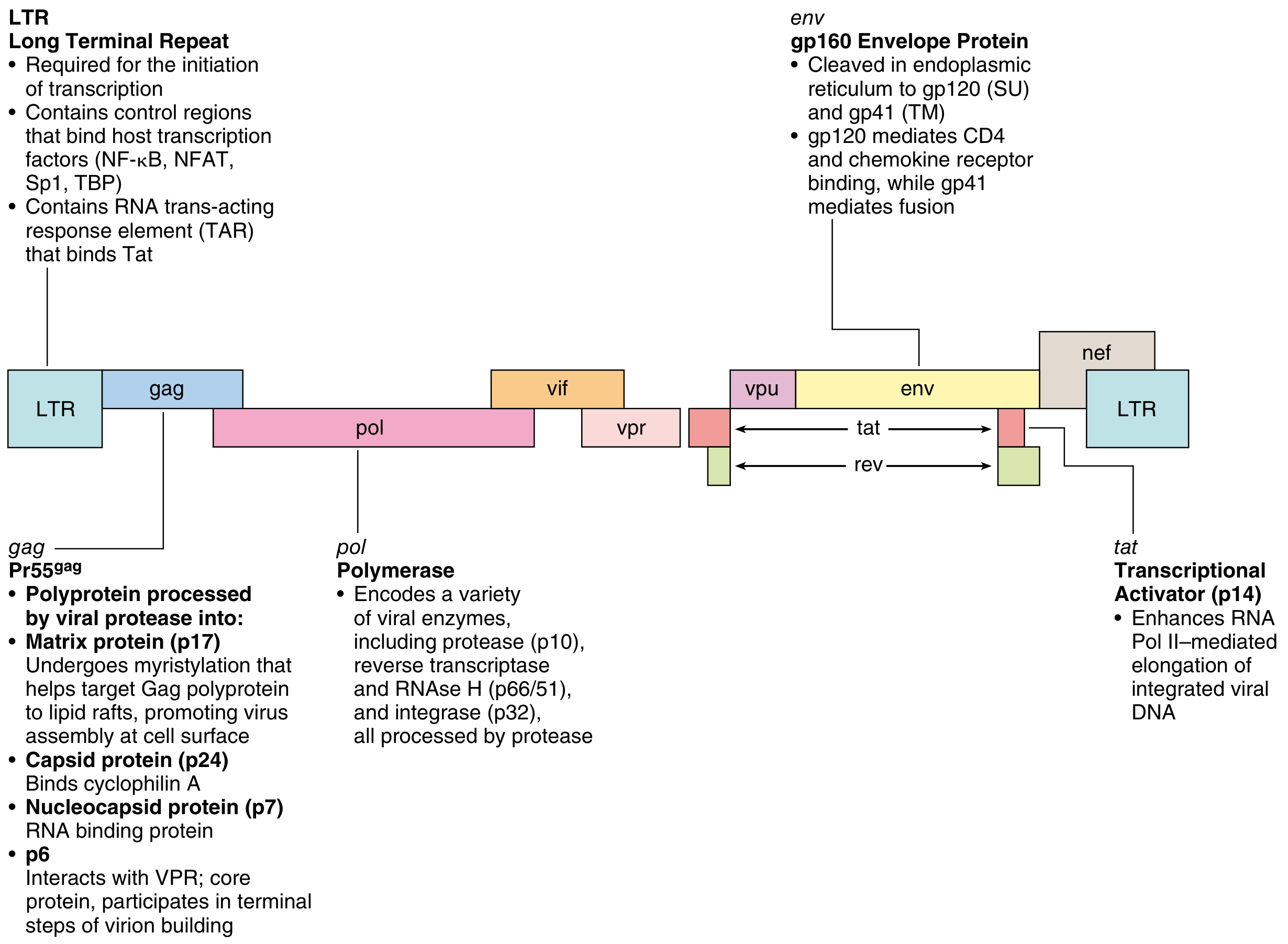
Key genes:
- gag: Encodes core proteins (p24 capsid, p17 matrix, p7 nucleocapsid)
- pol: Encodes enzymes - reverse transcriptase (p66/51), integrase (p32), protease (p10)
- env: Encodes envelope glycoproteins gp160 → cleaved to gp120 (binds CD4) + gp41 (fusion)
- LTR: Long terminal repeats - contain NF-κB, NFAT, Sp1 binding sites; required for transcription
- tat: Transcriptional activator (essential for replication)
- rev: Regulates export of viral mRNA
- nef: Downregulates CD4, MHC-I on infected cells; enhances viral infectivity
- vif: Degrades APOBEC3G (cellular antiviral enzyme)
HIV Life Cycle (EASY TO DRAW)
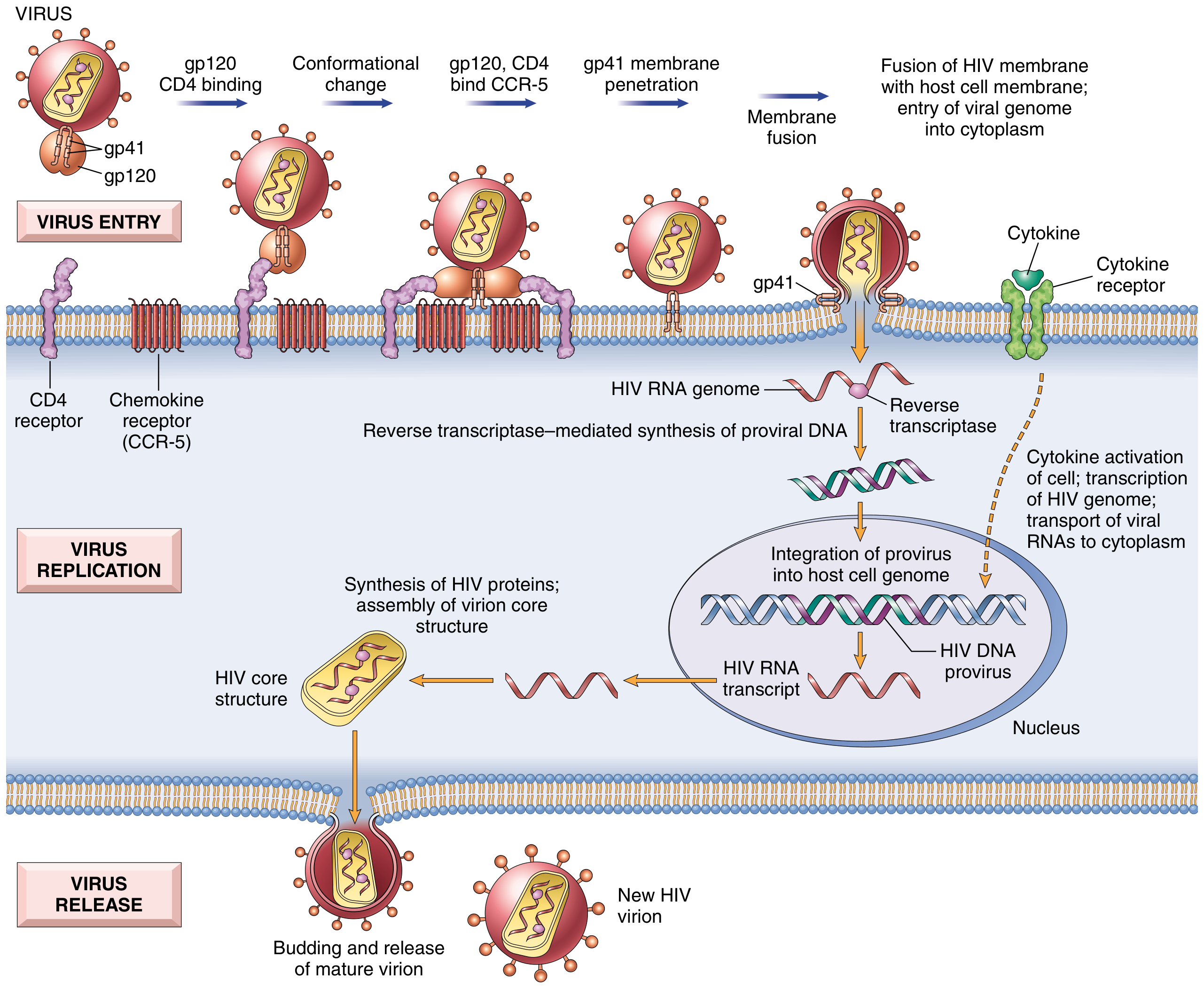
┌─────────────────────────────────────────────────────────────────────┐
│                    HIV LIFE CYCLE                                   │
└─────────────────────────────────────────────────────────────────────┘

STEP 1 - ATTACHMENT/ENTRY:
  HIV gp120 → binds CD4 receptor (T cells, macrophages, DCs)
       ↓ Conformational change in gp120
  gp120 → binds coreceptor CCR5 (M-tropic, macrophages - early)
                           or CXCR4 (T-tropic, T cells - late)
       ↓ Conformational change in gp41
  gp41 fusion peptide inserts into cell membrane
       ↓ Membrane fusion
  HIV RNA genome enters cytoplasm

STEP 2 - REVERSE TRANSCRIPTION:
  HIV RNA → (Reverse Transcriptase) → dsDNA (proviral DNA)

STEP 3 - INTEGRATION:
  Proviral DNA → (Integrase) → integrates into HOST genome
  → LATENT INFECTION (may persist for months/years)

STEP 4 - TRANSCRIPTION (Activation required):
  Cell activation (cytokines, infections) → NF-κB activation
  → Transcription of HIV genes via Tat protein
  → HIV mRNA → cytoplasm

STEP 5 - TRANSLATION:
  HIV proteins synthesized (Gag, Pol, Env)
  → Core proteins assemble

STEP 6 - BUDDING AND MATURATION:
  Immature virus buds from cell membrane
  → Viral Protease cleaves polyproteins
  → Mature infectious HIV virion released
  → CD4+ T cell dies
Drugs targeting each step:
- Entry: Maraviroc (CCR5 antagonist), Enfuvirtide (fusion inhibitor)
- Reverse transcriptase: NRTI (Zidovudine), NNRTI (Nevirapine)
- Integrase: Raltegravir
- Protease: Lopinavir
Etiopathogenesis of AIDS
HIV = Human Immunodeficiency Virus
- Family: Retroviridae, genus Lentivirus
- Types: HIV-1 (worldwide), HIV-2 (West Africa)
- Transmission: Sexual, parenteral (blood/needles), mother-to-child (vertical)
┌─────────────────────────────────────────────────────────────────┐
│            MECHANISMS OF CD4+ T CELL DEPLETION                  │
└─────────────────────────────────────────────────────────────────┘

DIRECT EFFECTS:
• Productive viral replication → cell lysis
• Accumulation of unintegrated viral DNA → toxic
• HIV-induced apoptosis (via gp120 cross-linking of CD4)
• Formation of syncytia (gp120 on infected cell binds CD4 on uninfected cell)

INDIRECT EFFECTS:
• Immune-mediated killing of HIV-infected T cells by CD8+ CTLs
• Chronic immune activation → activation-induced cell death (AICD)
• Destruction of CD4+ T cell precursors in thymus and bone marrow
• Macrophage and DC dysfunction → impaired antigen presentation

ROLE OF MACROPHAGES AND DCs:
• HIV infects macrophages via CCR5 (early in infection)
• Macrophages are RESERVOIRS of HIV (not killed, virus persists)
• Infected DCs carry virus to lymph nodes → amplify infection

VIRAL LOAD AND CD4 COUNT:
• Normal CD4 = 500-1500 cells/μL
• AIDS defined as CD4 < 200 cells/μL OR AIDS-defining illness
• Viral load correlates with rate of disease progression
Laboratory Diagnosis of HIV
SCREENING TESTS (highly sensitive):
┌─────────────────────────────────────────────────────────┐
│  ELISA (4th generation = detects HIV Ag + Ab together)  │
│  • Detects HIV p24 antigen + anti-HIV antibodies        │
│  • Window period: ~18 days for 4th gen                  │
│  • Sensitivity: >99.9%                                  │
└─────────────────────────────────────────────────────────┘

CONFIRMATORY TESTS:
┌─────────────────────────────────────────────────────────┐
│  WESTERN BLOT                                           │
│  • Gold standard confirmatory test                      │
│  • Detects antibodies to specific viral proteins        │
│  • Positive if bands at: p24, gp41, gp120/160          │
└─────────────────────────────────────────────────────────┘

MONITORING TESTS:
• CD4+ T cell count: Immunological status, when to start ART
• HIV RNA viral load (RT-PCR): Treatment response, infectivity
• HIV resistance testing: Guide drug selection
• p24 antigenemia: Early infection, infants

TESTS FOR INFANTS (<18 months):
• HIV DNA PCR or HIV RNA PCR (maternal antibodies persist, ELISA unreliable)
• Positive at 4 weeks → confirmatory test at 4 months

RAPID TESTS:
• Point-of-care, result in 20-30 min
• Used in resource-limited settings, labor/delivery
Clinical Stages of HIV Infection / AIDS
WHO Clinical Staging:
STAGE 1: ACUTE HIV INFECTION (2-4 weeks after exposure)
━━━━━━━━━━━━━━━━━━━━━━━━━━━━━━━━━━━━━━━━━━━━━━━━━━━━━━━
• Acute retroviral syndrome (mononucleosis-like illness)
• Fever, lymphadenopathy, pharyngitis, rash, myalgia
• High viral load, transient CD4 fall
• Lasts 2-4 weeks, resolves spontaneously
• Anti-HIV antibodies appear (seroconversion)

STAGE 2: CHRONIC ASYMPTOMATIC PHASE (Clinical Latency)
━━━━━━━━━━━━━━━━━━━━━━━━━━━━━━━━━━━━━━━━━━━━━━━━━━━━━━━
• Typically asymptomatic (may have persistent generalized lymphadenopathy)
• Low-level viral replication in lymph nodes
• Gradual CD4 decline (~50 cells/μL/year)
• Duration: Untreated = 8-10 years (without ART)
• Persistent Generalized Lymphadenopathy (PGL)

STAGE 3: SYMPTOMATIC HIV DISEASE
━━━━━━━━━━━━━━━━━━━━━━━━━━━━━━━━━━━━━━━━━━━━━━━━━━━━━━━
• Constitutional symptoms: Weight loss, fever, night sweats, diarrhea
• CD4 200-500 cells/μL
• Minor opportunistic infections
• Oral candidiasis, oral hairy leukoplakia (EBV), shingles (VZV)
• Bacterial pneumonias (recurrent)

STAGE 4: AIDS (Full-blown AIDS)
━━━━━━━━━━━━━━━━━━━━━━━━━━━━━━━━━━━━━━━━━━━━━━━━━━━━━━━
• CD4 < 200 cells/μL OR AIDS-defining illness
• Major opportunistic infections (see below)
• Constitutional: Wasting syndrome (HIV wasting > 10% weight loss)

WHO STAGES: 1 → 2 → 3 → 4 (correlates with CDC classification)
CDC: Category A (acute/asymptomatic) → B (symptomatic) → C (AIDS)
AIDS-Defining Opportunistic Infections by CD4 Count
CD4 < 500: Candida (oral), Kaposi sarcoma, TB reactivation
CD4 < 200: PCP (Pneumocystis jirovecii pneumonia) - MOST COMMON OI
           Toxoplasma encephalitis, Cryptococcal meningitis
CD4 < 100: Cryptosporidiosis, Microsporidiosis
CD4 < 50:  CMV retinitis, Disseminated MAC (Mycobacterium avium complex)
           PML (Progressive Multifocal Leukoencephalopathy - JC virus)
Common Opportunistic Infections:
| System | Pathogen | Presentation |
|---|---|---|
| Lungs | Pneumocystis jirovecii (PCP) | Dry cough, dyspnea, bilateral infiltrates; "ground glass" on HRCT |
| CNS | Toxoplasma gondii | Ring-enhancing lesion on CT; fever, headache, focal neuro signs |
| CNS | Cryptococcus neoformans | Meningitis; India ink = positive; cryptococcal antigen |
| GI | Cryptosporidium | Profuse watery diarrhea |
| Eye | CMV | Retinitis ("pizza pie" appearance); CD4 < 50 |
| Mouth | Candida albicans | White plaques (thrush), pseudomembranous |
| Lungs | MAC (M. avium complex) | Disseminated; fever, weight loss, diarrhea |
| Brain | JC virus (PML) | White matter demyelination; cognitive decline |
| Esophagus | HSV, CMV | Ulcerations |
Neoplasms in AIDS Patients ⭐⭐
25-40% of untreated HIV-infected individuals develop malignancy. All are linked to oncogenic DNA viruses - because HIV-mediated immunosuppression fails to control these latent viruses.
┌──────────────────────────────────────────────────────────────────────┐
│                  NEOPLASMS IN AIDS PATIENTS                          │
├──────────────────┬───────────────┬─────────────────────────────────  ┤
│ TUMOR            │ VIRUS/CAUSE   │ FEATURES                         │
├──────────────────┼───────────────┼──────────────────────────────────┤
│ KAPOSI SARCOMA   │ HHV-8 (KSHV) │ MOST COMMON tumor in AIDS;       │
│ ⭐ (KS)          │               │ Vascular tumor; purple/red skin  │
│                  │               │ plaques; also visceral           │
├──────────────────┼───────────────┼──────────────────────────────────┤
│ HIGH-GRADE       │ EBV           │ 2nd most common; diffuse large   │
│ B-CELL LYMPHOMA  │               │ B cell or Burkitt lymphoma;      │
│                  │               │ CNS lymphoma (primary)           │
├──────────────────┼───────────────┼──────────────────────────────────┤
│ CERVICAL CANCER  │ HPV (16, 18)  │ In HIV+ women; aggressive course │
├──────────────────┼───────────────┼──────────────────────────────────┤
│ ANAL CANCER      │ HPV           │ In HIV+ men (MSM)                │
├──────────────────┼───────────────┼──────────────────────────────────┤
│ PRIMARY CNS      │ EBV           │ Ring-enhancing lesion; must      │
│ LYMPHOMA         │               │ distinguish from toxoplasma      │
├──────────────────┼───────────────┼──────────────────────────────────┤
│ PRIMARY          │ HHV-8 + EBV   │ Pleural/peritoneal effusion;     │
│ EFFUSION         │               │ Body cavity lymphoma             │
│ LYMPHOMA         │               │                                  │
├──────────────────┼───────────────┼──────────────────────────────────┤
│ MULTICENTRIC     │ HHV-8         │ B-cell lymphoproliferative;      │
│ CASTLEMAN'S      │               │ systemic lymphadenopathy         │
│ DISEASE          │               │                                  │
└──────────────────┴───────────────┴──────────────────────────────────┘
Kaposi Sarcoma (KS) - Details:
- Most common neoplasm in AIDS patients
- Caused by HHV-8 (human herpesvirus 8 = Kaposi's Sarcoma Herpesvirus, KSHV)
- Morphology: Spindle-shaped cells expressing endothelial + smooth muscle markers; slit-like vascular spaces; mixed inflammatory infiltrate
- Lesions: Purple-red skin plaques (face, extremities, genitals) → disseminates to lungs, GI, lymph nodes
- Incidence fell dramatically with antiretroviral therapy (ART)
- Polyclonal or oligoclonal in early stages (not a true malignant tumor)
- HHV-8 encodes: viral cyclin D homologue, inhibitors of p53 → promotes spindle cell survival
QUICK REVISION SUMMARY FOR MUHS
| Topic | Key Point to Remember |
|---|---|
| Type I HSR | IgE + Mast cells + Histamine + Leukotrienes; SRS-A = LTC4+D4+E4 |
| Type II HSR | IgG/IgM against cell surfaces; complement/phagocytosis |
| Type III HSR | Immune complexes; "lumpy-bumpy" IF; Arthus, serum sickness |
| Type IV HSR | T-cell mediated; DTH; delayed 48-72 hrs; PPD test |
| Graft rejection | Hyperacute = preformed Ab; Acute = T cells; Chronic = both |
| HLA | Class I = HLA-A,B,C (all cells, CD8+); Class II = HLA-DR,DQ,DP (APCs, CD4+) |
| Autoimmunity | Failure of self-tolerance; molecular mimicry; HLA genes |
| SLE | Anti-dsDNA (specific); ANA (sensitive); Wire loop; Libman-Sacks; Onion skin |
| HIV Receptor | gp120 + CD4 + CCR5/CXCR4; gp41 = fusion |
| HIV Stages | Acute → Latent (8-10 yr) → Symptomatic → AIDS (CD4 <200) |
| AIDS tumors | KS (HHV-8); B-cell lymphoma (EBV); Cervical/Anal Ca (HPV) |
| HIV labs | ELISA screen → Western blot confirm; CD4 count + viral load monitor |
Source: Robbins & Kumar Basic Pathology (10th Ed.) and Robbins, Cotran & Kumar Pathologic Basis of Disease (10th Ed.)
This is a shared conversation. Sign in to Orris to start your own chat.