
Scrotal swelling differential diagnosis
scrotal swelling differential diagnosis diagram
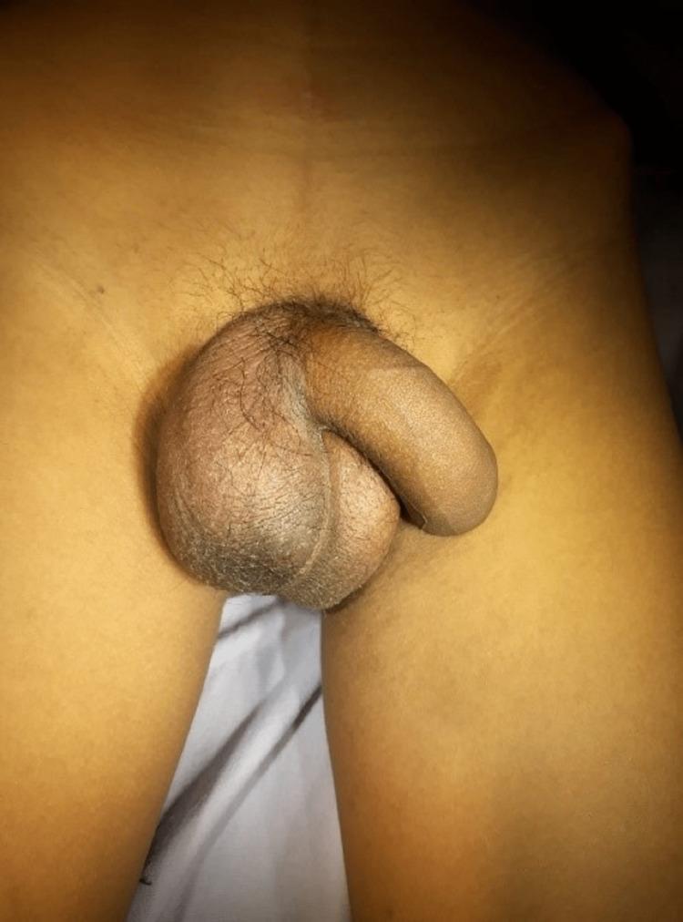
This clinical photograph displays the external genitalia of a male patient, focusing on the inguinal and scrotal region. The primary finding is significant asymmetrical scrotal swelling and enlargement, particularly on the patient's right side. The scrotal skin exhibits prominent rugae and a thickened, coarse texture with noticeable erythematous to dark-brown hyperpigmentation relative to the adjacent thigh skin. These visual signs are indicative of an 'acute scrotum,' a urological emergency. The differential diagnosis for such a presentation includes testicular torsion, epididymo-orchitis, or a strangulated inguinal hernia. The image illustrates the typical clinical manifestations of testicular ischemia or inflammatory processes within the scrotal sac, emphasizing the importance of rapid clinical evaluation and diagnostic imaging to differentiate between torsion and infectious etiologies.
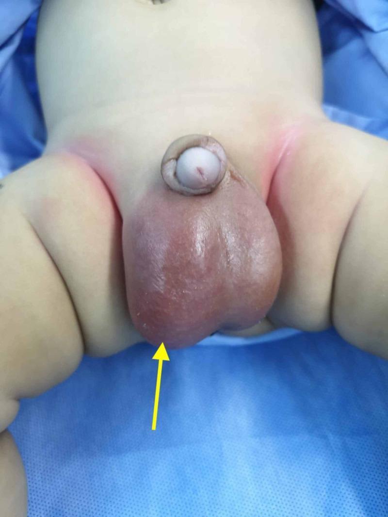
Clinical photograph of a pediatric patient's genital region demonstrating acute scrotal swelling. The image shows a significant, asymmetrical enlargement of the scrotum, predominantly affecting the right hemi-scrotum. The right scrotal sac is distended with a smooth, tense surface and exhibits a marked reddish-purple discoloration (erythema), indicated by a yellow arrow, which is highly suggestive of an underlying inflammatory or infectious process such as an infected hydrocele. The left hemi-scrotum also appears somewhat enlarged, indicating bilateral hydroceles, though it lacks the intense inflammatory color change seen on the right. Mild erythema is also visible in the bilateral inguinal folds. The penis is present and appears structurally normal but is slightly displaced superiorly and laterally by the large scrotal mass. This image serves as a clinical example of the differential diagnosis for an 'acute scrotum' in a pediatric setting, emphasizing the visual signs of secondary infection or inflammation complicating a simple hydrocele.
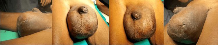
A series of four clinical photographs showing preoperative views of a massive, unilateral swelling of the left scrotum in an adult male. The swelling is approximately 25 x 15 cm, causing significant distortion and displacement of the penile shaft. The skin over the affected area is tense and shiny with a notable loss of normal scrotal rugosities (wrinkles), indicating severe distension. A visible cutaneous scar is present on the inferior aspect of the left hemiscrotum, consistent with a history of previous aspiration. The swelling is well-circumscribed, and the overlying skin appears hyperpigmented compared to the surrounding inguinal and thigh regions. This image illustrates the clinical presentation of chronic massive scrotal swelling, requiring differential diagnosis between giant hydrocele, pyocele, or a testicular neoplasm. The educational focus is on the physical examination findings of scrotal pathology, including changes in skin texture and topographical anatomy.
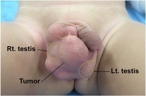
Clinical photograph of the perineal region of a pediatric patient showing significant scrotal swelling. The image features yellow-outlined areas identifying the anatomical positions of the bilateral testes, which are displaced by a large, centrally located, lobulated intrascrotal mass. The tumor is visibly larger than the testes, causing marked distension of the scrotal sac. The overlying skin appears intact and smooth without erythema or ulceration. Clinical markers indicate the tumor is independent of the testicles, localized within the right side of the scrotum. This visual demonstrates a typical presentation of an intrascrotal lipoblastoma, a rare benign mesenchymal tumor occurring in young children. The image serves to teach the differential diagnosis of pediatric scrotal masses, emphasizing the importance of identifying the relationship between the mass and normal testicular tissue during clinical examination and preoperative planning.
testicular torsion bell clapper deformity anatomy
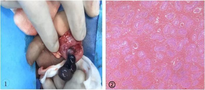
This dual-panel image demonstrates the surgical and pathological findings of testicular torsion associated with a bell clapper deformity. Panel 1 is an intraoperative clinical photograph of an inguinal/scrotal exploration. It shows a dark, cyanotic, and necrotic-appearing testis and epididymis held by gloved fingers. The tunica vaginalis is seen completely encircling the testis and distal spermatic cord, which characterizes the bell clapper deformity and allows the testis to rotate freely within the sac, leading to intravaginal torsion. Panel 2 is a histopathological micrograph (H&E stain) of the testicular tissue. The image reveals extensive coagulative necrosis of the seminiferous tubules and significant interstitial hemorrhage, indicated by the diffuse pink staining and loss of cellular and nuclear detail. This content illustrates a critical urologic emergency and is intended for medical professionals and students to recognize the gross and microscopic manifestations of non-viable testicular tissue following severe torsion.
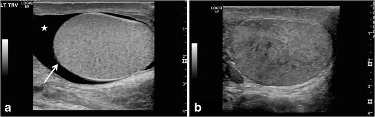
Two-panel gray-scale scrotal ultrasound images demonstrating sonographic features of testicular torsion in adolescent patients. Image (a) is a transverse view of the left scrotum showing a globular, enlarged testis with an abnormal horizontal lie (indicated by a white arrow). A surrounding anechoic area represents a reactive hydrocele (marked with a star). This orientation is characteristic of the 'bell clapper' deformity predisposing to torsion. Image (b) is a longitudinal view of a different patient's right testis showing a markedly heterogeneous parenchymal echotexture with mixed hyperechoic and hypoechoic areas. Such significant heterogeneity is a critical diagnostic sign of late-stage torsion, suggesting tissue congestion and potential nonviability. These diagnostic images illustrate key morphological and parenchymal changes used in pediatric radiology to differentiate testicular torsion from other causes of acute scrotum, such as epididymitis.
Scrotal Swelling - Differential Diagnosis
Painful Scrotal Swelling - "The Acute Scrotum"
1. Testicular Torsion (Spermatic Cord Torsion)
- Surgical emergency - orchidectomy risk rises sharply after 6 hours
- Peak incidence: neonates and adolescents
- Caused by "bell-clapper deformity" (abnormal proximal fixation of tunica vaginalis)
- Key features: sudden onset, severe pain, nausea/vomiting, high-riding testis with transverse lie, absent cremasteric reflex, no fever early
- Epidemiology: 20-30% of acute scrotum cases (excluding neonates)
2. Torsion of Testicular Appendage (Appendix Testis / Appendix Epididymis)
- Most common cause of acute scrotum in children (40-60%)
- Prepubertal peak; more indolent onset than cord torsion
- Tenderness localized to superior pole; "blue dot sign" may be visible through skin
- Managed conservatively; no surgical urgency
3. Epididymitis / Epididymo-orchitis
- 5-15% of acute scrotum cases
- Adolescents/young adults: sexually transmitted (Neisseria gonorrhoeae, Chlamydia trachomatis)
- Older men: coliforms, Pseudomonas
- Key features: gradual onset, dysuria, fever, epididymal tenderness (posterior), urethral discharge, elevated WBC, positive UA
- Prehn's sign (pain relief on elevation) was classically taught for epididymitis but is NOT reliable for distinguishing from torsion
- Park's Textbook of Preventive & Social Medicine
4. Orchitis
- Usually from epididymo-orchitis (secondary spread)
- Mumps orchitis: most common viral cause - unilateral; associated with parotitis and lymphadenopathy
- Also: TB, brucellosis, syphilis
5. Fournier's Gangrene (Necrotizing Fasciitis of Genitalia)
- Rapidly progressive, polymicrobial necrotizing fasciitis of genitalia and perineum
- Life-threatening - mortality up to 67% in some series
- Features: severe pain, scrotal swelling, skin discoloration → frank necrosis, crepitus (gas in tissues), foul-smelling discharge
- Requires urgent surgical debridement + broad-spectrum antibiotics - Schwartz's Principles of Surgery
6. Incarcerated / Strangulated Inguinal Hernia
- Bowel loops descend into scrotal sac via patent processus vaginalis
- Presents as painful, irreducible scrotal mass; no cough impulse
- Strangulation leads to ischemia - surgical emergency
- Risk factors: prematurity, age <1 year
7. Trauma / Hematocele
- Blunt scrotal trauma → hematocele (blood in tunica vaginalis) or testicular rupture
- Urgent US and surgical exploration if rupture suspected
8. Idiopathic Scrotal Edema (Acute)
- Rare, diagnosis of exclusion
- Scrotal wall edema without intrascrotal pathology; often in children
- Self-limiting
9. Reactive Hydrocele (secondary to inflammation)
- Can accompany epididymitis, orchitis, torsion, or tumor
- Pain from underlying pathology, not the hydrocele itself
Painless (or Mildly Discomforting) Scrotal Swelling
| Condition | Key Features |
|---|---|
| Hydrocele | Fluid in tunica vaginalis; transilluminates; smooth; non-tender. Communicating type fluctuates with Valsalva. Common in neonates (resolves by age 1) |
| Varicocele | Dilated pampiniform plexus veins; "bag of worms" feel; does NOT transilluminate; 15% of adolescent males. Mostly left-sided (90° drainage angle). Right-sided = must exclude intra-abdominal mass |
| Spermatocele | Epididymal cyst containing dead sperm; separate from testis; at head of epididymis; transilluminates |
| Epididymal cyst | Similar to spermatocele but contains clear fluid; separate from testis |
| Testicular tumor | Firm, non-tender testicular mass; does NOT transilluminate; associated with cryptorchidism. Rule out with US + AFP/hCG/LDH |
| Inguinal hernia (reducible) | Bowel/omentum descends into scrotum; cough impulse present; reducible; cannot "get above" the swelling |
| Lymphoedema | Can affect scrotal skin - filariasis (tropical), malignancy, post-radiation |
| Sebaceous cyst / epidermal cyst | Skin-level lesion; separate from testis; may have central punctum |
| Lipoma of cord | Soft, compressible cord-level mass |
| Undescended testis complications | Empty hemiscrotum + palpable inguinal mass |
Comparison Table: The Three Key Painful Diagnoses
| Feature | Testicular Torsion | Epididymitis | Appendage Torsion |
|---|---|---|---|
| Peak age | Neonates, adolescents | Adolescents, young adults | Prepubertal |
| Pain onset | Sudden | Gradual, progressive | Variable |
| Nausea/vomiting | More likely | Less likely | Less likely |
| Dysuria | Less likely | More likely | Less likely |
| Fever | Less likely | More likely | Less likely |
| Tender location (early) | Testis (diffuse) | Epididymis (posterior) | Superior pole |
| Cremasteric reflex | Absent | May be present | May be present |
| Testis position | High-riding, transverse | Normal, vertical | Normal, vertical |
Diagnostic Approach
- History - onset, duration, trauma, sexual activity, age, fever, urinary symptoms
- Exam - testis position/lie, cremasteric reflex, transillumination, "blue dot" sign, reducibility, cough impulse
- Doppler US - first-line imaging; assesses blood flow (absent in torsion), epididymal enlargement, hydrocele, tumor
- Labs - urine microscopy/culture, STI swab, CBC (infection), tumour markers (AFP, β-hCG, LDH) if mass found
- Surgical exploration - if torsion cannot be excluded, explore without delay; "time is testis"
Key Rule: Any acute scrotal pain in a child or adolescent must be treated as testicular torsion until proven otherwise. Do not wait for imaging if clinical suspicion is high - Campbell-Walsh Wein Urology
Painless testicular swellings
hydrocele varicocele spermatocele testicular swelling clinical
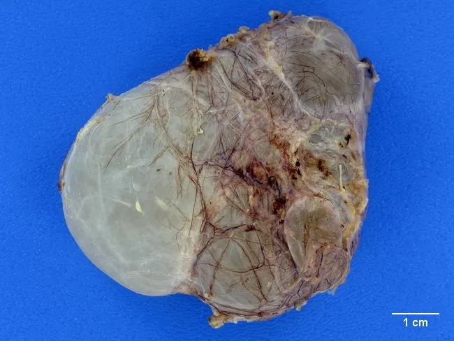
This gross pathology photograph depicts a scrotal adnexal cystic structure consistent with a spermatocele arising from dilatation of efferent ductules near the rete testis or the head of the epididymis. The specimen shows a thin-walled, translucent (glass-like) cyst attached to posterior testicular/epididymal tissue, with variable lobulation indicating possible multilocular configuration. A single or multiple cysts may be present, filled with clear, straw-colored fluid. The fluid composition may include motile or non-motile spermatozoa, a hallmark distinguishing spermatocele from hydrocele, which contains serous fluid without sperm. The surrounding testicular parenchyma appears unremarkable; vascular and stromal tissues may show marginal adhesions. The scale bar indicates 1 cm for size estimation; overall dimensions correspond to a small to moderate cystic lesion, typically a few millimeters to a few centimeters in diameter. Pathophysiology involves dilation of efferent ductules or proximal epididymal ducts with preserved testicular architecture, absence of malignant features, and minimal inflammatory infiltrate. Clinically, spermatocele presents as a painless scrotal swelling, often anterior or superior to the testis, distinct from hydrocele by fluid content. This image supports education in gross anatomy of the male reproductive tract, differential diagnosis of scrotal cystic lesions, and correlates with radiologic and clinical findings used to guide conservative versus surgical management, including epididymal-sparing excision when indicated.
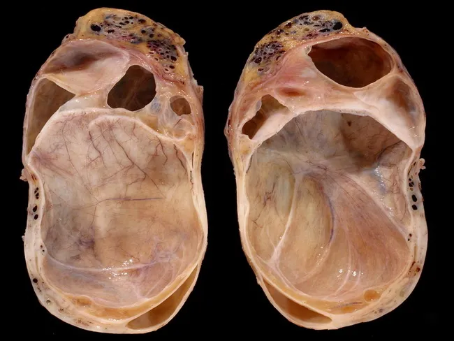
Gross pathology photograph of a scrotal/testicular specimen with a large hydrocele and an adjacent spermatocele. The bilateral testes are opened and presented in frontal orientation, allowing assessment of the hydrocele sac and contained parenchyma. A prominent hydrocele capsule surrounds the testis, producing marked compression and resultant pressure atrophy of the underlying testicular parenchyma. The residual functional testicular tissue is visible as a thin crescent-shaped rim occupying the inferior pole of each testis. At the superior pole, a small saccular structure corresponds to a spermatocele within the tunica vaginalis. The tunica albuginea is intact, with no discrete intratesticular mass identified on gross inspection. The cut surfaces show pale, trabeculated parenchyma with attenuated vascularity; the surrounding fluid appears clear or faintly opalescent. Overall morphology favors a chronic hydrocele with secondary testicular atrophy rather than neoplasia. The image highlights the anatomic relationship between hydrocele, residual testicular tissue, and epididymal cyst formation. Clinically relevant implications include potential impairment of spermatogenesis and testicular function, consideration for hydrocelectomy or surveillance, and the need for histopathologic confirmation to exclude occult malignancy. This photograph is a valuable teaching aid for pathology, urology, and medical education focusing on hydrocele-associated testicular atrophy and spermatocele. Educationally relevant for clinical correlation teaching.
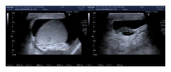
Two grayscale ultrasonographic images of the left hemiscrotum showing a hydrocele and an epididymal cyst. The image on the left displays the left testis ('Lt Testis') with a normal, homogeneous echotexture, partially surrounded by a hypoechoic to anechoic fluid collection consistent with a hydrocele. The image on the right focuses on the paratesticular structures, revealing a well-defined, rounded, anechoic structure within the epididymis, characteristic of an epididymal cyst (spermatocele). There is no significant internal septation or debris visible within the fluid collections. Measurement markers are visible at the bottom of the scans, indicating clinical assessment of testicular and cystic dimensions. This diagnostic imaging illustrates common benign scrotal findings often evaluated in the context of scrotal swelling or urological symptoms like hematuria and flank pain.
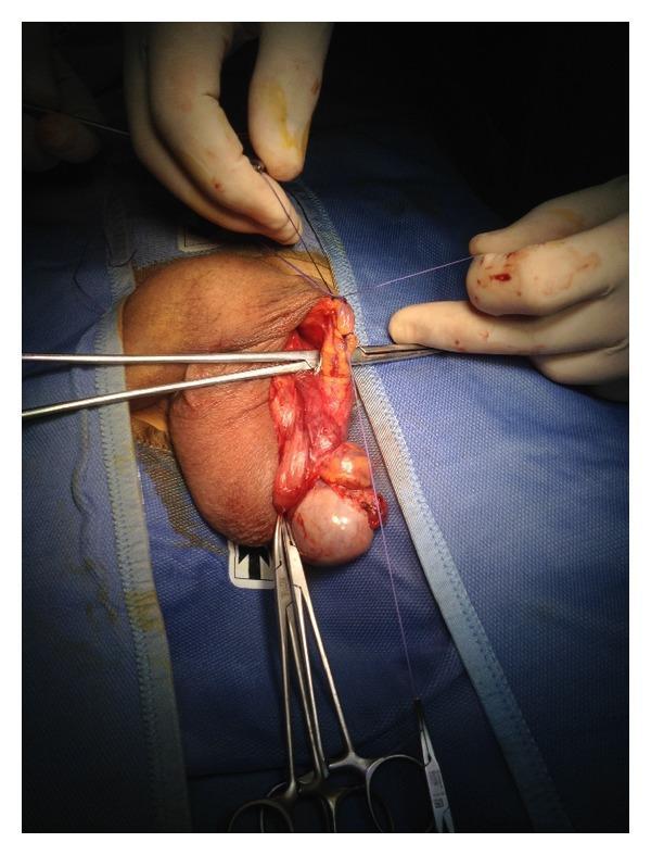
This clinical photograph shows an intraoperative view of a male surgical procedure, specifically the ligation of the anterior spermatic venous plexus for the treatment of varicocele. The surgical field is established through a scrotal approach, with the patient draped in a sterile blue cloth. The exposed anatomical structures include the left testis, tunica vaginalis, and the spermatic cord. Multiple surgical instruments are in use, including Farabeuf retractors and hemostatic forceps, which are being utilized to retract tissue and isolate vascular structures. A surgeon's hands are visible, demonstrating the application of a blue, absorbable suture (likely 2.0 Vicryl) to perform an en bloc ligation of the venous plexus near the external inguinal ring. The educational focus of this image is to illustrate the surgical anatomy of the male reproductive system and the technique of proximal spermatic cord dissection to preserve testicular vascularization during varicocelectomy or hydrocele repair.
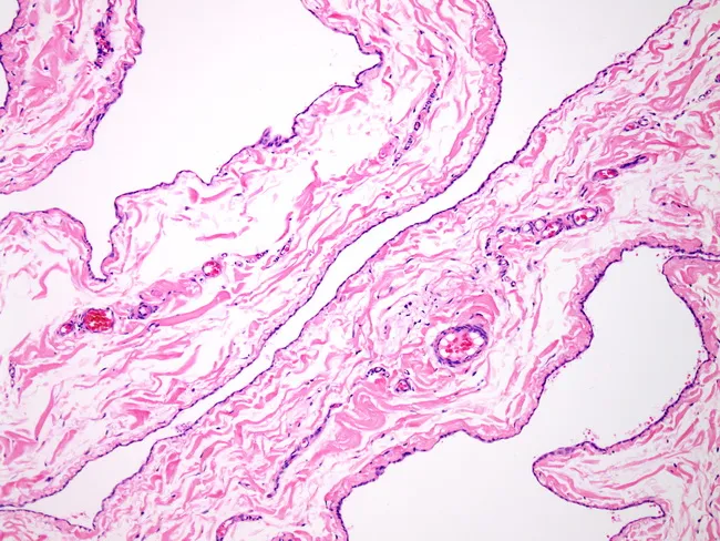
This light-microscopy histology image displays the wall of a spermatocele arising from the epididymal duct. The cyst wall is thin and delicate, composed of loose fibroconnective tissue that forms a simple capsule around a fluid-filled lumen. The epithelial lining is cuboidal to flattened and often ciliated, consistent with epididymal origin. The luminal contents are not the focus here, but the lining and wall architecture are characteristic of a benign epididymal cyst. Surrounding stroma is mild and non-destructive, with minimal inflammatory change and no epithelial atypia or invasion. Collectively these features support a diagnosis of epididymal cyst/spermatocele rather than a neoplasm. Clinically, spermatocele management favors simple excision, with recurrences uncommon; rare cases may be complicated by testicular torsion. The image is prepared as a paraffin-embedded tissue section, stained with Hematoxylin and Eosin, and viewed at low to mid magnification, enabling assessment of cyst wall thickness, epithelial morphology, and stromal context. In diagnostic practice, this histology aids differentiation from hydrocele (epididymal cyst without sperm content), tunica albuginea changes, or malignant epididymal neoplasms. Relevant clinical correlations include infertility considerations, testicular pain, and scrotal swelling. This representation reinforces the benign, excretory-epididymal nature of the lesion and supports surgical planning.
testicular germ cell tumor seminoma ultrasound
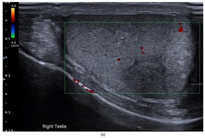
This color Doppler ultrasound image of the right testis demonstrates a solitary intratesticular mass, highly characteristic of a primary testicular germ cell tumor such as a seminoma. On grayscale imaging, the lesion appears as a predominantly hypoechoic and homogeneous mass with well-defined but slightly irregular margins. There is a notable absence of macroscopic calcifications or cystic components within the visible portion of the tumor. The color Doppler overlay shows internal vascularity, evidenced by scattered red flow signals within the mid-portion and periphery of the mass. Additionally, prominent vascularity is noted along the inferior boundary of the testis. This visual evidence supports the diagnostic workup for testicular malignancies in patients presenting with scrotal masses or secondary metastatic findings, such as retroperitoneal lymphadenopathy. The homogeneity and low echogenicity are typical sonographic features used by clinicians to differentiate seminomatous from non-seminomatous germ cell tumors, the latter of which tend to be more heterogeneous.
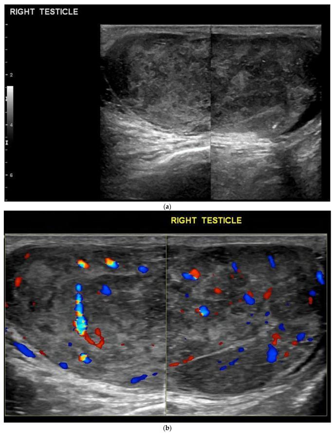
Two-panel diagnostic ultrasound of the right testicle showing a primary testicular tumor. (a) B-mode (grayscale) ultrasound reveals a large, heterogeneous, predominantly hypoechoic mass that infiltrates and expands the testicular parenchyma. The mass contains internal echogenic foci consistent with macrocalcifications and small anechoic cystic areas. (b) Color Doppler ultrasound demonstrates significant hypervascularity within the lesion, characterized by multiple red and blue flow signals distributed throughout the mass. This appearance is characteristic of germ cell tumors, such as seminoma or embryonal cell carcinoma, which typically present as hypervascular, ill-defined solid masses. The imaging highlights the role of ultrasound in distinguishing solid neoplastic lesions from avascular pathologies like hematomas or simple cysts. The case is representative of urological oncology teaching materials for identifying signs of testicular malignancy.
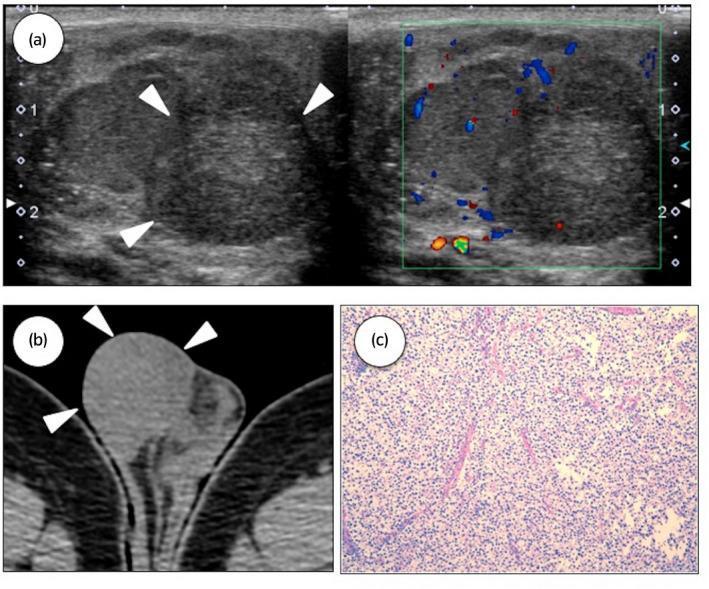
This composite educational image illustrates the multimodal diagnostic features of a testicular seminoma. Panel (a) presents scrotal ultrasound images of the right testicle. The grayscale view reveals a heterogeneous, predominantly hypoechoic intratesticular mass (white arrowheads) measuring approximately 2 cm. The adjacent color Doppler image demonstrates internal vascularity within the lesion. Panel (b) shows a computed tomography (CT) scan of the scrotum in the axial plane, identifying a well-circumscribed, slightly heterogeneous mass within the testicle (arrowheads). Panel (c) is a histopathological micrograph using Hematoxylin and Eosin (H&E) staining. It depicts the classic architectural and cellular characteristics of a pure seminoma, featuring a uniform population of large, polygonal cells with clear cytoplasm and distinct cell borders. These tumor cells are organized into nests or lobules separated by thin fibrous septa, which contain a characteristic lymphocytic infiltrate. This sequence demonstrates the clinical progression from initial radiological detection via ultrasound and CT to definitive pathological diagnosis through biopsy, highlighting key features of germ cell tumors in the urological specialty.
Painless Testicular / Scrotal Swellings
- Does it transilluminate? (fluid-filled vs. solid)
- Can you get above it? (intrascrotal vs. inguinoscrotal)
- Is the testis separate from or involved in the swelling?
1. Hydrocele
Types (classified by processus vaginalis anatomy):
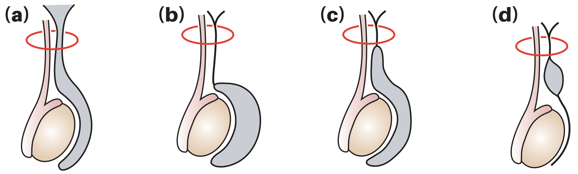
| Type | Mechanism | Key Feature |
|---|---|---|
| Communicating (congenital) | Patent processus vaginalis; peritoneal fluid enters scrotum | Swelling fluctuates - enlarges on crying/straining, smaller in morning; associated with indirect inguinal hernia |
| Non-communicating (vaginal) | Processus closed; fluid from imbalanced production/absorption by tunica | Fixed size; most common in adults |
| Infantile hydrocele | Mid-processus patent, proximal open | Swelling extends to external ring |
| Hydrocele of the cord | Mid-processus patent, both ends closed | Smooth oval swelling above testis near cord; moves down when testis pulled downward |
Clinical features:
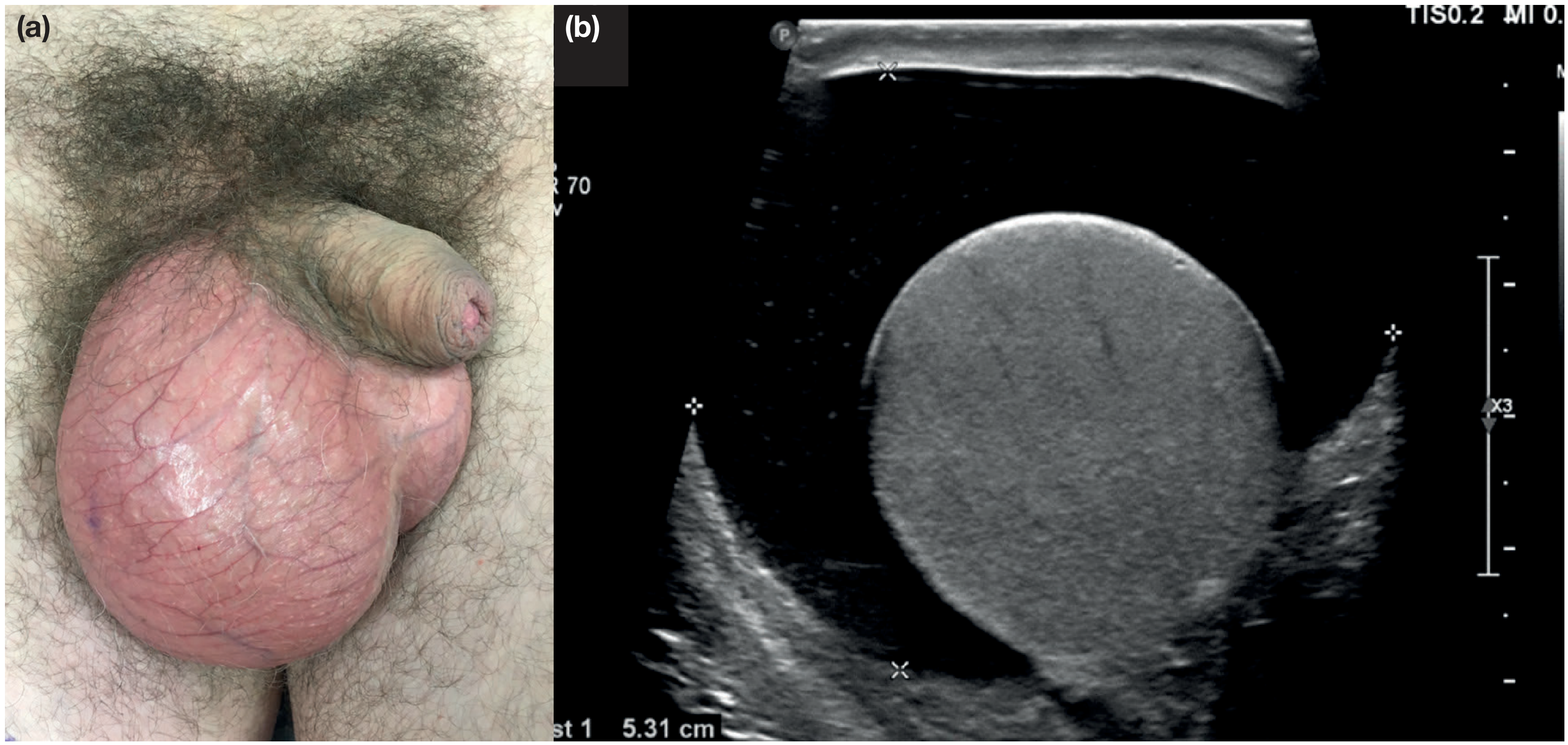
- Painless, smooth, transilluminates brightly (can see hand through it)
- Testis is WITHIN the swelling - cannot palpate testis separately
- You CAN get above the swelling
- "Secondary" hydrocele (in men >40) may indicate underlying tumor, infection, or torsion - do not aspirate without US to exclude tumor (risk of malignant needle-track seeding)
- Filarial hydrocele: major cause in tropics (Wuchereria bancrofti); fluid may contain chyle (chylocele)
Treatment:
- Neonates: expectant (most resolve by 18 months)
- Adults: surgical if large/symptomatic
- Lord's operation - plication of sac (thin-walled hydroceles)
- Jaboulay's procedure - eversion of sac behind testis
2. Varicocele
Grading:
| Grade | Description |
|---|---|
| Grade 0 (subclinical) | Only seen on imaging; not palpable |
| Grade I | Palpable only with Valsalva/exertion |
| Grade II | Palpable at rest; collapses when supine |
| Grade III | Visibly bulging; does NOT collapse when supine |
Key features:
- "Bag of worms" feel - soft, compressible, tortuous above the testis
- Does NOT transilluminate
- Decreases in size when supine (Grade I and II)
- Vague dragging/aching discomfort, especially after prolonged standing
- Left-sided in >90% of cases - left spermatic vein drains at 90° into left renal vein (longer, higher pressure)
- Right-sided varicocele is a red flag - must exclude retroperitoneal tumor (e.g., right renal cell carcinoma) compressing the IVC
Clinical significance:
- Infertility - venous pooling raises scrotal temperature and accumulates toxic metabolites; responsible for up to 40% of male infertility presentations
- Testicular atrophy - especially in adolescents; monitor with annual US if volume discrepancy >20%
Treatment:
- Surgical ligation (inguinal, subinguinal, laparoscopic) or radiological embolization
- Microsurgical varicocelectomy preserves lymphatics, reducing hydrocele risk to <1%
3. Epididymal Cysts
Clinical features:
- Common in middle age; often multiple and bilateral
- Lie posterior to and separate from the testis - this is the key distinguishing feature from hydrocele
- Feel like "tiny bunches of grapes" posterior to the testis
- Transilluminate
- Usually asymptomatic; confirmed by ultrasound
Treatment:
- Conservative if small/asymptomatic
- Excision if large or uncomfortable; multilocular cysts may require partial/total epididymectomy
- Aspiration is ineffective (usually multilocular) - Bailey & Love's Surgery
4. Spermatocele
Clinical features:
- Located in the epididymal head, above and behind the upper pole of the testis
- Usually softer and laxer than epididymal cysts
- Separate from the testis; transilluminates
- Usually small and asymptomatic
Distinguishing from epididymal cyst:
| Feature | Spermatocele | Epididymal Cyst |
|---|---|---|
| Fluid | Contains spermatozoa ("barley water") | Clear fluid, no sperm |
| Loculation | Unilocular | Often multilocular |
| Consistency | Softer, laxer | Tense |
| Location | Epididymal head | Any part of epididymis |
5. Testicular Tumour
Clinical features:
- Firm, non-tender intratesticular mass
- Does NOT transilluminate
- May have an associated secondary hydrocele (present in ~10-20% - this can mask the tumor)
- Painless in ~80% of cases at presentation; dull ache in ~20%
- History of cryptorchidism (undescended testis) is the single biggest risk factor - 3-5x increased risk even after orchidopexy
Classification - Germ Cell Tumours (95% of testicular tumours):
| Type | Peak Age | Markers | Notes |
|---|---|---|---|
| Seminoma | 30-40 yrs | PLAP, β-hCG (10%) | Homogeneous on US; radiosensitive |
| Non-seminomatous GCT (NSGCT) | 20-30 yrs | AFP, β-hCG, LDH | Heterogeneous on US; includes embryonal, yolk sac, choriocarcinoma, teratoma |
Tumour markers:
- AFP (α-fetoprotein) - elevated in yolk sac tumour, embryonal carcinoma; never elevated in pure seminoma
- β-hCG - elevated in choriocarcinoma; may be mildly elevated in seminoma
- LDH - correlates with tumour bulk
- PLAP (placental alkaline phosphatase) - seminoma
Management:
- Urgent scrotal US if any clinical suspicion
- Tumour markers (AFP, β-hCG, LDH) BEFORE orchidectomy
- Inguinal orchidectomy (never trans-scrotal biopsy - risk of lymphatic spread to inguinal nodes rather than para-aortic)
- Staging CT thorax/abdomen/pelvis
6. Inguinal Hernia (Scrotal Extension)
- Reducible bowel/omentum descends into scrotum via patent processus vaginalis
- Painless when reducible; cough impulse present
- Cannot get above the swelling (extends up into inguinal canal)
- Bowel sounds may be audible
- Becomes painful if incarcerated or strangulated
Quick Reference: Examination Summary
| Swelling | Transilluminates | Separate from Testis | Can Get Above | Key Feature |
|---|---|---|---|---|
| Hydrocele | ✅ Yes | ❌ No (surrounds) | ✅ Yes | Bright transillumination; testis within |
| Epididymal cyst | ✅ Yes | ✅ Yes | ✅ Yes | Posterior/separate from testis; multilocular |
| Spermatocele | ✅ Yes | ✅ Yes | ✅ Yes | Epididymal head; barley-water fluid |
| Varicocele | ❌ No | ✅ Yes | ✅ Yes | "Bag of worms"; reduces supine; left >>right |
| Testicular tumour | ❌ No | ❌ No (within testis) | ✅ Yes | Firm, solid, irregular; DO NOT MISS |
| Inguinal hernia | ❌ No | ✅ Yes | ❌ No | Cough impulse; reducible; can't get above |
Golden Rule: Any painless solid intratesticular mass = testicular cancer until proven otherwise. Arrange urgent scrotal ultrasound. - Goldman-Cecil Medicine
Anal fistula surgeries
anal fistula types classification Parks surgery diagram
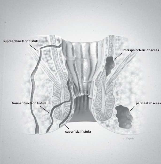
**Imaging Modality:** Medical illustration/Anatomical diagram. **Anatomical Region:** Coronal cross-section of the anorectal region, including the anal canal, rectum, internal and external anal sphincters, and surrounding perianal soft tissues. **Observed Pathology:** The illustration classifies various types of anorectal fistulas and abscesses based on their anatomical trajectory and location. **Characteristic Visual Features:** * **Fistula Tracts:** Three distinct epithelial-lined tracks are labeled. A **superficial fistula** is shown confined to the perianal skin and distal anal canal. A **transsphincteric fistula** is depicted crossing both the internal and external sphincter muscles into the ischioanal fossa. A **suprasphincteric fistula** is illustrated looping above the puborectalis muscle before descending to the skin surface. * **Abscess Formations:** Two localized collections of fluid/pus are identified. An **intersphincteric abscess** is positioned within the space between the internal and external sphincter muscles. A **perineal (perianal) abscess** is shown as a larger pocket located in the subcutaneous tissue adjacent to the anal verge. **Key Diagnostic Features:** The diagram emphasizes the relationship between inflammatory tracts and the sphincter complex, following the Parks classification system for fistula-in-ano. Suitable for clinical education on perianal sepsis and surgical planning.
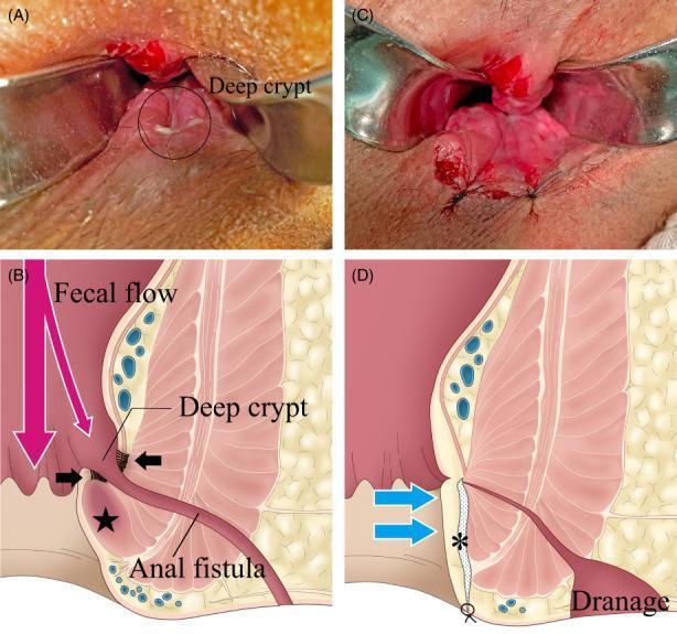
A composite image consisting of clinical photographs and pathophysiology diagrams illustrating the anatomical changes during anal fistula surgery. Panel A shows a preoperative clinical photograph of the anal canal with a 'deep crypt' circled, appearing as a cave-like invagination. Panel B is a corresponding cross-sectional diagram showing how inflammatory fibrosis (indicated by black arrows) and a hardened internal sphincter (star) create a receptacle for fecal flow, leading into an anal fistula tract. Panel C presents the postoperative clinical view using an anal speculum, demonstrating the resolution of the deep crypt. Panel D provides a postoperative diagram illustrating the surgical effect: dissection of the fistula and fibrous tissue (asterisk) and internal sphincterotomy result in the flattening of the anal canal (blue arrows) and establishment of a drainage pathway. This set demonstrates the 'Surgical Incision of Fistula Tract and Internal Sphincterotomy' (SIFT-IS) technique, focusing on eradicating the deep crypt to prevent persistent fecal entry and chronic inflammation in transsphincteric fistulas.
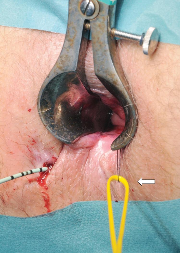
This clinical photograph captures an intraoperative view of a surgical procedure for a complex fistula-in-ano. A metallic Parks or similar anal retractor is positioned to provide exposure of the anal canal. The surgical field demonstrates the management of a fistula track using specialized instrumentation. On the left side of the image, a urological guidewire with black and white markings is seen entering the external fistula opening, which shows minor associated hemorrhage. A yellow vascular sling (indicated by a white arrow) is being threaded through the track using a 1/0 nylon suture loop. The educational focus of the image is the technique of passing a vascular sling from the internal opening to the external opening to serve as a loose seton. This procedure is critical in colorectal surgery for maintaining drainage of the fistulous track while preventing abscess formation and protecting the anal sphincter muscles. The image is intended for intermediate to advanced medical learners focusing on proctological surgical techniques.
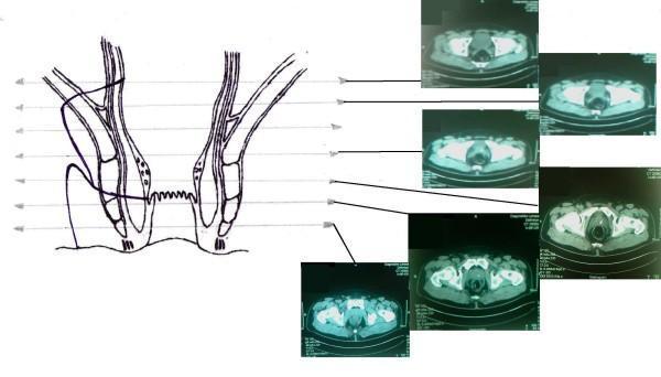
This composite educational image features a Comparison Chart and Diagnostic Image set correlating perianal anatomy with radiological findings. On the left, a coronal anatomical diagram of the rectum and anal canal illustrates two fistulous tracts originating from a shared internal orifice in the rectal wall. One tract demonstrates an extrasphincteric path, while the other shows the placement of a surgical seton. The diagram highlights key landmarks including the internal and external anal sphincters, the levator ani muscle, and the dentate line. Horizontal reference lines extend from different anatomical levels of the schematic to a series of six axial CT scans (labeled as MRI in context) on the right. These cross-sectional diagnostic images display the pelvic floor, rectal lumen, and surrounding soft tissues at sequential heights, facilitating a 3D understanding of the fistula's complex trajectory relative to the pelvic musculature. This material is designed for advanced medical education in colorectal surgery and radiology, focusing on the diagnosis and management of complex anal fistulas.
?