
Methhemoglobinemia
methemoglobinemia
methemoglobinemia cyanosis blood color chocolate brown
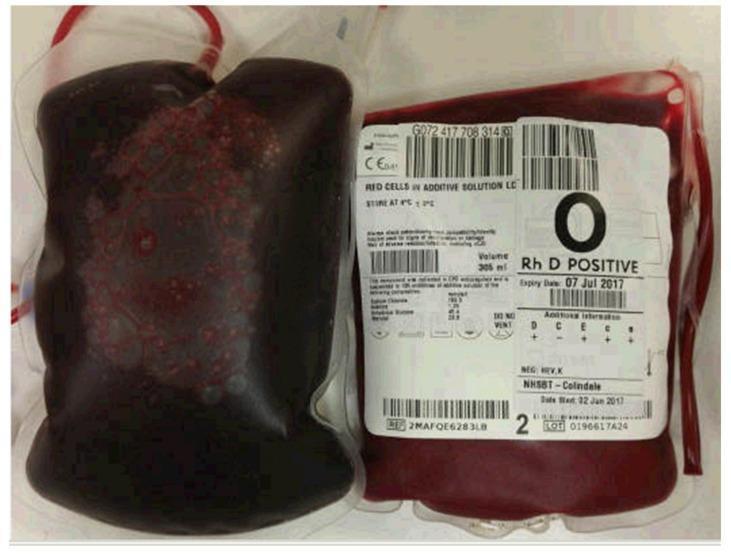
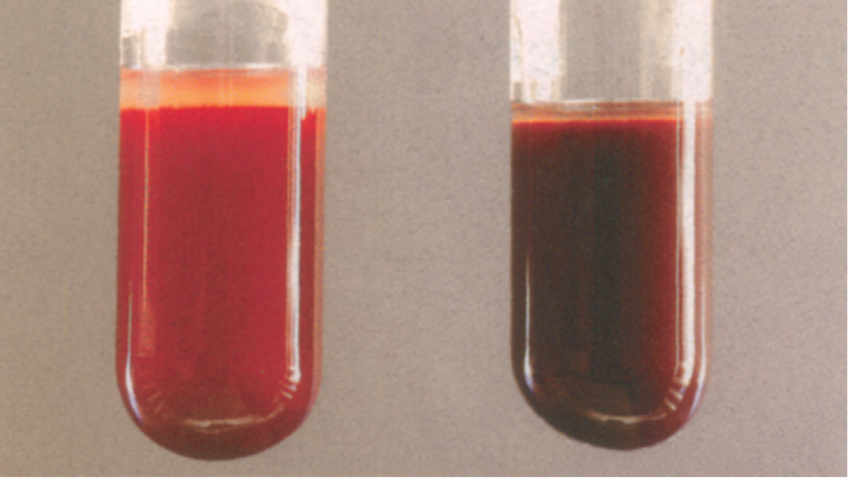
Clinical photograph showing a side-by-side comparison of two blood bags, illustrating a striking color discrepancy indicative of specific hemoglobinopathies. On the left is a bag of venesected patient blood characterized by a dark, 'chocolate brown' or muddy hue, which is a classic diagnostic sign of sulfhemoglobinemia or methemoglobinemia. Unlike normal deoxyhemoglobin, this discoloration does not revert to bright red upon exposure to oxygen. Small, dark red aggregations are visible within the brownish fluid. On the right is a standard donor unit of 'RED CELLS IN ADDITIVE SOLUTION' for comparison, exhibiting the expected bright red color of healthy, oxygenated blood. The donor bag label identifies it as Type O Rh D Positive. This image serves as a visual teaching tool for hematology and emergency medicine, demonstrating the clinical presentation of 'chocolate-colored blood' which often correlates with central cyanosis and low pulse oximetry readings despite normal arterial oxygen tension (PaO2).
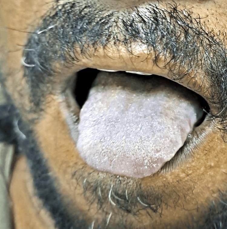
This clinical photograph shows a close-up view of a patient's open mouth, focusing on the dorsal surface of the tongue protruding slightly. The tongue exhibits a prominent bluish-slate discoloration, a classic clinical sign of central cyanosis. The surface texture of the tongue appears normal with visible lingual papillae, but the diffuse blue-grey hue indicates a systemic reduction in oxygenated hemoglobin or the presence of abnormal hemoglobin variants. In this clinical context, the presentation is characteristic of methemoglobinemia, a condition where hemoglobin is oxidized to methemoglobin, which cannot bind oxygen. The surrounding perioral skin and lips show a natural brown pigmentation and dark facial hair, providing a stark color contrast to the abnormal hue of the mucous membrane. This image serves as an educational example for identifying central cyanosis and differentiating it from peripheral cyanosis, emphasizing the importance of examining highly vascularized mucosal surfaces in patients with suspected hypoxemia or toxic ingestions.
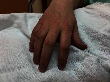
A clinical photograph of a patient's hand resting on a white hospital drape, demonstrating generalized peripheral cyanosis. The dorsal surface of the hand, knuckles, and all fingers exhibit a distinct, pervasive dusky bluish-purple or slate-gray hue. This discoloration is notably uniform across the skin and extends to the nail beds, which appear darkly cyanotic. The image serves as a clinical illustration of central or peripheral cyanosis, specifically associated with congenital methemoglobinemia, a condition where elevated levels of methemoglobin reduce the blood's oxygen-carrying capacity, resulting in characteristic skin and extremity discoloration despite normal arterial oxygen tension. The photograph highlights the visual presentation of 'chocolate-colored' blood manifesting as cutaneous cyanosis in a clinical setting.
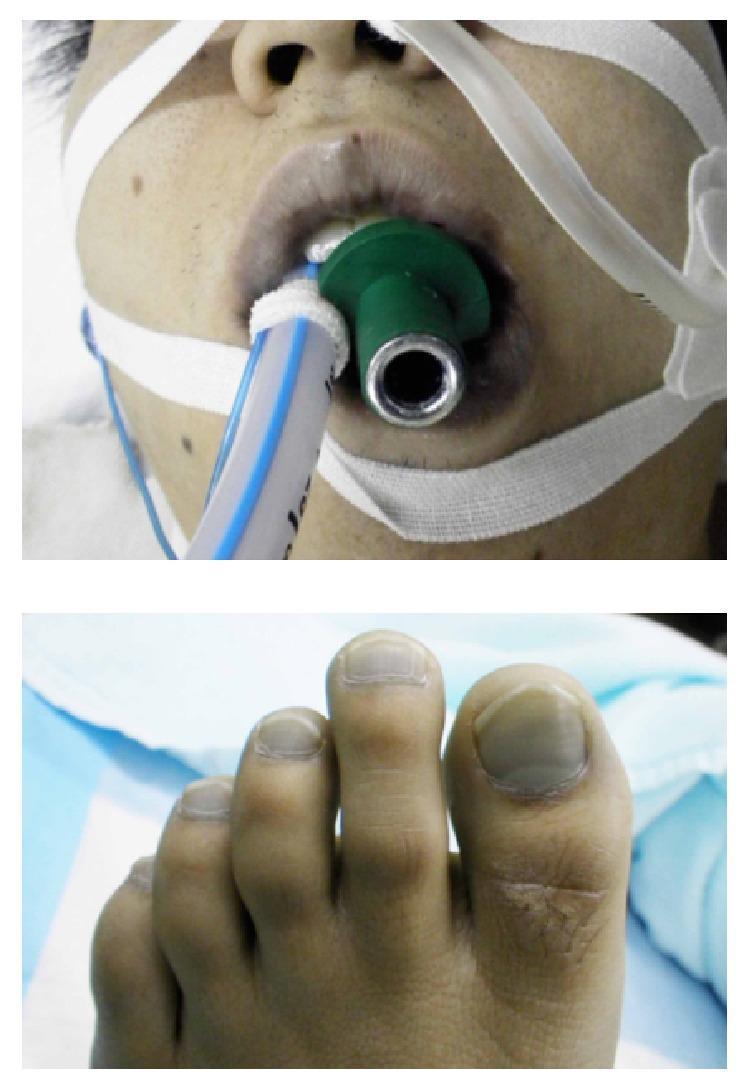
This composite clinical photograph illustrates severe central and peripheral cyanosis in a patient. The top image shows the perioral region and lower face, characterized by a profound blue-gray discoloration of the lips and surrounding skin. A green bite block and an endotracheal tube are visible, secured with white medical tape, indicating emergency airway management and mechanical ventilation. The bottom image displays the patient's foot, where the nail beds of the toes exhibit a matching slate-blue or dusky hue, characteristic of peripheral cyanosis. These visual findings are consistent with methemoglobinemia, a condition where an abnormal level of methemoglobin in the blood reduces its oxygen-carrying capacity, leading to 'chocolate-colored' blood and apparent cyanosis that does not improve with supplemental oxygen. The educational focus is on identifying clinical signs of hypoxia or hemoglobinopathies in an emergency critical care setting.
Methemoglobinemia
Definition
Pathophysiology
Normal Reduction Pathways
-
Primary pathway (NADH-dependent) - Cytochrome b5 reductase uses NADH (from glycolysis) to reduce cytochrome b5, which then reduces MetHb back to Hb. This accounts for ~95% of MetHb reduction under normal circumstances.
-
Secondary pathway (NADPH-dependent) - NADPH-methemoglobin reductase uses NADPH (from the hexose monophosphate shunt via G6PD). Normally responsible for <5% of reduction, but this is the pathway activated by methylene blue (the antidote).
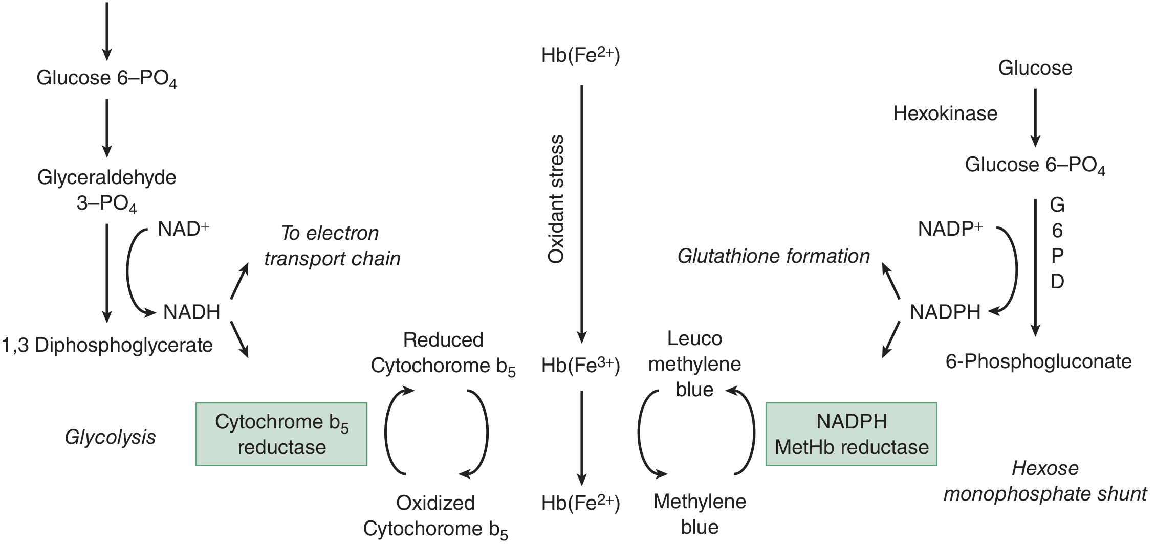
Why MetHb is Worse than Equivalent Anemia
- Reduces oxygen-carrying capacity (like anemia)
- Causes a leftward shift of the oxyhemoglobin dissociation curve in the remaining normal hemoglobin - meaning hemoglobin holds oxygen more tightly and releases less to tissues at any given PO₂
Types
| Type | Cause |
|---|---|
| Acquired (toxic) | Most common; drugs, chemicals, dietary nitrates |
| Congenital | Cytochrome b5 reductase deficiency (autosomal recessive) |
| Hemoglobin M disease | Rare hemoglobin variant; iron permanently in Fe³⁺ state |
Causes (Acquired)
| Category | Agents |
|---|---|
| Local anesthetics | Benzocaine (most common), prilocaine, lidocaine (rare), dibucaine |
| Analgesics | Phenazopyridine (commonly reported), phenacetin |
| Antimicrobials | Dapsone (hydroxylamine metabolite; cimetidine inhibits formation), sulfamethoxazole, antimalarials |
| Nitrates/Nitrites | Amyl nitrite, isobutyl nitrite ("poppers"), sodium nitrite, nitroglycerin, silver nitrate, ammonium nitrate, contaminated well water |
| Other | Rasburicase, aniline dyes, naphthalene |
Key pearl: Benzocaine is the local anesthetic most commonly associated with MetHb. Dapsone is notable because its hydroxylamine metabolite is the culprit, and this can be partially prevented by cimetidine. - Tintinalli's Emergency Medicine, p. 1372
Infants are especially vulnerable to nitrate-induced MetHb from well water: oral nitrates are converted to nitrites by GI bacteria, and fetal hemoglobin is more readily oxidized. - Rosen's Emergency Medicine, p. (nitrates section)
G6PD Deficiency and MetHb
Clinical Features by MetHb Level
| MetHb % | Symptoms |
|---|---|
| <3% | Normal (physiologic) |
| Up to 15% | Graying/ashen skin discoloration |
| 15-20% | Cyanosis (central) - often the first alarming sign |
| 15%+ | "Chocolate-brown" blood on venipuncture (classic sign) |
| 20-45% | Fatigue, dyspnea, weakness, dizziness, headache, tachycardia |
| 45-70% | Drowsiness, syncope, seizures, metabolic acidosis, dysrhythmias |
| >70% | Coma, death |
Key Diagnostic Clues
- Cyanosis that does NOT improve with supplemental O₂ - hallmark finding
- "Chocolate-colored" arterial blood - does not turn bright red when exposed to air
- Pulse oximetry reads ~85% regardless of actual MetHb level (SpO₂ is falsely "normal" or fixed near 85% because the pulse oximeter cannot distinguish MetHb from OxyHb/DeoxyHb)
- ABG shows normal PaO₂ (oxygen is dissolved normally in plasma)
- Saturation gap: SpO₂ (pulse ox) differs markedly from SaO₂ (co-oximetry)
Clinical Photos
Diagnosis
- Co-oximetry (multi-wavelength pulse oximetry or ABG co-oximeter) is the definitive test - directly measures MetHb %
- Standard 2-wavelength pulse oximetry is unreliable - reads falsely ~85% or is artifact-prone
- ABG: PaO₂ normal, but calculated SaO₂ is falsely high if not directly measured by co-oximetry
- CBC, G6PD level (before treating with methylene blue), hemolysis workup if indicated
Treatment
Algorithm
- MetHb >30% (regardless of symptoms), OR
- MetHb >20% with symptoms (dyspnea, dysrhythmias, angina, seizures, altered mental status)
- Repeat dose of 1 mg/kg if symptoms persist or MetHb remains >30% after 30-60 minutes.
- Maximum total dose typically ~7 mg/kg (paradoxically causes MetHb at high doses)
How Methylene Blue Works
Special Situations
| Situation | Management |
|---|---|
| G6PD deficiency | Methylene blue is relatively contraindicated (causes hemolysis). Use ascorbic acid (vitamin C) - slower, repeat doses needed |
| Pregnancy | Risk-benefit analysis; consider C-section, hyperbaric oxygen, or exchange transfusion |
| Refractory/Severe | Exchange transfusion (removes MetHb-containing RBCs and replaces with functional ones) |
| Asymptomatic, MetHb <20% | Remove offending agent only; MetHb normalizes within 24-36 hours |
Disposition
- MetHb >15% or any symptomatic patient: admit for observation
- MetHb <15%, asymptomatic, offending agent removed: may discharge with close follow-up
Methemoglobinemia vs. Sulfhemoglobinemia
| Feature | Methemoglobinemia | Sulfhemoglobinemia |
|---|---|---|
| Mechanism | Hb-Fe²⁺ → Fe³⁺ (oxidation) | Sulfur incorporated into porphyrin ring |
| Reversibility | Reversible (enzymatically or with methylene blue) | Irreversible for life of RBC |
| Blood color | Chocolate-brown | Greenish |
| Treatment | Methylene blue | Supportive; exchange transfusion |
| Key clue | Responds to methylene blue | Fails to respond to methylene blue |
If a patient's "methemoglobin" level is elevated but they fail to respond to methylene blue, consider sulfhemoglobinemia. - Rosen's Emergency Medicine
Summary Points for Exams
- MetHb = Hb-Fe²⁺ oxidized to Fe³⁺ - cannot carry O₂
- Primary reduction enzyme: cytochrome b5 reductase (NADH-dependent)
- Antidote (methylene blue) works via NADPH/G6PD pathway
- G6PD deficient: no increased MetHb risk, but high hemolysis risk; methylene blue is contraindicated
- Classic triad: cyanosis + normal PaO₂ + chocolate-brown blood
- Pulse oximetry reads ~85% regardless of actual MetHb level
- Most common local anesthetic cause: benzocaine; most common antimicrobial: dapsone
- Infants at high risk from nitrate-contaminated well water
- Sulfhemoglobinemia = methylene blue non-responder