
inflammation complete topic i need every detail
Reading File
Inflammation is a protective response to injury/infection involving vascular changes, cellular events, and chemical mediators. It can be acute (short-lived, neutrophil-dominated) or chronic (prolonged, lymphocyte/macrophage-dominated). Key mediators include histamine, prostaglandins, cytokines, and complement.
Searching sources...
Finding Sources
Finding Sources
Finding Sources
Finding Sources
inflammation complete topic i need every detail in defence and immunity mainly from abbas book
Reading File
Searching sources...
Finding Sources
Finding Sources
Finding Sources
Finding Sources
Finding Sources
Now I have a clear map. Let me read all the key inflammation sections in parallel.
Reading File
Reading File
Excellent content retrieved. Let me now read the systemic consequences, functional responses, and the cytokine sections in detail.
Reading File
Reading File
Reading File
Reading File
Reading File
Loading Image
Loading Image
Reading File
I now have all the key content. Let me also get the IL-1, IL-6, and the macrophage repair / antiviral response sections.
Reading File
Reading File
Loading Image
Here is the complete, detailed coverage of Inflammation from Cellular and Molecular Immunology by Abbas, Lichtman & Pillai (9780323757485):
INFLAMMATION - Complete Topic
Cellular and Molecular Immunology - Abbas, Lichtman & Pillai
1. DEFINITION AND OVERVIEW
Inflammation is the accumulation of leukocytes, plasma proteins, and fluid derived from the blood at an extravascular tissue site of infection or injury. It is the principal way by which the innate immune system deals with infections and tissue injury.
- Leukocytes and plasma proteins normally circulate in blood and must be recruited to extravascular sites of infection/injury
- They perform effector functions: kill microbes, clear dead cells, and begin tissue repair
- Acute inflammatory responses begin with recognition of microbial PAMPs (pathogen-associated molecular patterns) or DAMPs (damage-associated molecular patterns) by tissue sentinel cells
Sentinel cells include: macrophages, dendritic cells (DCs), and mast cells. These cells respond by secreting mediators that act on small blood vessels to promote:
- Increased blood flow
- Delivery of plasma proteins
- Migration of leukocytes into tissues
2. INITIATION: SENTINEL CELLS AND PATTERN RECOGNITION
The inflammatory response is triggered when sentinel cells in normal tissues use:
- Toll-like receptors (TLRs)
- Cytosolic innate pattern recognition receptors (NLRs, cGAS, etc.)
...to sense microbes and injured cells.
Mast cells respond to PAMPs/DAMPs by secreting:
- Histamine - causes vasodilation and increased capillary permeability
- Prostaglandins - also cause vasodilation and increased permeability
This increases blood flow and promotes movement of plasma proteins (complement proteins, pentraxins, collectins, antibodies) out of blood vessels into tissues.
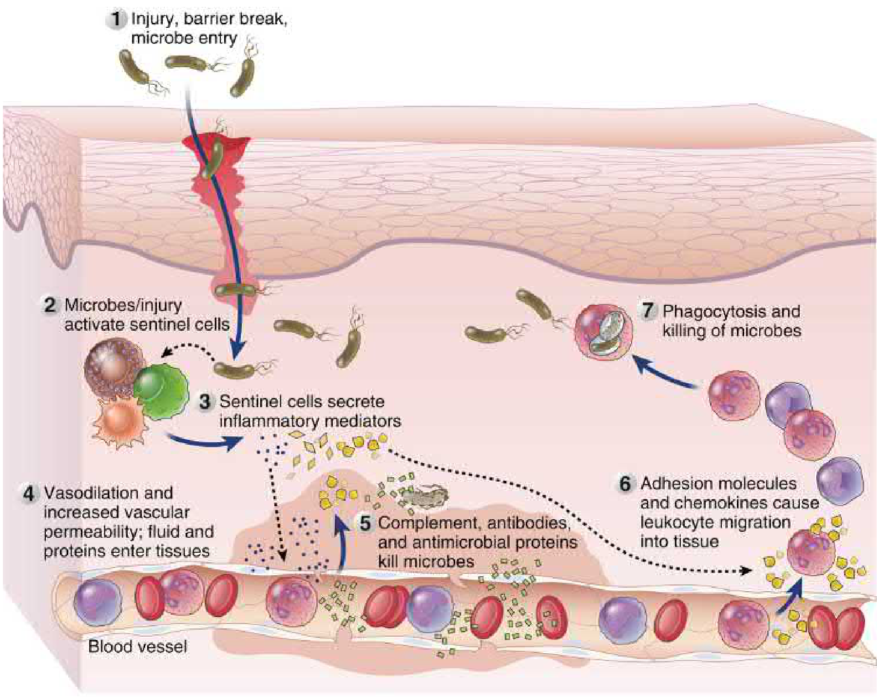
Step-by-step sequence (as shown in Figure 4.14 above):
- Injury, barrier break, or microbe entry
- Microbes/injury activate sentinel cells (macrophages, DCs, mast cells)
- Sentinel cells secrete inflammatory mediators (cytokines, histamine, prostaglandins)
- Vasodilation and increased vascular permeability → fluid and proteins enter tissues
- Complement, antibodies, and antimicrobial proteins kill microbes
- Adhesion molecules and chemokines cause leukocyte migration into tissue
- Phagocytosis and killing of microbes
3. THE MAJOR PROINFLAMMATORY CYTOKINES OF INNATE IMMUNITY
One of the earliest innate responses to infection/damage is secretion of cytokines by tissue cells. Key properties of these cytokines:
- Produced mainly by tissue macrophages and DCs (also mast cells, endothelial cells, some epithelial cells)
- Usually act paracrine (on nearby cells); in severe infections, may enter circulation and act endocrine (at a distance)
- May have overlapping or unique actions; one cytokine may stimulate production of others (cytokine cascades)
- Serve multiple roles: induce inflammation, inhibit viral replication, promote T cell responses, and limit innate immune responses
TABLE 4.5 - Key Cytokines of Innate Immunity
| Cytokine | Size | Principal Source | Principal Targets and Effects |
|---|---|---|---|
| TNF | 17 kD; 51 kD homotrimer | Macrophages, T cells | Endothelial cells: activation (inflammation, coagulation); Neutrophils: activation; Liver: acute-phase proteins; Hypothalamus: fever; Muscle/fat: catabolism (cachexia); Many cells: apoptosis |
| IL-1 | 17 kD mature; 33 kD precursor | Macrophages, endothelial, epithelial cells | Endothelial: activation; Hypothalamus: fever; Liver: acute-phase proteins; T cells: Th17 differentiation |
| Chemokines | 8-10 kD | Macrophages, endothelial, T cells, fibroblasts, platelets | Leukocytes: chemotaxis, activation, migration into tissues |
| IL-12 | Heterodimer 35+40 kD | Macrophages, DCs | T cells: Th1 differentiation; NK + T cells: IFN-γ synthesis, increased cytotoxic activity |
| Type I IFNs (IFN-α/β) | 15-21 kD | Macrophages, plasmacytoid DCs, fibroblasts | Antiviral resistance; NK cell activation; Adaptive immunity enhancement |
| IL-6 | - | Macrophages, endothelial cells | Liver: acute-phase proteins; B cells: growth/differentiation; T cells: Th17 differentiation |
| IL-10 | - | Macrophages, T cells | Macrophages/DCs: inhibit cytokine production (anti-inflammatory) |
3A. TUMOR NECROSIS FACTOR (TNF)
- Also called TNF-α (to distinguish from TNF-β/lymphotoxin)
- Produced mainly by macrophages; also DCs and mast cells
- Originally named for causing necrosis of tumors (inflammation and thrombosis of tumor blood vessels)
- Structure: synthesized as a non-glycosylated type II membrane homotrimeric protein
- Cleaved by a membrane metalloproteinase → released polypeptide fragment
- Three polypeptide chains polymerize into a triangular pyramid-shaped circulating protein
- Receptor-binding sites are at the base of the pyramid → can simultaneously bind three receptor molecules
- Member of the TNF superfamily (all form homotrimers)
- Primary signal: TLR and NLR activation → NF-κB and AP-1 transcription factors
Key actions of TNF in inflammation:
- Activates endothelial cells → upregulates adhesion molecules (E-selectin, ICAM-1, VCAM-1) → promotes leukocyte recruitment
- Stimulates neutrophil and monocyte activation
- Works with IL-1 to stimulate chemokine secretion (CXCL8, CCL2)
- Induces acute-phase protein synthesis in liver
- Acts on hypothalamus → fever (endogenous pyrogen)
3B. INTERLEUKIN-1 (IL-1)
- Two forms: IL-1α and IL-1β (less than 30% homologous, but same receptors and same biologic activities)
- Main active secreted form: IL-1β
- Sources: activated macrophages (major), neutrophils, DCs, epithelial cells (keratinocytes), endothelial cells
- Actions similar to TNF
Two-signal requirement for IL-1β production:
- Signal 1: TLR, NLR, or KLR signaling → activates NF-κB → transcription and production of 33-kD pro-IL-1β
- Signal 2: Inflammasome activation → caspase-1 is activated → proteolytically cleaves pro-IL-1β to generate 17-kD mature IL-1β
Neither IL-1α nor IL-1β has a hydrophobic signal sequence for conventional secretion. IL-1β may be secreted through membrane pores formed by gasdermin D.
IL-1 Receptor signaling:
- Binds type I IL-1 receptor (expressed on endothelial cells, epithelial cells, leukocytes)
- Receptor has extracellular Ig domain + cytosolic TIR domain (same as TLRs)
- Signaling: activates NF-κB and AP-1
- Type II IL-1 receptor: decoy receptor - cannot activate signals, limits IL-1 responses
3C. INTERLEUKIN-6 (IL-6)
- Produced by macrophages and endothelial cells (stimulated by TNF and IL-1)
- Stimulates liver to produce acute-phase proteins (CRP, SAP, fibrinogen)
- Promotes B cell growth and differentiation
- Promotes Th17 T cell differentiation
- Part of the cytokine cascade: TNF → IL-1 → IL-6 → acute-phase response
4. SEQUENCE OF EVENTS IN INFLAMMATION: VASCULAR CHANGES AND LEUKOCYTE RECRUITMENT
4A. Vascular Changes
Triggered by mast cell-derived histamine and prostaglandins:
- Vasodilation → increased blood flow → more leukocytes delivered to affected area
- Increased capillary permeability → plasma proteins (complement, pentraxins, collectins, antibodies) enter tissues
- Changes blood flow characteristics → enhance physical interactions of circulating leukocytes with vessel walls
4B. Leukocyte Recruitment
TNF, IL-1, IL-6, and chemokines (produced by sentinel cells at infection/injury sites) have multiple effects:
Step 1 - Endothelial Activation:
- Postcapillary venule endothelial cells increase surface expression of adhesion molecules:
- E-selectin (induced by TNF and IL-1 via NF-κB)
- ICAM-1 (intercellular adhesion molecule 1) - ligand for integrins
- VCAM-1 (vascular cell adhesion molecule 1) - ligand for integrins
- P-selectin - stored in cytoplasmic granules, mobilized to surface by thrombin from coagulation cascade
Step 2 - Chemokine-Mediated Migration:
- TNF and IL-1 stimulate cells to secrete chemokines:
- CXCL8 (IL-8) → binds receptors on neutrophils
- CCL2 (MCP-1) → binds receptors on monocytes
- Chemokines increase affinity of leukocyte integrins for their ligands
- Chemokines stimulate directional movement (chemotaxis) of leukocytes
- Result: increased neutrophil and monocyte adhesion to endothelium → transmigration through vessel wall
Step 3 - Leukocyte Accumulation:
- Leukocytes accumulate in tissues → form the inflammatory infiltrate
- Neutrophils arrive first (most abundant leukocyte in blood, most rapid responder to chemotactic signals)
- Monocytes (become macrophages in tissue) become increasingly prominent over time; may be dominant in some reactions
- If TNF is absent or blocked (e.g., in patients treated with anti-TNF drugs, or TNF knockout mice) → failure to contain infections
Step 4 - Bone Marrow Stimulation:
- TNF, IL-1, and IL-6 from inflammatory sites enter the blood → delivered to bone marrow
- Work with colony-stimulating factors → enhance production of neutrophils from progenitors
- Promote release of mature neutrophils into blood
- Increases supply of cells that can be recruited and replace consumed leukocytes
5. PHAGOCYTOSIS AND KILLING OF MICROBES
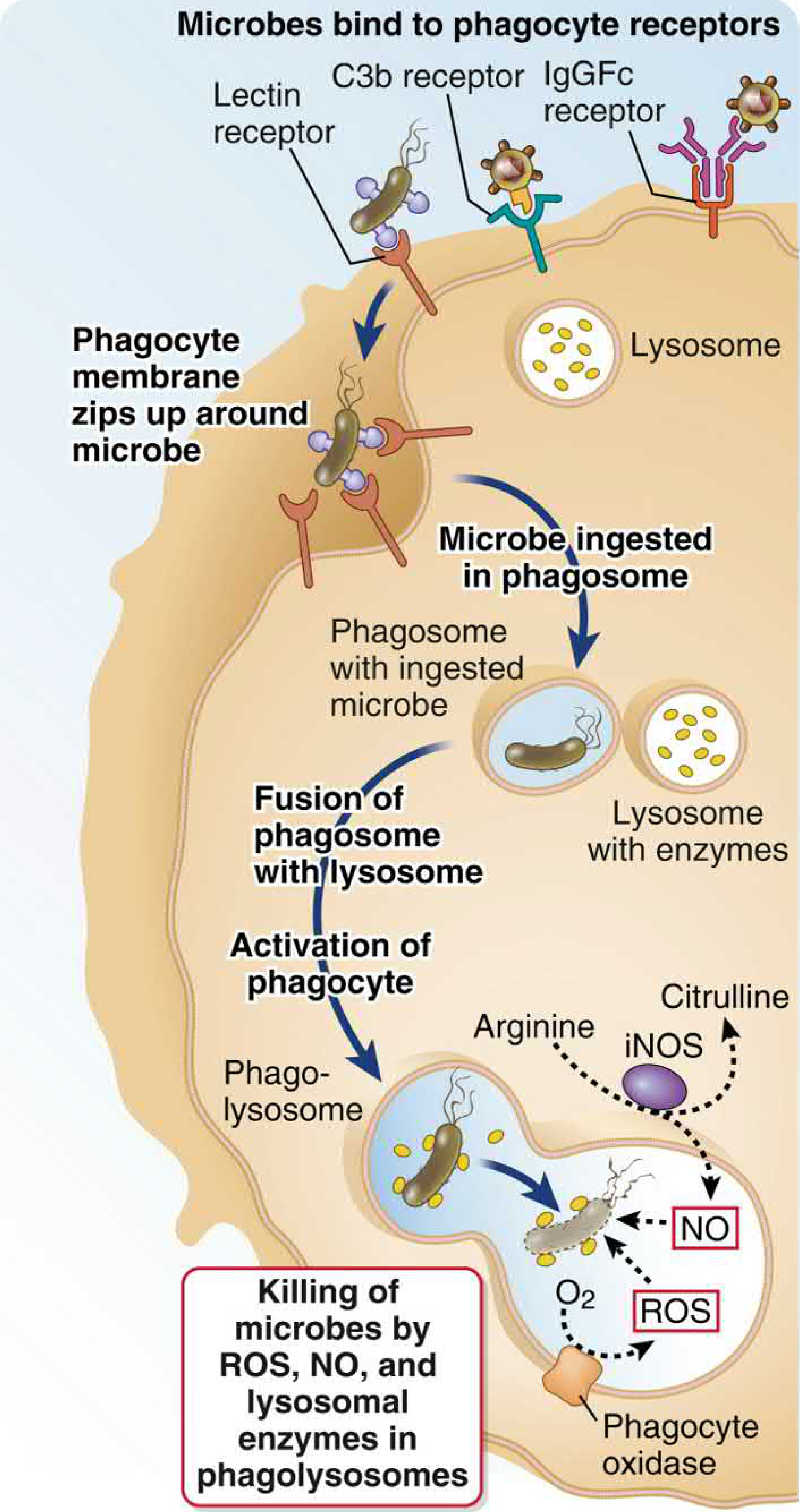
Neutrophils and macrophages recruited to infection sites ingest microbes by phagocytosis and destroy them.
5A. Phagocytosis - Definition
- Active, energy-dependent process of engulfment of large particles (> 0.5 μm diameter) into vesicles
- Phagocytic vesicles fuse with lysosomes → ingested particles are destroyed
- Killing mechanisms are isolated from the rest of the cell inside the phagolysosome
5B. Receptors for Phagocytosis
Phagocytes express several receptors:
- Pattern recognition receptors (C-type lectins, scavenger receptors): bind specific molecular patterns (e.g., mannose receptor binds mannose residues on microbes)
- Fc receptors (FcγRI): high-affinity receptor for IgG → phagocytosis of antibody-opsonized microbes
- Complement receptors (C3b receptor): bind complement-opsonized microbes
- Plasma lectin receptors
Opsonization: Coating microbes with antibodies (IgG) is the most efficient system. The IgG Fc end interacts with FcγRI on phagocytes → efficient phagocytosis. This illustrates the link between innate and adaptive immunity - antibodies (adaptive) engage phagocytes (innate) to perform protective functions.
5C. Mechanism of Phagocytosis
- Microbe binds to receptor on phagocyte
- Plasma membrane invaginates and extends a cup-shaped projection around the microbe
- Membrane cup extends beyond the diameter of the particle, then closes and pinches off
- Forms a phagosome (inside-out intracellular vesicle containing the foreign particle)
- Receptor signals activate microbicidal activities of the phagocyte
- Phagosome fuses with lysosome → phagolysosome
5D. Microbicidal Mechanisms (inside the phagolysosome)
1. Reactive Oxygen Species (ROS):
- Generated by phagocyte oxidase (NADPH oxidase) in the phagolysosome membrane
- O₂ is reduced to superoxide (O₂⁻), which is converted to H₂O₂ and then to hypochlorous acid (HOCl)
- HOCl (the principal microbicidal reactive oxygen intermediate) kills microbes
- Patients with Chronic Granulomatous Disease (CGD): phagocyte oxidase mutations → cannot generate ROS → susceptible to recurrent bacterial/fungal infections
2. Nitric Oxide (NO):
- Generated by inducible nitric oxide synthase (iNOS)
- Uses arginine as substrate → produces NO + citrulline
- NO combines with ROS to form peroxynitrite and other reactive nitrogen species → toxic to microbes
- iNOS is especially important in macrophages (induced by IFN-γ from T cells and NK cells)
3. Lysosomal Enzymes:
- Acid proteases, phospholipases, and other hydrolases in lysosomes
- Digest killed microbes
- Also contribute to killing in the phagolysosome environment (low pH)
6. ROLE OF MACROPHAGES IN TISSUE REPAIR
Acute inflammation is associated with significant host cell death (from microbes or collateral damage from microbicidal activities of leukocytes). Once offending agents are eliminated, damaged tissue must be repaired. Macrophages play a critical role:
- Clear dead cells (efferocytosis)
- Secrete growth factors that promote regeneration and angiogenesis
- Secrete TGF-β and other cytokines → stimulate collagen synthesis by fibroblasts → scar tissue formation
Two macrophage activation states:
- Classically activated macrophages (M1): microbicidal, promote inflammation - dominant early in the reaction
- Alternatively activated macrophages (M2): promote tissue repair - dominant later in the reaction
7. SYSTEMIC AND PATHOLOGIC CONSEQUENCES OF INFLAMMATION
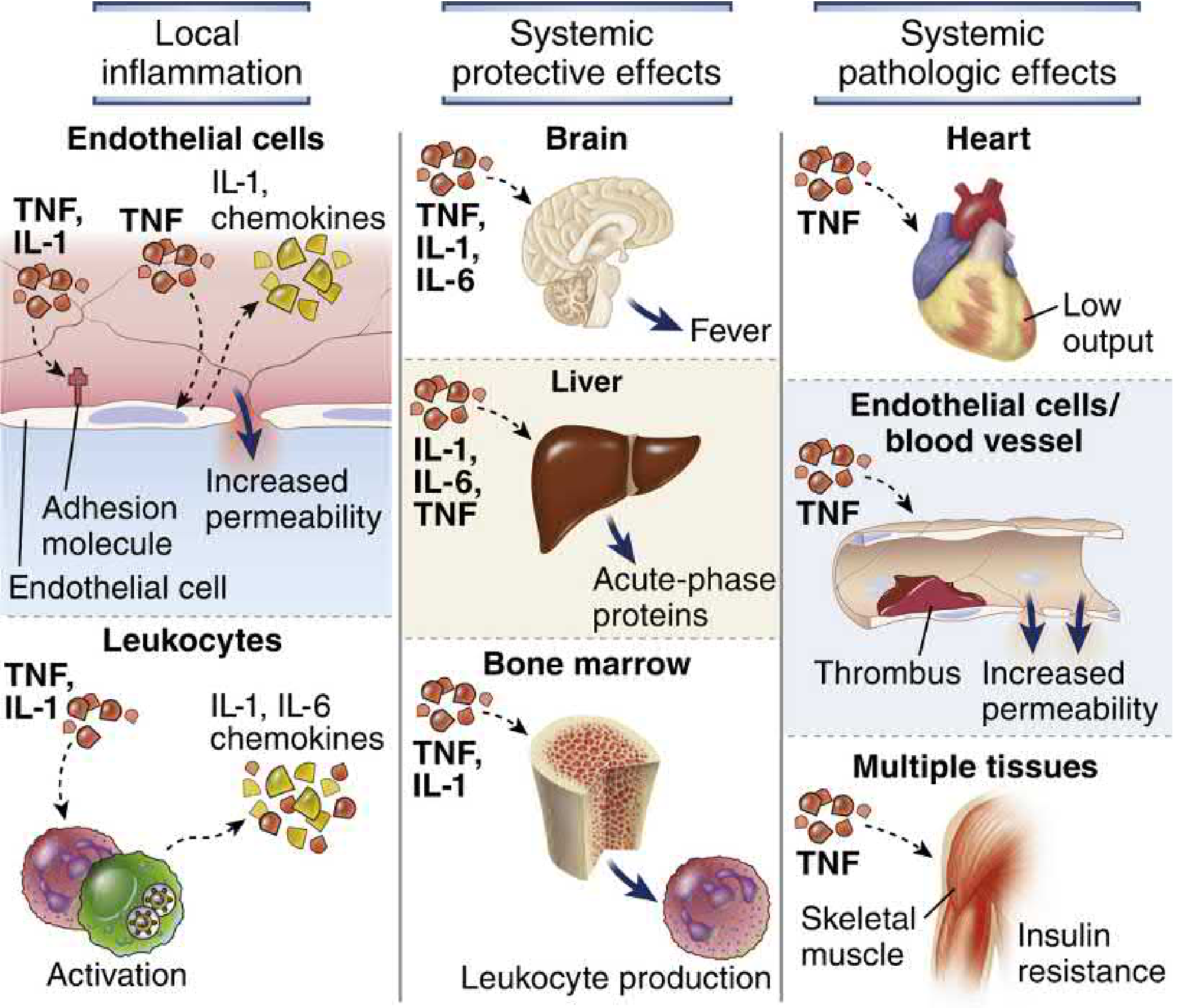
TNF, IL-1, and IL-6 produced during innate immune responses to infection/tissue damage have systemic effects that contribute to host defense and cause clinical manifestations.
7A. Protective Systemic Effects
| Effect | Mediator(s) | Mechanism |
|---|---|---|
| Fever | TNF, IL-1 (endogenous pyrogens) | Act on hypothalamus → increase prostaglandin synthesis → prostaglandins stimulate neurotransmitters that raise body temperature (reduce heat loss via vasoconstriction + increase heat generation) |
| Leukocytosis | TNF, IL-1, IL-6 + colony-stimulating factors | Circulate to bone marrow → promote release of neutrophils and monocytes + stimulate production of new cells |
| Acute-phase response | TNF, IL-1, IL-6 | Induce hepatocytes to produce CRP, SAP, fibrinogen → secreted into blood |
Fever and aspirin: Prostaglandin synthesis inhibitors (aspirin, NSAIDs) reduce fever by blocking TNF/IL-1-induced prostaglandin synthesis in hypothalamic cells.
Acute-phase proteins:
- CRP (C-reactive protein) and SAP (serum amyloid P) - pentraxins that play protective roles in infection (opsonization, complement activation)
- Fibrinogen - precursor of fibrin; contributes to hemostasis and tissue repair
- Elevated plasma levels of these proteins are clinically used as markers of infection/inflammation (ESR elevated by fibrinogen; CRP is a direct acute-phase marker)
7B. Pathologic Effects of Excess TNF (Sepsis)
When TNF is produced in very large amounts (as in severe infections), it enters the bloodstream and causes:
| Pathologic Effect | Mechanism |
|---|---|
| Shock (decreased blood pressure) | TNF inhibits myocardial contractility and vascular smooth muscle tone |
| Intravascular thrombosis (DIC) | TNF stimulates endothelial expression of tissue factor (activates coagulation) + inhibits expression of thrombomodulin (anticoagulant) + neutrophil activation → vascular plugging |
| Cachexia (wasting of muscle and fat) | TNF suppresses appetite + reduces synthesis of lipoprotein lipase (needed to release fatty acids from circulating lipoproteins for tissue use) → prolonged production |
7C. Sepsis Syndrome
A systemic complication of severe (usually bacterial or fungal) infection:
- Clinically: fever, fast heart rate, fast respiratory rate, metabolic abnormalities, mental disturbances
- Most often initiated by LPS (lipopolysaccharide/endotoxin) from gram-negative bacteria activating TLR4
- LPS triggers massive TNF production → septic shock
- The cytokine storm (TNF + IL-1 + IL-6 + other mediators) causes the organ dysfunction seen in sepsis
8. FUNCTIONAL RESPONSES OF INNATE IMMUNITY: THREE PILLARS
Abbas organizes the functional outcomes of innate immune activation into three categories:
8A. Inflammation
(covered in detail above)
8B. Cell Death (Programmed Death Pathways)
- Apoptosis of infected cells helps eliminate viral replication sites
- Virally infected cells are hypersensitive to TNF-induced apoptosis
- TNF receptor engages both proinflammatory AND pro-apoptotic pathways; viral infection shifts balance toward apoptosis
- Misfolded viral proteins → unfolded protein response → may culminate in apoptosis
- Pyroptosis: inflammatory cell death mediated by gasdermin D pores (formed downstream of inflammasome → caspase-1 → gasdermin D cleavage)
8C. Antiviral Response (Type I Interferons)
Type I IFNs (IFN-α, IFN-β) are produced in response to viral infection:
- IFN-α: mainly from macrophages and plasmacytoid DCs (the specialist IFN-α-producing cell)
- IFN-β: mainly from fibroblasts and other cell types
Principal antiviral activities of type I IFN (working in concert):
- Induce antiviral resistance in uninfected cells (block viral replication pathways)
- Activate NK cells (which kill virally infected cells)
- Enhance adaptive immune responses
Clinical relevance:
- Patients with severe COVID-19 often have defects in type I IFN:
- ~10% produce autoantibodies against their own type I IFN (may predate infection)
- 3.5-4% have inherited mutations affecting type I IFN production or signaling
- IFN-α is in clinical use as antiviral in viral hepatitis; also used in some tumors (boosts CTL activity or inhibits proliferation)
- IFN-β is therapy for multiple sclerosis (mechanism not fully known)
9. STIMULATION OF ADAPTIVE IMMUNITY BY INFLAMMATION
Innate immune inflammatory responses do not just defend against infection - they set in motion the adaptive immune response.
Two-Signal Hypothesis for Lymphocyte Activation
Activation of lymphocytes requires two distinct signals:
- Signal 1 (Antigen): Ensures the response is specific
- Signal 2 (Innate-derived molecules): Ensures adaptive responses are triggered only when there is a dangerous infection (not when lymphocytes recognize harmless/self antigens)
Signal 2 molecules produced during innate immune reactions:
- Costimulators (e.g., B7 molecules on APCs) → for T cells
- Cytokines (IL-1, IL-2, IL-6, IL-12, etc.) → for both T and B cells
- Complement breakdown products (C3d) → for B cells
The nature of signal 2 not only enhances the magnitude of the adaptive response but also influences its nature/quality (type of T cell differentiation: Th1, Th2, Th17, CTL, etc.).
10. SUMMARY: KEY MEDIATORS AND THEIR ROLES
| Mediator | Source | Key Inflammatory Role |
|---|---|---|
| Histamine | Mast cells | Vasodilation, increased vascular permeability (immediate) |
| Prostaglandins | Mast cells, many cells | Vasodilation, increased permeability, fever |
| TNF | Macrophages, DCs, mast cells | Endothelial activation (adhesion molecules), fever, acute-phase response, cachexia, shock (excess) |
| IL-1 | Macrophages, DCs, epithelial cells | Same as TNF; also Th17 differentiation; requires inflammasome for processing |
| IL-6 | Macrophages, endothelial cells | Acute-phase proteins, leukocyte production, Th17 differentiation |
| CXCL8 (IL-8) | Macrophages, endothelium | Chemotaxis of neutrophils |
| CCL2 (MCP-1) | Macrophages, endothelium | Chemotaxis of monocytes |
| E-selectin / P-selectin | Endothelial cells | Rolling of leukocytes on endothelium |
| ICAM-1 / VCAM-1 | Endothelial cells | Firm adhesion of leukocytes before transmigration |
| IL-12 | Macrophages, DCs | Th1 differentiation, NK cell activation, IFN-γ production |
| Type I IFNs | pDCs, macrophages, fibroblasts | Antiviral resistance, NK activation |
| TGF-β | Macrophages (M2) | Collagen synthesis, tissue repair, scar formation |
| ROS / NO | Neutrophils, macrophages | Intraphagolysosomal killing of microbes |
Key Points to Remember (Abbas perspective)
- Inflammation is fundamentally an innate immune response - the principal defensive action of the innate system
- The key cellular sentinels initiating inflammation are macrophages, DCs, and mast cells
- The three master proinflammatory cytokines are TNF, IL-1, and IL-6 - they act locally AND systemically
- Neutrophils arrive first; monocytes/macrophages follow and dominate later
- Phagocytes kill microbes using ROS (phagocyte oxidase), NO (iNOS), and lysosomal enzymes inside phagolysosomes
- Systemic inflammation causes fever, leukocytosis, and the acute-phase response - all protective
- Excess TNF in sepsis causes shock, DIC, and cachexia - pathologic consequences
- Inflammation connects innate to adaptive immunity via the two-signal hypothesis (Signal 2 comes from innate reactions)
- M1 macrophages (classically activated) fight infection early; M2 macrophages (alternatively activated) repair tissue later
- Type I IFNs are the antiviral arm of innate immunity, distinct from the pro-inflammatory cytokines
Source: Cellular and Molecular Immunology, Abbas, Lichtman & Pillai (9th Ed.) - Chapter 4 (Innate Immunity), pp. 260-295That is the complete inflammation topic as covered in Abbas's Cellular and Molecular Immunology, organized into 10 comprehensive sections with all textbook figures. Here's a quick orientation to what was covered:
What's included (all from Abbas):
| Section | Content |
|---|---|
| 1 | Definition - what inflammation is and why it exists |
| 2 | Initiation - sentinel cells, PAMPs/DAMPs, mast cells, histamine/prostaglandins |
| 3 | Three master cytokines - TNF, IL-1, IL-6 (structure, production, signaling, actions) - with full Table 4.5 |
| 4 | Step-by-step leukocyte recruitment - selectins, integrins, ICAM-1, VCAM-1, chemokines (CXCL8, CCL2), neutrophil vs monocyte timing, bone marrow stimulation |
| 5 | Phagocytosis - receptors, mechanism, killing by ROS / NO / lysosomal enzymes (with CGD explanation) |
| 6 | Tissue repair - M1 vs M2 macrophages, TGF-β, fibrosis |
| 7 | Systemic consequences - fever, leukocytosis, acute-phase response (CRP, SAP, fibrinogen) + pathologic TNF effects in sepsis (shock, DIC, cachexia) |
| 8 | Three functional outcomes of innate immunity: inflammation, cell death (pyroptosis/apoptosis), antiviral (Type I IFNs + COVID-19 note) |
| 9 | Bridge to adaptive immunity - two-signal hypothesis |
| 10 | Master summary table of all mediators |
immunodeficiency : primary and secondary
Reading File
Searching sources...
Finding Sources
Finding Sources
I have the full map. Let me read all the key sections in parallel.
Reading File
Reading File
Reading File
Reading File
Reading File
Reading File
Reading File
Loading Image
Reading File
Loading Image
Your credits are exhausted. You can purchase a top-up or subscribe to a plan to continue using Orris. If you're already subscribed, your daily credits will reset tomorrow.
from abbas book
Your credits are exhausted. You can purchase a top-up or subscribe to a plan to continue using Orris. If you're already subscribed, your daily credits will reset tomorrow.
tumor immunology
Your credits are exhausted. You can purchase a top-up or subscribe to a plan to continue using Orris. If you're already subscribed, your daily credits will reset tomorrow.
B lymphocytes represent what percentage of total lymphocytes? A) 1-5% B) 5-15% C) 20-30% D) 40-50% E) 60-70%
Your credits are exhausted. You can purchase a top-up or subscribe to a plan to continue using Orris. If you're already subscribed, your daily credits will reset tomorrow.
This is a shared conversation. Sign in to Orris to start your own chat.