
Short note on peripheral vascular disease
peripheral vascular disease atherosclerosis limb ischemia
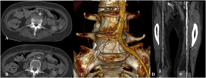
Multi-panel preoperative imaging of the arterial system demonstrating peripheral artery disease (PAD) and extensive atherosclerosis. Panels A and B are axial CT-angiography slices of the infrarenal abdominal aorta showing significant calcified atherosclerotic plaques and mural irregularities. Panel C provides a 3D volume-rendered reconstruction of the abdominal aorta and its bifurcation, highlighting severe vessel tortuosity, an uneven aortic surface, and high-grade stenosis or occlusion at the origin of the right common iliac artery. Panel D displays a coronal maximum intensity projection (MIP) or reformatted view of the bilateral femoral-popliteal axes, illustrating diffuse calcification of the arterial walls and luminal narrowing more pronounced on the right side. The set demonstrates the radiologic manifestations of multilevel atherosclerotic burden in the vascular system, relevant for surgical planning in cases of lower limb ischemia.
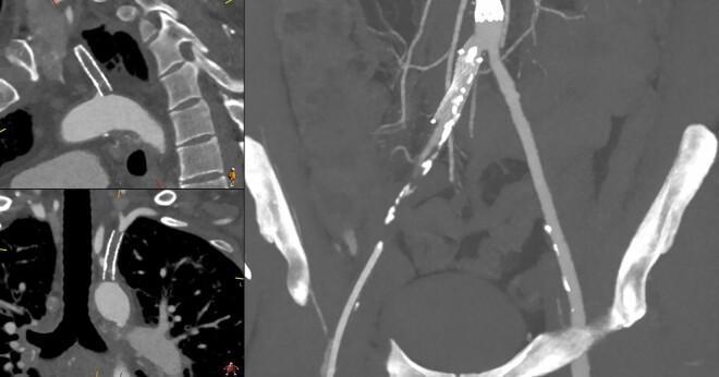
This diagnostic image consists of multi-planar Computed Tomography Angiography (CTA) views illustrating peripheral artery disease (PAD) in the upper and lower extremities. The left panels provide sagittal and coronal reconstructions of the aortic arch and proximal great vessels, highlighting an occluded or severely stenosed left subclavian artery characterized by a truncated contrast column and endovascular stent markers. The larger right panel shows a Maximum Intensity Projection (MIP) of the abdominal aorta and iliofemoral arteries. This view demonstrates significant atherosclerotic burden, evidenced by extensive, irregular mural calcifications (bright white foci) and luminal narrowing along the common and superficial femoral arteries. There is a visible discrepancy in contrast opacification between the patent proximal vessels and the diseased, calcified distal segments. This visual aid is intended for medical education regarding the diagnostic evaluation of systemic atherosclerosis, vascular imaging interpretation, and the clinical presentation of chronic limb-threatening ischemia.
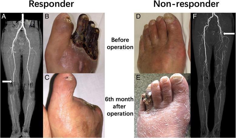
A multi-panel clinical composite comparing treatment outcomes in critical limb ischemia (CLI) between a 'Responder' and a 'Non-responder' using diagnostic imaging and clinical photographs. The Responder case (left) illustrates thromboangiitis obliterans (Buerger's disease). Preoperative CTA (A) shows an arterial occlusion at the popliteal artery level (white arrow). The corresponding clinical photo (B) displays advanced dry gangrene of the second, third, and fourth toes. At 6 months post-operation (C), the foot shows complete wound healing following debridement and cell transplantation. The Non-responder case (right) illustrates atherosclerosis obliterans. Preoperative CTA (F) reveals a proximal superficial femoral artery occlusion (white arrow). Initial clinical photography (D) shows only mild cyanosis and skin changes. However, 6 months post-treatment (E), the condition has progressed to distal gangrene of the lateral toes with diffuse desquamation and xerosis of the dorsal foot skin. This comparison highlights the prognostic variability in CLI based on etiology (TAO vs. ASO) and arterial occlusion level, serving as an educational tool for evaluating peripheral vascular disease progression and therapeutic response.
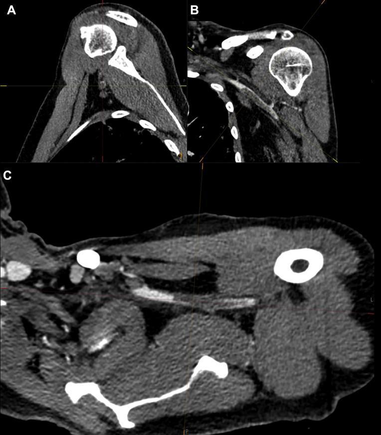
This diagnostic image set consists of three Computed Tomography Angiography (CTA) views of the left upper extremity and shoulder region, demonstrating vascular pathology in a clinical context of COVID-19-related thrombosis.
"peripheral artery disease"[MeSH Terms] AND management
Peripheral Vascular Disease (PVD)
Definition
Epidemiology
-
Prevalence of 3-10% in the general population, rising to 15-20% in persons over 70 years
-
Present in 50% of patients with diabetic foot ulcers
-
Smoking accounts for approximately half of all peripheral artery disease cases
-
Washington Manual of Medical Therapeutics, p. 908
Etiology & Risk Factors
| Risk Factor | Notes |
|---|---|
| Smoking | Strongest single modifiable risk factor; also increases bypass surgery failure rates |
| Diabetes mellitus | Risk increased by age, disease duration, and neuropathy |
| Hypertension | Accelerates atherogenesis |
| Dyslipidemia | Promotes plaque formation |
| Age >70 years | Exponentially increases prevalence |
Pathophysiology
- Adequate flow at rest, insufficient during exercise → intermittent claudication
- Insufficient flow at rest → rest pain
- Tissue breakdown → ulceration and gangrene
Clinical Features
- Intermittent claudication - reproducible cramping, aching, or heaviness in calf/thigh/buttock muscles provoked by walking and relieved by rest (typically from proximal disease)
- Rest pain - typically affects the foot, worsens at night, relieved by dependency (distal disease)
- Numbness, weakness, and heaviness in the limbs
- Coldness in the legs/feet
- Diminished or absent peripheral pulses
- Dependent rubor (dusky red when dependent) and pallor on elevation
- Absence of hair over the dorsum of the foot
- Dystrophic/thickened toenails
- Cool, dry, fissured skin
- Painful ischemic ulcers, typically at the toes and distal pressure points
- Calf muscle atrophy
Diagnosis
Ankle-Brachial Index (ABI)
ABI = Systolic BP at ankle / Systolic BP at arm
| ABI Value | Interpretation |
|---|---|
| >1.4 | Calcified/non-compressible vessels (common in diabetes) |
| 1.0-1.4 | Normal |
| 0.9-1.0 | Borderline |
| <0.9 | Diagnostic for PAD (95% sensitivity) |
| <0.4 | Severe ischemia, limb-threatening |
Other Investigations
- Duplex ultrasound - first-line imaging for anatomical localization
- CT angiography (CTA) - detailed roadmap for revascularization planning; demonstrates stenosis, calcification, multilevel disease
- MR angiography (MRA) - avoids radiation; useful in renal impairment
- Catheter angiography - gold standard, used when intervention is planned
- Toe-brachial index (TBI) - preferred in calcified vessels (diabetics)
- Segmental pressures and pulse volume recordings
Imaging Findings
Fontaine Classification
| Stage | Features |
|---|---|
| I | Asymptomatic |
| IIa | Mild claudication (walking distance >200 m) |
| IIb | Severe claudication (walking distance <200 m) |
| III | Rest pain |
| IV | Ulceration / gangrene |
Management
1. Risk Factor Modification
- Smoking cessation - single most effective intervention
- Glycemic control in diabetes
- Antihypertensive therapy (target BP <130/80 mmHg)
- Statin therapy (regardless of LDL level)
2. Medical Therapy
- Antiplatelet therapy: Clopidogrel 75 mg/day is preferred over aspirin in patients with diabetes and PVD
- Cilostazol (100 mg BD) - phosphodiesterase inhibitor; improves claudication walking distance; contraindicated in heart failure
- Statins: Reduce cardiovascular events and may slow disease progression
- ACE inhibitors/ARBs: Reduce cardiovascular risk
3. Exercise Rehabilitation
- Supervised exercise training for 30-45 minutes, 3x/week
- Improves claudication distance, quality of life, and collateral circulation
- First-line for intermittent claudication (before revascularization)
4. Revascularization
- Severe/disabling claudication not responding to conservative therapy
- Rest pain (Fontaine III)
- Tissue loss / gangrene (Fontaine IV)
- Percutaneous transluminal angioplasty (PTA) ± stenting
- Drug-eluting balloons for femoropopliteal disease
- Bypass grafting (aortobifemoral, femoropopliteal, femorodistal)
- Endarterectomy (for short aortoiliac disease)
5. Wound Care
Complications
- Tissue ulceration and non-healing wounds
- Gangrene and major limb amputation
- Acute limb ischemia (thrombosis or embolism)
- Increased risk of MI, stroke, and cardiovascular death
- In diabetics: accelerated neuropathy + PVD = highest amputation risk
Key Points to Remember
- ABI <0.9 is diagnostic (95% sensitivity)
- Smoking + Diabetes = strongest risk factors
- PVD = marker of systemic atherosclerosis; check for coronary/cerebrovascular disease
- Cilostazol for claudication; contraindicated in CHF
- Clopidogrel preferred over aspirin in diabetics with PVD
- Revascularization (endovascular preferred) for limb-threatening ischemia
- Diabetics may be asymptomatic despite severe disease due to concurrent neuropathy