
Pathogenesis of rheumatic heart disease
rheumatic heart disease pathogenesis Aschoff nodule mitral valve
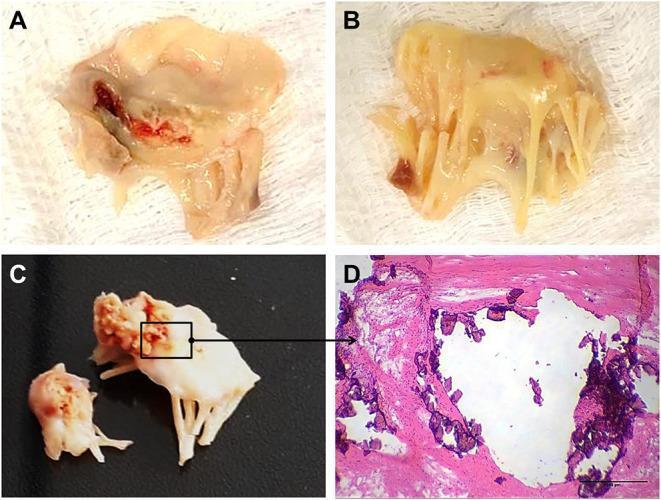
A multi-panel figure illustrating the gross and microscopic pathology of rheumatic heart valve disease (RHVD) in human mitral valves. Panels A and B present clinical photographs of excised mitral valve tissue from a 49-year-old female, showing marked leaflet thickening, retraction, and an opaque, yellowish appearance consistent with chronic fibrosis. Panel A specifically shows focal areas of reddish-dark discoloration, suggesting hemorrhage or tissue damage. Panel C displays gross pathology from a 61-year-old male, highlighting advanced calcification with rigid, cauliflower-like deposits on the valve structure. Panel D is a high-magnification light microscopy image (Hematoxylin and Eosin stain) of the anterior mitral valve leaflet. It demonstrates nodular calcification, appearing as irregular, dark purple hematoxylin-positive deposits embedded within the eosinophilic (pink) fibrotic collagenous matrix. A scale bar of 500 μm is provided. This figure serves as an educational resource for identifying the progression from chronic inflammation and fibrosis to end-stage valvular calcification in rheumatic heart disease.
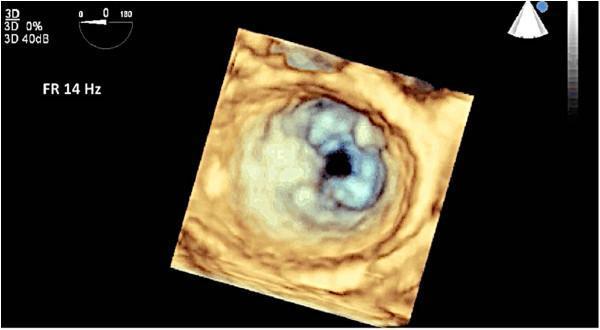
This diagnostic image is a 3D full-volume transesophageal echocardiogram (TEE) demonstrating a ‘surgeon’s view’ (en face from the left atrium) of the mitral valve. The image illustrates classic features of rheumatic heart disease. The mitral valve exhibits significant leaflet thickening and restricted mobility, resulting in a characteristic 'fish mouth' appearance of the valve orifice during diastole. This morphology is primarily caused by extensive commissural fusion, particularly involving the anterolateral commissure, and tethering of the leaflets. These visual findings are diagnostic markers for rheumatic mitral stenosis. The echocardiographic modality provides high spatial resolution (FR 14 Hz) necessary for pre-surgical planning and assessing the extent of valvular deformity. Key anatomical structures visible include the fused anterior and posterior mitral valve leaflets and the stenotic central orifice.
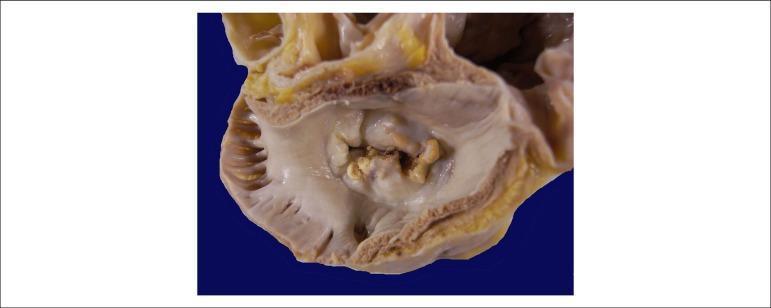
This clinical photograph shows a gross pathological specimen of a human heart, specifically an atrial view of the mitral valve. The image demonstrates hallmark signs of chronic rheumatic heart disease. Key features include significant thickening and fibrosis of the valve leaflets, resulting in a 'fish-mouth' or 'buttonhole' appearance of the valvular orifice. There is evidence of extensive commissural fusion and multiple focal areas of yellowish-white calcification along the leaflet margins and within the valve body. These structural changes characterize severe mitral stenosis, where the restricted opening of the valve impairs blood flow from the left atrium to the left ventricle. The surrounding atrial endocardium appears thickened, consistent with chronic pressure overload. This visual is highly representative for educational purposes in cardiology and pathology to illustrate the long-term sequelae of rheumatic fever on cardiac valves.
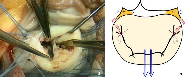
This figure presents a dual-panel comparison of a mitral valve commissurotomy procedure for rheumatic heart disease. Panel (a) is an intraoperative clinical photograph showing the surgical field through an atriotomy. The mitral valve leaflets are visible, appearing thickened and pale, consistent with rheumatic changes. Surgical instruments, including nerve hooks and forceps, are actively manipulating the anterior and posterior leaflets, while traction sutures are placed along the valve annulus to provide exposure. Panel (b) is a schematic illustration defining the surgical protocol for the commissurotomy. It highlights the trigones in orange and uses blue arrows to indicate the symmetrical traction applied by nerve hooks around the major chordae. Grey outlines identify the commissural leaflets (C1 and C2). Red dashed lines and bars quantify the safety margins for the incision, specifically stopping 3 to 5 mm from the annulus and 2 to 3 mm laterally from the trigones to preserve subvalvular structures and chordal integrity. The visual serves to bridge clinical practice with the precise anatomical landmarks required for successful valve repair.
Pathogenesis of Rheumatic Heart Disease
1. The Triggering Event: GAS Pharyngitis
- GAS undergoes phagocytosis by neutrophils, macrophages, and dendritic cells at the pharyngeal mucosa
- Processed antigens activate both humoral (B cell/antibody) and cellular (CD4+ T cell) arms of the adaptive immune system
- A latent period of 2-3 weeks follows the infection before symptoms of ARF appear - this delay reflects the time needed to generate a full adaptive immune response; by this point, streptococci are completely absent from the lesions
2. Molecular Mimicry: The Core Mechanism
| Streptococcal Antigen | Cross-reactive Host Target |
|---|---|
| M protein (S2, light meromyosin) | Cardiac myosin (heavy & light chains) |
| N-acetyl-β-D-glucosamine (GAS cell wall carbohydrate) | Laminin (extracellular matrix of valves), cardiac myosin epitopes |
| M protein epitopes | Tropomyosin |
| GAS neuraminidase | Neuronal gangliosides (Sydenham's chorea) |
3. Antibody-Mediated Valvular Injury
- Cross-reactive IgG antibodies bind to the valvular endothelium and basement membrane, particularly targeting laminin and cardiac myosin epitopes
- Antibody binding triggers complement activation and Fc receptor engagement on macrophages, generating an inflammatory signal
- This upregulates expression of adhesion molecules, particularly VCAM-1 (vascular cell adhesion molecule-1) on the valvular endothelium
- VCAM-1 interacts with VLA-4 (very late activation antigen-4) expressed on CD4+ T lymphocytes, facilitating their extravasation into valve tissue
- Additional molecules upregulated include ICAM, P-selectin, and several chemokines: CCL3/MIP-1α (myocardium), CCL1/I-309 and CXCL9/Mig (valvular tissue)
- T cells infiltrating the valve migrate predominantly along CXCL9/Mig chemokine gradients
4. T Cell-Mediated Injury
- Intralesional T cell clones from valve tissue of RHD patients show 63.2% reactivity against human light meromyosin (LMM) peptides - the major cardiac myosin autoantigen
- 34% of these T cell clones show cross-reactivity patterns: myosin + valve proteins; myosin + streptococcal M5 peptides; or all three
- CD4+ T cells activated by myosin epitopes can also trigger broad recognition of valvular proteins with structural similarity (bystander activation)
- Within the Aschoff body, cytokines from stimulated T cells lead to macrophage activation - producing the characteristic Anitschkow cells
| Cytokine Subset | Role |
|---|---|
| IFN-γ, TNF (Th1) | Pro-inflammatory; dominant in valve and myocardial infiltrates |
| IL-17, IL-23 (Th17) | Large numbers found in valves; drive autoimmunity |
| IL-4, IL-10 (Th2) | Regulatory; very low in valvular tissue - this imbalance (lack of IL-4) may contribute to progressive valvular lesion |
| Tregs (CD4+CD25+FoxP3+) | Immunosuppressive; present but insufficient to halt tissue injury |
5. The Aschoff Body (Pathognomonic Lesion)
- Central zone of fibrinoid necrosis
- Surrounded by lymphocytes (primarily T cells), scattered plasma cells
- Anitschkow cells (pathognomonic): plump activated macrophages with abundant cytoplasm and nuclei showing centrally condensed chromatin in a slender, wavy ribbon pattern - the "caterpillar cells"
- Aschoff bodies can occur in any of the three cardiac layers during ARF, hence pancarditis
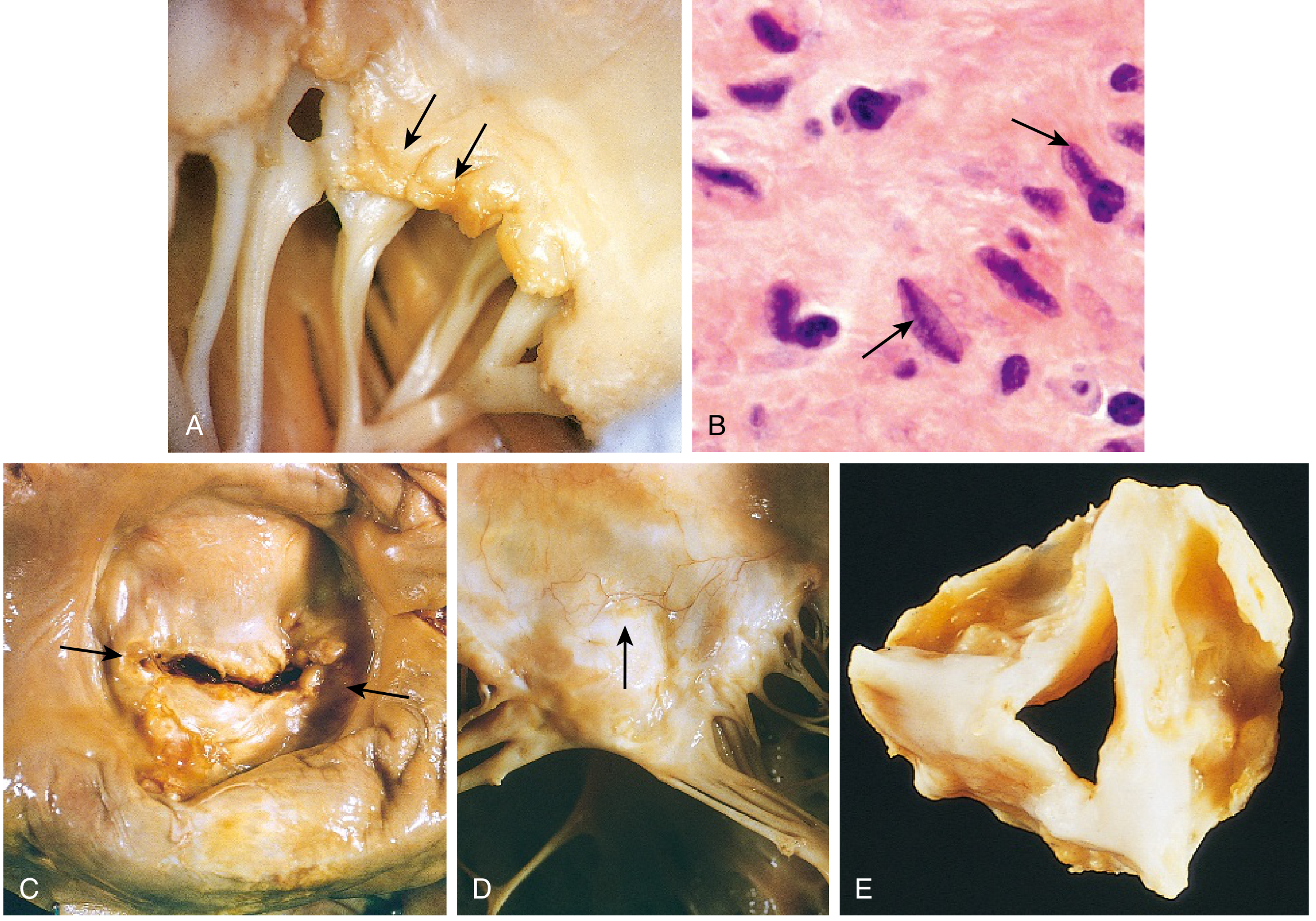
6. Acute Rheumatic Carditis - Layer by Layer
| Layer | Acute Finding | Outcome |
|---|---|---|
| Pericardium | Fibrinous exudate | Usually resolves without sequelae |
| Myocardium | Scattered Aschoff bodies in interstitial connective tissue | May cause cardiomegaly and functional MR in severe cases |
| Endocardium/Valves | Fibrinoid necrosis + fibrin deposition along valve closure lines → 1-2 mm verrucae (sterile vegetations) | Progressive scarring with repeated episodes |
- Valvular involvement (valvulitis) follows myocarditis
- The mitral and aortic valves are most severely affected due to the higher mechanical stress they endure
7. Progression to Chronic RHD
- Aschoff bodies are replaced by fibrous scar in chronic disease
- Valve leaflets become permanently thickened and retracted
- Commissural fusion narrows the valve orifice
- Chordae tendineae thicken, shorten, and fuse
- Neovascularization occurs in the leaflets (normally avascular)
- Calcification superimposes on fibrosis, creating the classic "fishmouth" or "buttonhole" stenosis of the mitral valve
- Upregulated: vimentin, lumican, apolipoprotein-A1
- Downregulated: collagen-VI, haptoglobin-related protein, prolargin, biglycan, cartilage oligomeric matrix protein
8. Valve Distribution and Consequences
| Valve | Frequency |
|---|---|
| Mitral alone | ~70% |
| Mitral + Aortic | ~25% |
| Tricuspid | Less frequently and less severely |
| Pulmonary | Almost always spared |
- Left atrial dilation → atrial fibrillation → mural thrombus formation → systemic emboli
- Pulmonary venous hypertension → pulmonary congestion → right ventricular hypertrophy and failure
9. Mechanism Summary (Flowchart)
GAS pharyngitis
       ↓
Innate immune activation (macrophages, dendritic cells, neutrophils)
       ↓
Antigen presentation → CD4+ T cell & B cell activation
       ↓
MOLECULAR MIMICRY
  ├── Antibodies: GAS carbohydrate (N-acetylglucosamine) ↔ laminin, myosin
  ├── T cells: GAS M protein ↔ cardiac myosin LMM, valve proteins
       ↓
Antibodies bind valve endothelium → complement activation
→ VCAM-1/VLA-4 upregulation → CD4+ T cell infiltration
       ↓
T cell cytokines (IFN-γ, TNF, IL-17) → macrophage activation → Aschoff bodies
       ↓
Fibrinoid necrosis of valve → verrucae (acute phase)
       ↓
Repeated episodes of ARF → organizing inflammation → fibrosis
       ↓
Commissural fusion + chordal shortening + calcification
       ↓
CHRONIC RHD: Mitral/aortic stenosis & regurgitation
Key Points
- The mechanism is autoimmune, not direct bacterial invasion; organisms are absent from cardiac lesions at the time of symptoms
- Only pharyngeal GAS (not skin) triggers ARF - the pharynx's lymphoid environment is uniquely permissive for the cross-reactive immune response
- Genetic susceptibility determines who among infected individuals develops ARF (estimated 3-6% of the population is susceptible)
- The deforming fibrotic lesions result from healing and scarring after acute inflammation, explaining why repeated episodes cause cumulative valve damage
- Secondary prophylaxis with penicillin to prevent recurrent GAS pharyngitis is the most effective intervention to halt progression