
External ear anatomy make it easy for examination
Reading File
This is a visual anatomy question - I'll search the medical library for external ear anatomy with images.
Searching sources...
Finding Sources
Finding Sources
Reading File
Reading File
Now let me get the detailed anatomy of the auricle (pinna) with its surface landmarks, and search for images.
Reading File
Finding Sources
Finding Sources
Reading File
Reading File
Excellent! I have the key images. Let me fetch the most important anatomical diagrams and also get the external acoustic meatus/EAC anatomy details.
Loading Image
Loading Image
Reading File
I now have excellent images and comprehensive content. Let me compile the full answer.
External Ear Anatomy - Easy Reference for Examination
The external ear has two parts: the auricle (pinna) and the external acoustic meatus (EAC). Here is a complete breakdown structured for clinical examination.
PART 1: The Auricle (Pinna)
The auricle is a flap of elastic cartilage covered by skin. It has a characteristic pattern of ridges (elevations) and depressions that are essential to know for examination.
Labeled Diagram
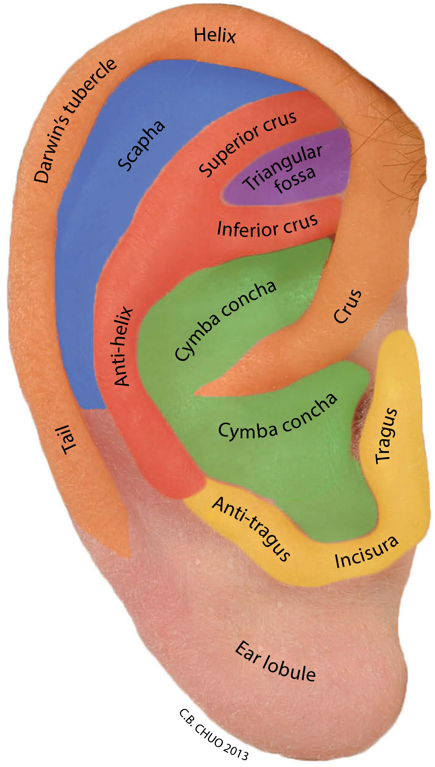
Color-coded surface anatomy of the lateral auricle - Scott-Brown's Otorhinolaryngology
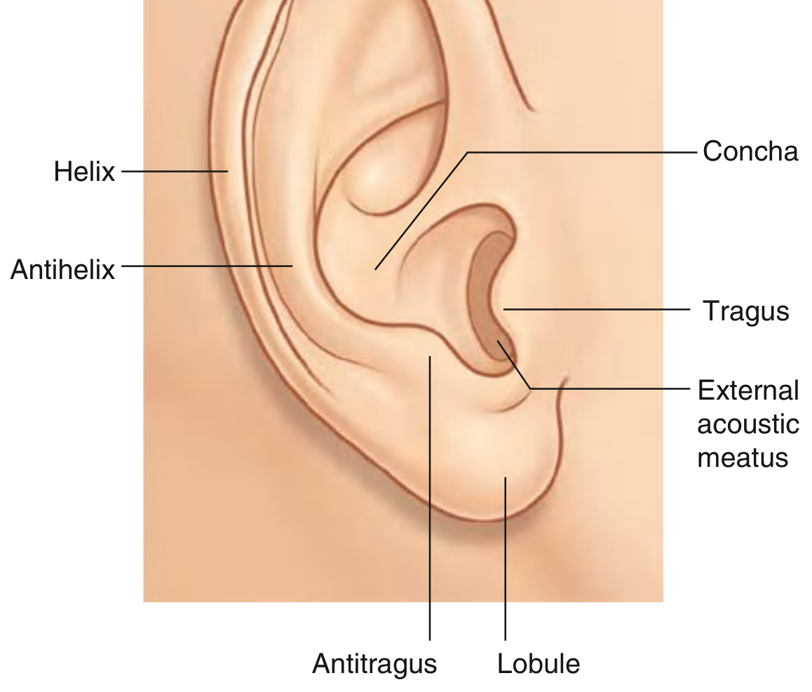
Classic labeled diagram of the auricle - Gray's Anatomy for Students
RIDGES (Elevations) - Easy Memory: "H-A-T-AT"
| Structure | Description | Exam Tip |
|---|---|---|
| Helix | Large outer rim/folded margin of the auricle; ends inferiorly at the lobule | The outermost curved fold - trace it from top to lobule |
| Antihelix | Smaller curved rim parallel and anterior to the helix; separates helix from concha | The inner Y-shaped fold; splits superiorly into two crura |
| Tragus | Prominent quadrangular cartilaginous projection anterior to the EAM opening; points posteriorly | Press it gently - tenderness = otitis externa |
| Antitragus | Small elevation opposite the tragus, above the lobule, at the end of the antihelix | Smaller bump directly opposite the tragus |
DEPRESSIONS (Hollows) - Easy Memory: "SCOTT"
| Structure | Description | Exam Tip |
|---|---|---|
| Scapha | Narrow, shallow groove between helix and antihelix | The long groove running alongside the antihelix |
| Concha | Deepest and widest depression, bracketed by antihelix; leads into the EAM | The "bowl" of the ear - deepest central part |
| Cymba conchae | Upper portion of the concha above the crus of the helix | Above the helical crus |
| Cavum conchae | Lower portion of the concha below the crus of the helix | Below the helical crus; leads directly into EAM |
| Triangular fossa | Depression between the superior and inferior crura of the antihelix | The "V" space at the top between the two crura |
Other Named Landmarks
| Structure | Description |
|---|---|
| Lobule | Inferior fleshy part of the ear - the only part with NO cartilage (only fat/connective tissue) |
| Incisura (intertragic notch) | The notch between the tragus and antitragus |
| Darwin's tubercle | Small prominence on the posterior upper helix; a vestigial structure |
| Crus of the helix | The root/origin of the helix that slopes across the top of the concha |
| Superior crus | Upper limb of the bifurcating antihelix |
| Inferior crus | Lower limb of the bifurcating antihelix |
PART 2: External Acoustic Meatus (EAC / Ear Canal)
The EAC extends from the external auditory meatus (visible opening) to the lateral surface of the tympanic membrane (TM). It measures approximately 2.5 cm in adults.
Two Distinct Portions
| Outer 1/3 (Cartilaginous) | Inner 2/3 (Bony) | |
|---|---|---|
| Skeleton | Cartilage | Tympanic part of temporal bone |
| Skin | Thick with hair follicles, sebaceous + apocrine glands | Thin, no subcutaneous layer, no glands or hair |
| Cerumen | Produced here (hydrophobic, pH 6.0-6.5) | None produced |
| Special features | Fissures of Santorini (allow infection/tumour spread to parotid) | Foramen of Huschke (allows spread to parotid deep lobe) |
Key EAC Landmarks
- Isthmus - narrowest point at the junction of bony and cartilaginous portions
- Fissures of Santorini - transverse cartilaginous slits; pathway for infection to spread to surrounding soft tissues
- Foramen of Huschke - bony defect from incomplete ossification in the anterior canal; communicates with parotid
Self-Cleansing Mechanism
The sloughed keratinous layer of the TM migrates outward (centrifugally) toward the canal, where it mixes with glandular secretions and is extruded as cerumen. This is why you should never insert objects into the canal - it disrupts this process.
PART 3: Innervation (Clinical Importance)
The auricle receives sensory supply from multiple sources - which is why referred otalgia is so common:
| Nerve | Area Supplied |
|---|---|
| Great auricular nerve (C2, C3 - cervical plexus) | Anterior/posterior inferior auricle, lobule |
| Lesser occipital nerve (C2 - cervical plexus) | Posterosuperior auricle |
| Auriculotemporal nerve (V3 - mandibular division) | Anterior auricle, tragus, anterior EAC |
| Facial nerve [VII] | Small area of EAC (Ramsay Hunt zone) |
| Vagus nerve [X] (Arnold's nerve) | Posterior EAC wall, part of auricle - stimulation causes cough reflex |
Clinical pearl: The vagal branch (Arnold's nerve) in the posterior EAC explains why syringing/inserting objects can trigger coughing or even fainting (vasovagal).
PART 4: Auricle Position (Quick Reference for Exam)
- Longitudinal axis tilted 15-30 degrees posteriorly from vertical
- Upper border of ear is level with the lateral eyebrow in ~85% of individuals
- Located approximately one ear-length posterior to the lateral orbital margin
- Conchal-mastoid angle: ~30 degrees (important in prominent ear/otoplasty assessment)
Quick Examination Checklist
- Inspect the auricle - helix, antihelix, tragus, antitragus, lobule, scapha, concha
- Tragal pressure test - press on tragus; pain = otitis externa
- Pinna traction - pull pinna up and back (adults) / down and back (children) to straighten EAC before otoscopy
- EAC inspection - note hair, wax (cerumen), skin changes, foreign bodies
- Postauricular sulcus - look/feel behind the ear; obliteration = mastoiditis
- Preauricular area - check for lymph nodes, pits (preauricular sinus), tags
Sources: Gray's Anatomy for Students, p. 1094; Scott-Brown's Otorhinolaryngology Head & Neck Surgery; Cummings Otolaryngology Head and Neck Surgery, p. 2648
This is a shared conversation. Sign in to Orris to start your own chat.