
Bleed in small brain
cerebellar hemorrhage CT scan brain bleed
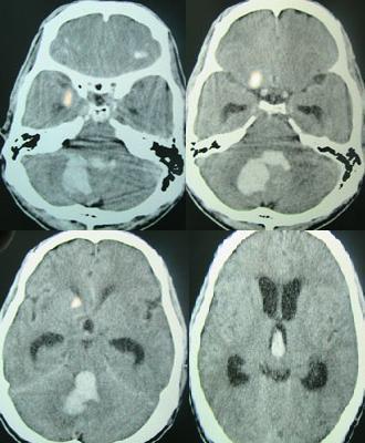
This diagnostic image consists of four axial non-contrast CT scan slices of the brain, demonstrating acute intracranial pathology. The primary finding is a large, irregularly shaped hyperdense area in the posterior fossa, consistent with an acute cerebellar hemorrhage. This hematoma exerts significant mass effect on the fourth ventricle and brainstem. Secondary findings include intraventricular hemorrhage (IVH), visible as hyperdense material settled within the lateral and third ventricles. There is evidence of obstructive hydrocephalus, characterized by the enlargement of the frontal horns of the lateral ventricles and the temporal horns. Smaller hyperdense foci are also noted in the supratentorial region, which may represent secondary parenchymal extension or hypertensive bleeds. This clinical case illustrates the radiographic presentation of a hypertensive posterior fossa bleed leading to secondary ventricular obstruction and acute hydrocephalus, a critical neurosurgical emergency.
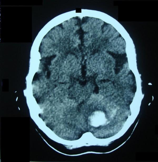
An axial non-contrast computed tomography (CT) scan of the brain demonstrating an acute hypertensive intracranial hemorrhage. A prominent, well-circumscribed hyperdense (bright) lesion is visible in the left deep cerebellar hemisphere, specifically involving the region of the dentate nuclei. The lack of surrounding hypodense vasogenic edema suggests a very acute onset, approximately within a few hours of the scan. In the contralateral (right) cerebellar hemisphere, there is a subtle area of decreased density/slit-like space indicating a resolved previous hematoma. The supratentorial structures, including the ventricles and cortical sulci, appear relatively normal in size and configuration for the patient's age. This image is clinically significant for demonstrating recurrent spontaneous intraparenchymal hemorrhage in a patient with hypertension, highlighting the typical locations and the appearance of acute versus chronic blood products on CT.
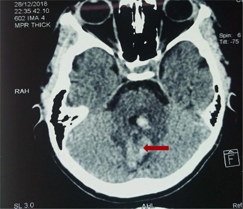
This diagnostic axial CT scan of the brain demonstrates an acute intracranial hemorrhage. The primary finding, indicated by a red arrow, is a well-defined area of hyperdensity within the left cerebellar hemisphere, consistent with an intraparenchymal hematoma. The hyperdense signal indicates acute clotted blood, which shows increased attenuation compared to the surrounding cerebellar parenchyma. A faint rim of hypodensity is visible around the hemorrhage, representing perihematomal edema. Additional hyperdense foci are noted near the midline, possibly involving the cerebellar vermis. Anatomical landmarks such as the temporal lobes, orbits, and bony skull structures are visible, providing spatial orientation. This image serves as an educational example of spontaneous posterior fossa hemorrhage, which in this clinical context is associated with systemic coagulopathy following an elapid or viperid snake bite. It illustrates the role of non-contrast CT in identifying acute hemorrhagic stroke and evaluating for secondary mass effect or brainstem compression.
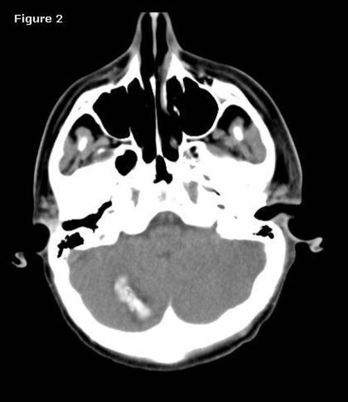
This diagnostic image is an axial non-contrast computed tomography (CT) scan of the brain at the level of the posterior fossa and skull base. The primary finding is an area of hyperdensity in the right cerebellar hemisphere, measuring approximately 15 x 20 mm, which is consistent with an acute intracerebellar hemorrhage. The hemorrhage appears somewhat linear and is localized within the cerebellar folia. Evaluation of the surrounding brain tissue reveals diffuse decreased parenchymal density and effacement of the sulcal spaces and cisterns, suggestive of significant cerebral edema. Visible anatomical landmarks include the sphenoid sinus, ethmoid air cells, and the bony structures of the skull base. This scan illustrates a potential postoperative complication (remote cerebellar hemorrhage) following supratentorial neurosurgery. The target audience for this educational material includes neurosurgery residents and radiology students studying postoperative complications and intracranial pathology.
Cerebellar Hemorrhage ("Bleed in the Small Brain")
Epidemiology
- Accounts for ~5-10% of all intracerebral hemorrhages (ICH)
- Represents ~10% of all cerebellar strokes
- About 75% of patients have hypertension as the underlying cause
Causes
| Cause | Notes |
|---|---|
| Hypertension (most common) | Rupture of distal SCA (superior cerebellar artery) branches near the dentate nucleus |
| Cerebral amyloid angiopathy (CAA) | Especially in elderly; tends to be superficial |
| AVM / cavernous malformations | Tend to be more superficial |
| Anticoagulant / thrombolytic therapy | Drug-related coagulopathy |
| Hemorrhagic metastases | Melanoma, renal cell, choriocarcinoma |
| Trauma | Posterior fossa impact |
| Coagulopathies | Thrombocytopenia, DIC |
- Localization in Clinical Neurology, 8e, p. 1392
Clinical Features
Appendicular ataxia + Horizontal gaze palsy + Peripheral facial palsy (all ipsilateral to the bleed)
| Symptom | Frequency |
|---|---|
| Vomiting | 77-81% |
| Headache (occipital) | 67-80% |
| Dizziness/Vertigo | 59-60% |
| Gait/truncal ataxia | 56% |
| Dysarthria | 22-42% |
| Drowsiness | 42% |
| Altered consciousness | 60-73% |
| Brainstem findings | ~100% in severe cases |
- Inability to stand or walk (truncal ataxia)
- Nystagmus, skew deviation, ocular bobbing
- Ipsilateral horizontal gaze paresis (from pontine compression)
- Ipsilateral facial paresis (CN VII - from pontine compression)
- Small (miotic) but reactive pupils
- Absent/asymmetric corneal reflexes
- Babinski sign (~50%)
- Nuchal rigidity (~50%)
- Plum and Posner's Diagnosis and Treatment of Stupor and Coma, p. 289
- Bradley and Daroff's Neurology in Clinical Practice, p. 1027
Why Is It Dangerous?
- Compress the brainstem (pons/medulla) - causing coma and cardiorespiratory failure
- Obstruct the 4th ventricle - causing acute obstructive hydrocephalus
- Tonsillar herniation downward (through foramen magnum)
- Upward tentorial herniation
"There is a notorious tendency for abrupt deterioration to coma and death after a period of clinical stability under hospital observation." - Bradley and Daroff's Neurology
CT Scan Imaging
Diagnosis
- Urgent non-contrast CT head (first-line)
- CBC, platelets, INR, PTT, BMP, glucose
- ECG (sympathetic overactivity can cause arrhythmias)
- If no clear hypertensive etiology: CTA or MRA to rule out AVM/aneurysm
- Consider vascular anomaly workup if: age <65, female, no hypertension history, cocaine use, nonsmoker
Red Flags for Poor Outcome / Urgent Surgery
| Indicator | Significance |
|---|---|
| Hematoma ≥ 3 cm diameter | High surgical urgency |
| Neurologic deterioration | Immediate surgery |
| Brainstem compression signs | Immediate surgery |
| Hydrocephalus on CT | Needs drainage |
| Coma at presentation | 100% mortality without surgery |
| Effacement of quadrigeminal cistern | Impending herniation |
Treatment
Medical Management
- ICU admission for all patients
- Reverse anticoagulation immediately (Vitamin K, FFP, PCC for warfarin; specific reversal agents for NOACs)
- Hold antiplatelet agents; give platelets if thrombocytopenic
- Blood pressure control:
- Systolic 150-220 mmHg → lower to 140 mmHg acutely
- Systolic >220 mmHg → IV antihypertensives (labetalol, nicardipine)
- Airway protection if GCS is low
Surgical Management
- Hemorrhage > 3 cm in diameter
- Neurologic deterioration
- Brainstem compression
- Obstructive hydrocephalus
- Suboccipital craniectomy with clot evacuation (definitive)
- Ventriculostomy (EVD) for hydrocephalus management
"Cerebellar hemorrhages can compress vital brainstem structures and surgical evacuation can be lifesaving." - Symptom to Diagnosis, 4th Ed.
- Localization in Clinical Neurology, 8e, p. 1392
- Symptom to Diagnosis, 4th Ed.
- Plum and Posner's, p. 290
Key Takeaway
Prognosis?
cerebellar hemorrhage prognosis outcome
Prognosis of Cerebellar Hemorrhage
The Four Clinical Grades (Plum & Posner Classification)
| Grade | Hematoma Size | Presentation | Prognosis |
|---|---|---|---|
| I - Minor | <1.5-2 cm | Mild unilateral cerebellar signs, headache, self-limited | Excellent; often resolves without surgery |
| II - Moderate | <3 cm | Occipital headache, gradual cerebellar/oculomotor signs, mild drowsiness | Can recover spontaneously with careful observation |
| III - Severe | Any; progressive | Acute/subacute headache + vomiting, progressive deficits, ipsilateral ataxia, gaze paresis, obtundation | Poor without surgery; good if operated promptly |
| IV - Catastrophic | Large (5 cm typical) | Sudden loss of consciousness, pinpoint pupils, absent oculovestibular responses, quadriplegia | Very poor; ~20% of all cases; high mortality even with surgery |
"About one-fifth of patients with cerebellar hemorrhage develop early pontine compression with sudden loss of consciousness, respiratory irregularity, pinpoint pupils, absent oculovestibular responses, and quadriplegia." - Plum and Posner's, p. 291
Key Prognostic Factors
1. Hematoma Size - The Most Important Factor
- <3 cm diameter: Spontaneous recovery possible; conservative management may suffice
- ≥3 cm diameter: High risk of fatal deterioration; surgical evacuation strongly recommended
- >5 cm: Very high mortality; often fatal even with surgery (as in Patient Vignette 4.3)
2. Level of Consciousness
- Alert/drowsy at admission: Good prognosis if treated promptly
- Obtunded: Urgent surgery required; prognosis fair
- Stupor/coma: Very poor; "patients almost always do poorly if one waits until coma develops to initiate surgical treatment" - Plum and Posner's
- Coma without surgery: 100% mortality - Bradley and Daroff's Neurology
3. Brainstem Compression Signs
- Ipsilateral gaze palsy (CN VI/PPRF)
- Ipsilateral facial palsy (CN VII)
- Extensor plantar responses
- Loss of oculocephalic reflexes
4. CT Features (Poor Prognosis)
- Hematoma ≥3 cm
- Hydrocephalus (4th ventricle obstruction)
- Effacement of the quadrigeminal cistern
- Intraventricular extension of blood (correlates with fever and worse outcome)
5. Speed of Deterioration
"Deterioration from alertness or drowsiness to stupor often occurs over a few minutes, and even brief delays to carry out radiographic procedures can prove fatal." - Plum and Posner's, p. 291
Long-Term Functional Outcomes
-
Cerebellar ataxia (persistent limb or gait ataxia)
-
Dysarthria
-
Oculomotor deficits
-
Cerebellar Cognitive Affective Syndrome (CCAS): impaired executive function, spatial cognition, language deficits, blunted or disinhibited affect - especially with posterior hemisphere damage
-
These cognitive and behavioral sequelae may persist even after the hematoma resolves
-
Plum and Posner's, p. 291
Surgical vs. Conservative: What the Evidence Shows
- Surgery vs. conservative management showed no significant difference in death or disability at 6 months for supratentorial bleeds (74% vs 76% poor outcome at 6 months)
- Cerebellar hemorrhage is an exception - surgery is more clearly beneficial here because the confined posterior fossa means brainstem compression is life-threatening and reversible if treated early
-
Small (<3 cm), neurologically stable patients: conservative management is reasonable
-
Ventriculostomy alone is NOT sufficient - it relieves hydrocephalus but does NOT decompress the brainstem, and may actually worsen upward transtentorial herniation
-
Bradley and Daroff's Neurology, p. 1401-1403
Summary: Prognosis at a Glance
| Situation | Expected Outcome |
|---|---|
| Small bleed (<3 cm), alert, no hydrocephalus | Good; often full recovery |
| Moderate bleed, early surgery before coma | Fair to good |
| Large bleed (≥3 cm) with hydrocephalus, prompt surgery | Guarded; survival possible |
| Coma at presentation, no surgery | ~100% mortality |
| Coma at presentation, emergency surgery | High mortality; poor functional outcome |
| Pontine compression signs present | Emergency surgery - delay = death |