
Basic Principles of color Doppler 10 marks PG Obs and Gynec exam
I'll read the medical library skill first, then research color Doppler principles comprehensively.
Reading File
Now let me search the library and images in parallel.
Finding Sources
Finding Sources
Searching Images
color Doppler ultrasound principle waveform obstetrics
Now let me read the key sections - I'll search specifically for Obs & Gynec Doppler content and the echocardiography textbook's color Doppler sections in parallel.
Finding Sources
Reading File
Searching Images
Doppler shift frequency blood flow velocity ultrasound diagram
Finding Sources
Reading File
Finding Sources
Finding Sources
Finding Sources
Loading Image
Now let me read the key Creasy & Resnik MFM textbook content on Doppler technique in obstetrics:
Reading File
Reading File
I now have excellent material. Let me read a bit more from Creasy & Resnik on the indices formulas and clinical application, plus fetch the waveform image:
Reading File
I now have comprehensive data from authoritative sources. Let me compile the full 10-mark PG exam answer.
Basic Principles of Color Doppler
PG Obstetrics & Gynaecology Examination - 10 Marks
1. The Doppler Effect (Physical Basis)
The Doppler effect, described by Austrian physicist Christian Johann Doppler (1842), states that the frequency of a wave changes when there is relative motion between the source and the observer. In medical ultrasound, the "source" is the transducer emitting sound waves, and the "moving object" is the column of red blood cells (RBCs) acting as moving reflectors.
The Doppler equation is:
Fd = (2 × Ft × V × cos θ) / c
Where:
- Fd = Doppler frequency shift
- Ft = transmitted frequency
- V = velocity of blood flow
- θ (theta) = angle of insonation (angle between the ultrasound beam and the direction of flow)
- c = speed of sound in tissue (~1540 m/s)
- Factor 2 = accounts for the double transit (transducer → target → transducer)
Key implication: The angle of insonation must be kept less than 60°; at 90° (perpendicular), cos 90° = 0 and no Doppler shift is detected. This is the angle dependence of color Doppler - Textbook of Clinical Echocardiography.
2. Types of Doppler Ultrasound
| Type | Principle | Use in Obs/Gynec |
|---|---|---|
| Continuous Wave (CW) | Transmits and receives simultaneously; measures all velocities along beam | High-velocity flows (not range specific) |
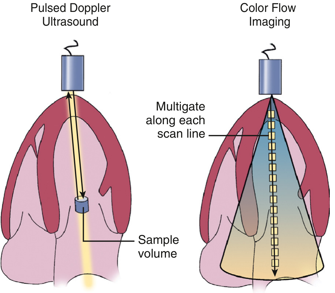
| Pulsed Wave (PW) | Pulses transmitted; only one sample volume at specific depth is interrogated | Specific vessel interrogation (e.g., uterine, umbilical arteries) |
| Color Flow Doppler | Multi-gate PW across entire 2D image plane; velocities color-coded | Real-time flow mapping in fetoplacental circulation |
| Power Doppler | Displays amplitude/energy of Doppler signal (not direction/velocity) | Low-velocity, small-vessel flow (e.g., trophoblastic invasion) |
3. Principles of Color Doppler Flow Imaging
Color Doppler is an extension of pulsed Doppler applied simultaneously to hundreds of sample volumes across a 2D scan plane. The following are its core principles:
(a) Multi-gate Sampling
Rather than interrogating a single sample volume (as in PW Doppler), multiple sample gates are placed along each scan line across the entire 2D field of interest. This is repeated across adjacent scan lines to build a real-time 2D color flow map superimposed on the gray-scale B-mode image.
(b) Autocorrelation Technique
- To process signals from hundreds of gates rapidly enough for real-time display, autocorrelation (a statistical method) is used instead of Fast Fourier Transform (FFT).
- Along each scan line, multiple ultrasound pulses are fired (typically 8 bursts = "burst length"). The back-scattered signals from these 8 bursts at each depth are autocorrelated to estimate mean velocity at that point.
- This gives a mean velocity estimate (not the full spectral velocity distribution) - hence color Doppler shows average flow, not peak velocity.
(c) Color Assignment (Color Map)
The standard convention:
- Red = flow toward the transducer
- Blue = flow away from the transducer
- Brightness/shade = velocity (brighter = higher velocity, up to the Nyquist limit)
- Green/variance color = turbulence or disturbed flow (when variance option is on)
Mnemonic: BART - Blue Away, Red Toward.
(d) Pulse Repetition Frequency (PRF) and Nyquist Limit
- PRF = number of ultrasound pulses emitted per second; determined by the maximum imaging depth.
- The Nyquist limit = PRF/2, which is the maximum velocity that can be measured without aliasing.
- Velocity must be sampled at least twice per cycle (Nyquist theorem) to avoid ambiguity.
(e) Aliasing
- When flow velocity exceeds the Nyquist limit, aliasing occurs: the color "wraps around" from maximum red to maximum blue (or vice versa), giving a mosaic or "flame" appearance.
- In obstetrics, aliasing can be used diagnostically - e.g., the PISA (Proximal Isovelocity Surface Area) method and identifying high-velocity turbulent jets.
4. Indices Derived from Doppler Waveforms
After identifying a vessel with color Doppler, spectral (pulsed wave) Doppler is used to measure the blood flow velocity waveform (FVW). Three mathematically derived indices quantify downstream vascular resistance - Creasy & Resnik's Maternal-Fetal Medicine:
| Index | Formula | Notes |
|---|---|---|
| S/D Ratio (Systolic-Diastolic ratio) | Peak Systolic Velocity / End-Diastolic Velocity | Simplest; cannot be calculated when EDV = 0 |
| Resistance Index (RI) (Pourcelot Index) | (PSV - EDV) / PSV | Range 0-1; independent of angle |
| Pulsatility Index (PI) | (PSV - EDV) / Mean Velocity | Can be calculated even with absent/reversed EDV |
Normal trend: As gestation advances, placental resistance falls → EDV increases → S/D ratio and PI decrease (normal fetoplacental maturation).
5. Key Obstetric Applications
| Vessel | Normal Finding | Abnormal Finding | Significance |
|---|---|---|---|
| Umbilical Artery | Forward diastolic flow; PI decreases with gestation | Absent/Reversed EDV | Placental insufficiency, IUGR |
| Uterine Artery | High diastolic flow by 24 weeks; notching disappears | Persistent diastolic notch; high RI | Preeclampsia risk, uteroplacental insufficiency |
| Middle Cerebral Artery (MCA) | High resistance (low diastolic) | Low PSV → raised PSV in fetal anemia | Brain-sparing, fetal anemia (PSV >1.5 MoM) |
| Ductus Venosus | Positive "a" wave | Absent/Reversed "a" wave | Severe cardiac compromise, acidosis |
6. Power Doppler
Power Doppler maps the amplitude (energy) of the Doppler signal rather than direction or velocity:
- 3-5x more sensitive than color Doppler for detecting slow/low-volume flow
- Less angle-dependent (cos θ does not factor in)
- Cannot show direction of flow
- Applications: trophoblast invasion mapping, tumor vascularity (e.g., gestational trophoblastic disease), testicular and ovarian blood flow
7. Instrument Controls in Color Doppler
| Control | Effect |
|---|---|
| Color gain | Amplifies signal; excessive gain → noise artifact |
| PRF / Velocity scale | Adjust to match expected flow velocity |
| Wall filter | Eliminates low-frequency noise from vessel wall motion |
| Color box size/position | Smaller box = higher frame rate |
| Angle correction | Required for accurate velocity measurement (keep <60°) |
8. Safety Considerations
Color Doppler deposits more acoustic energy than B-mode. Safety is assessed using:
- Thermal Index (TI): risk of tissue heating (TI < 1 is generally accepted for obstetrics)
- Mechanical Index (MI): risk of cavitation (MI < 0.7 in obstetrics)
- Apply the ALARA principle (As Low As Reasonably Achievable): minimize exposure time, power output, and color box size, especially in the first trimester.
- Spectral Doppler should be used in the first trimester only when clinical benefit outweighs risk - Creasy & Resnik's Maternal-Fetal Medicine.
Summary / Examiner Points
- Doppler effect: frequency shift proportional to velocity × cos θ
- Color Doppler = multi-gate PW Doppler with autocorrelation; displays mean velocity as color
- BART convention (Blue Away, Red Toward); brightness = velocity
- Nyquist limit = PRF/2; aliasing occurs when exceeded
- Three indices: S/D, RI (Pourcelot), PI - all reflect downstream resistance
- Key vessels in Obs: umbilical artery, uterine artery, MCA, ductus venosus
- Power Doppler: amplitude-based, more sensitive, angle-independent, no directional info
- Safety: ALARA principle; TI <1, MI <0.7 in obstetrics
Sources: Textbook of Clinical Echocardiography (Otto); Creasy & Resnik's Maternal-Fetal Medicine, 8th Ed.; Campbell Walsh Wein Urology
This is a shared conversation. Sign in to Orris to start your own chat.