
Valves and TEL in ent head and neck for 20 marker
Reading File
Finding Sources
Finding Sources
Finding Sources
Finding Sources
Finding Sources
Finding Sources
Reading File
Finding Sources
Finding Sources
Reading File
Reading File
Searching PubMed
"tracheoesophageal puncture" voice prosthesis
A note before the answer: "TEL" doesn't correspond to any standard ENT abbreviation in the library. Given the pairing with "Valves in ENT/Head and Neck," this is almost certainly TEP – Tracheoesophageal Puncture (with voice prosthesis), which is the classic paired topic in PG ENT exams. I've answered on that basis - let me know if you meant something else (e.g., TEF - tracheoesophageal fistula).
Valves in ENT & Head-Neck, and TEP (Tracheoesophageal Puncture) — 20 Marks
Introduction
A "valve" in the aerodigestive tract is any anatomical structure that permits unidirectional flow of air, food, or secretions while preventing reflux/regurgitation. Several such valves exist along the ENT and head-neck axis, and understanding them is clinically important for nasal obstruction surgery, swallowing/airway protection, and voice rehabilitation after laryngectomy (where the valve principle is surgically recreated - TEP).
A. Valves in ENT and Head & Neck
1. Nasal Valve (external and internal)
The nasal valve is the narrowest segment of the nasal airway and the single most important site for physiological airflow resistance.
- External nasal valve: bounded by the septum, the alar rim (lower lateral crus, sesamoid complex, fibrofatty tissue), and the nasal sill.
- Internal nasal valve: bounded medially by the septum, laterally by the caudal edge of the upper lateral cartilage and the head of the inferior turbinate, and inferiorly by the nasal floor. Normal angle: 10-15°.
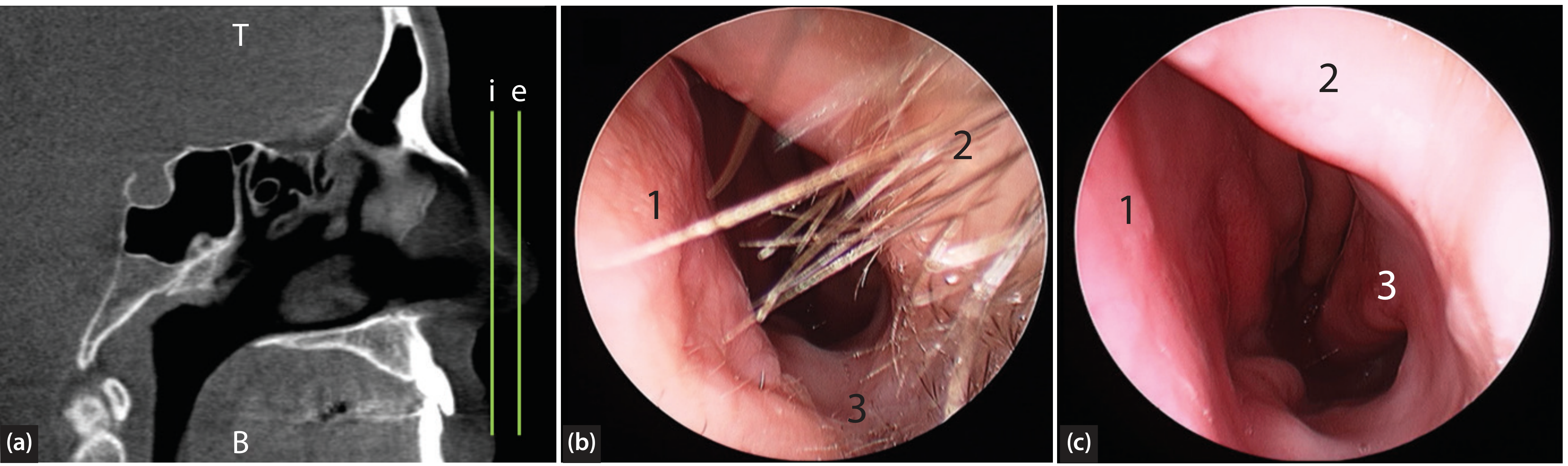
Clinical relevance: collapse of either valve causes nasal obstruction; assessed clinically by the Cottle maneuver (lateral traction on the cheek widens the valve and relieves obstruction) and modified Cottle test with a probe. Failure to address the nasal valve during septoplasty/rhinoplasty is a well-recognized cause of persistent post-operative nasal obstruction, corrected with spreader grafts, alar batten grafts, or a fascia lata sling (as in facial feminization/reconstructive rhinoplasty) - Scott-Brown's Otorhinolaryngology, p. 2097-2111; K J Lee's Essential Otolaryngology.
2. Laryngeal Valve (glottic sphincter)
The larynx functions as a three-tiered valve protecting the lower airway:
- Supraglottic level - epiglottis/aryepiglottic folds
- False cord level - vestibular (ventricular) folds
- True glottic level - vocal folds (the principal dynamic sphincter)
This sphincteric action closes the airway during swallowing (preventing aspiration), enables the Valsalva maneuver, effective coughing, and straining, and opens/closes rhythmically to generate voice - Gray's Anatomy for Students; K J Lee's Essential Otolaryngology.
3. Pharyngoesophageal (PE) Segment / Cricopharyngeus - the "upper esophageal sphincter"
The cricopharyngeus muscle and proximal cervical esophagus form the PE segment, which is tonically contracted at rest (preventing air from entering the esophagus and preventing reflux) and relaxes reflexively during swallowing to allow the bolus to pass - Yamada's Textbook of Gastroenterology, p. 766.
- Killian's dehiscence, the area of weakness between the thyropharyngeus and cricopharyngeus, is the site of Zenker's diverticulum when this valve fails to relax properly.
- This same segment becomes the neoglottis (vibratory sound source) after laryngectomy - central to esophageal speech and TE speech (below).
4. Eustachian Tube Valve Mechanism
Normally closed at rest, the Eustachian tube opens transiently during swallowing and yawning (via tensor veli palatini contraction), equalizing middle ear pressure while preventing reflux of nasopharyngeal secretions and sound/pressure transmission into the middle ear.
5. Velopharyngeal Valve
The soft palate and posterior/lateral pharyngeal walls act as a sphincter separating the naso- and oropharynx during speech (preventing hypernasality) and swallowing (preventing nasal regurgitation). Incompetence causes velopharyngeal insufficiency.
(The ileocecal valve is a true anatomical valve but belongs to the GI tract, not head-neck, and is excluded from ENT enumeration.)
B. TEP - Tracheoesophageal Puncture and Voice Prosthesis
Definition and Principle
TEP is a surgically created fistula/shunt between the posterior tracheal wall and the anterior wall of the cervical esophagus, into which a one-way silicone valve (voice prosthesis) is placed. On occlusion of the tracheostoma, pulmonary air is diverted through the prosthesis into the pharyngoesophageal (PE) segment, causing mucosal vibration that generates sound for tracheoesophageal (TE) speech - Cummings Otolaryngology, p. 2149.
TE speech is pulmonary-driven, so it is the closest of all alaryngeal methods to normal laryngeal voice: functional voice typically develops within 2 weeks, with a success rate of ~90%, making it the current gold standard for voice rehabilitation after total laryngectomy (superseding esophageal speech and the electrolarynx) - Cummings Otolaryngology, p. 2149; Schwartz's Principles of Surgery.
Timing
- Primary TEP: performed at the time of total laryngectomy.
- Secondary TEP: performed later (after healing/adjuvant therapy), endoscopically or by direct puncture under local/general anesthesia.
Types of Voice Prostheses
| Type | Examples | Who changes it |
|---|---|---|
| Non-indwelling | Blom-Singer duckbill, Blom-Singer low-resistance, Panje (with safety strap) | Patient self-removes/replaces |
| Indwelling | Groningen button, Provox, Blom-Singer indwelling | Trained clinician only |
Indwelling prostheses are sturdier, require less patient dexterity (maintained by brushing/flushing rather than removal), and are favored in Europe; non-indwelling devices dominate US practice and carry a ~20% risk of inadvertent dislodgment/aspiration in some series - Cummings Otolaryngology, p. 2150.
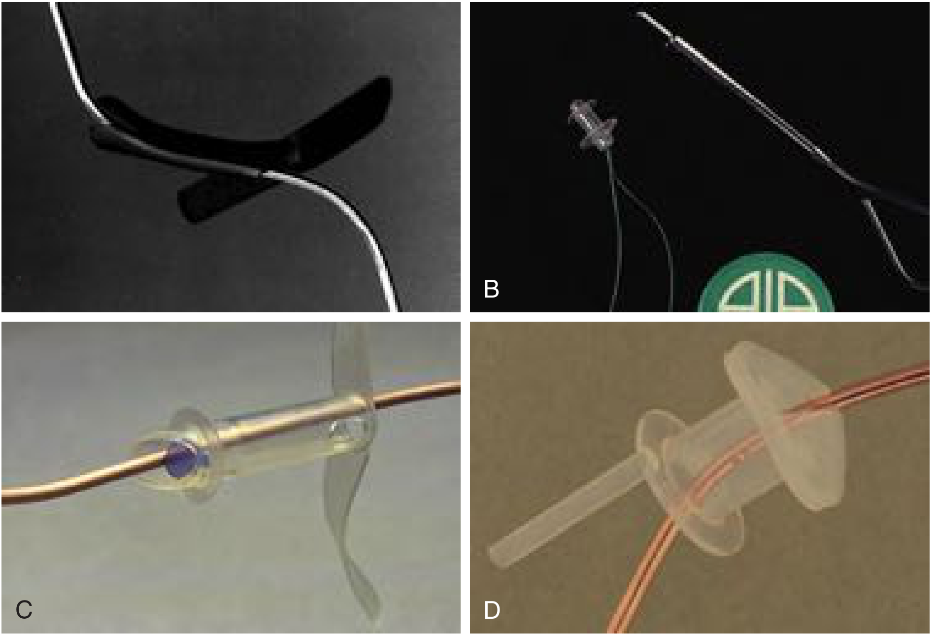
Airflow management for speech
The patient occludes the stoma manually (finger occlusion) or uses a hands-free tracheostoma valve (automatic valve that closes with expiratory airflow, often combined with a heat-and-moisture exchanger), diverting air through the prosthesis for phonation.
Complications
- Periprosthetic or transprosthetic leakage (through valve incompetence or around an enlarged tract) - the most common problem
- Candidal colonization/biofilm of the silicone valve causing leakage and reduced device life
- Granulation tissue at the puncture site
- Widening/enlargement of the TE tract needing resizing or surgical closure
- Stomal stenosis, dislodgement, and aspiration
- Cricopharyngeal (PE segment) spasm impairing voicing - managed with pharyngeal plexus neurectomy, cricopharyngeal myotomy, or botulinum toxin injection
Recent evidence (contextual update)
Recent systematic reviews support and refine current practice: a 2024 meta-analysis confirms good voice-related quality of life after total laryngectomy with TEP-based rehabilitation (PMID 34763996); a 2023 systematic review evaluated tissue-augmentation techniques for periprosthetic leakage around voice prostheses (PMID 37012583); and a 2024 review compared surgical techniques for closing an enlarged TE fistula (PMID 37659224). These don't contradict the classic teaching but reinforce that leakage and tract-widening remain the dominant long-term complications requiring individualized management.
Summary Table for Quick Recall
| Valve | Location | Key function/relevance |
|---|---|---|
| Nasal valve (int./ext.) | Nasal airway, narrowest point | Airflow resistance; obstruction, rhinoplasty |
| Laryngeal (glottic) valve | Larynx - 3 tiers | Airway protection, phonation, Valsalva |
| PE segment/cricopharyngeus | Pharyngoesophageal junction | Swallowing, Zenker's, TE speech source |
| Eustachian tube | Nasopharynx-middle ear | Pressure equalization, reflux prevention |
| Velopharyngeal valve | Soft palate/pharynx | Speech resonance, nasal regurgitation prevention |
| TEP + voice prosthesis | Trachea-esophagus (surgical) | One-way valve for post-laryngectomy voice restoration |
Diagram tip for exam: draw a lateral neck cross-section showing the tracheostome, TE puncture tract, voice prosthesis (duckbill valve), and PE segment with the airflow arrow diverted from trachea to esophagus - examiners award marks heavily for this labeled diagram.
This is a shared conversation. Sign in to Orris to start your own chat.