
Parkinsons disease
"Parkinson disease"[MeSH Terms] AND management
Parkinson's Disease
Overview
Pathophysiology
The Dopamine-Acetylcholine Imbalance
- Inhibitory dopaminergic neurons (from substantia nigra)
- Excitatory cholinergic neurons (within neostriatum)
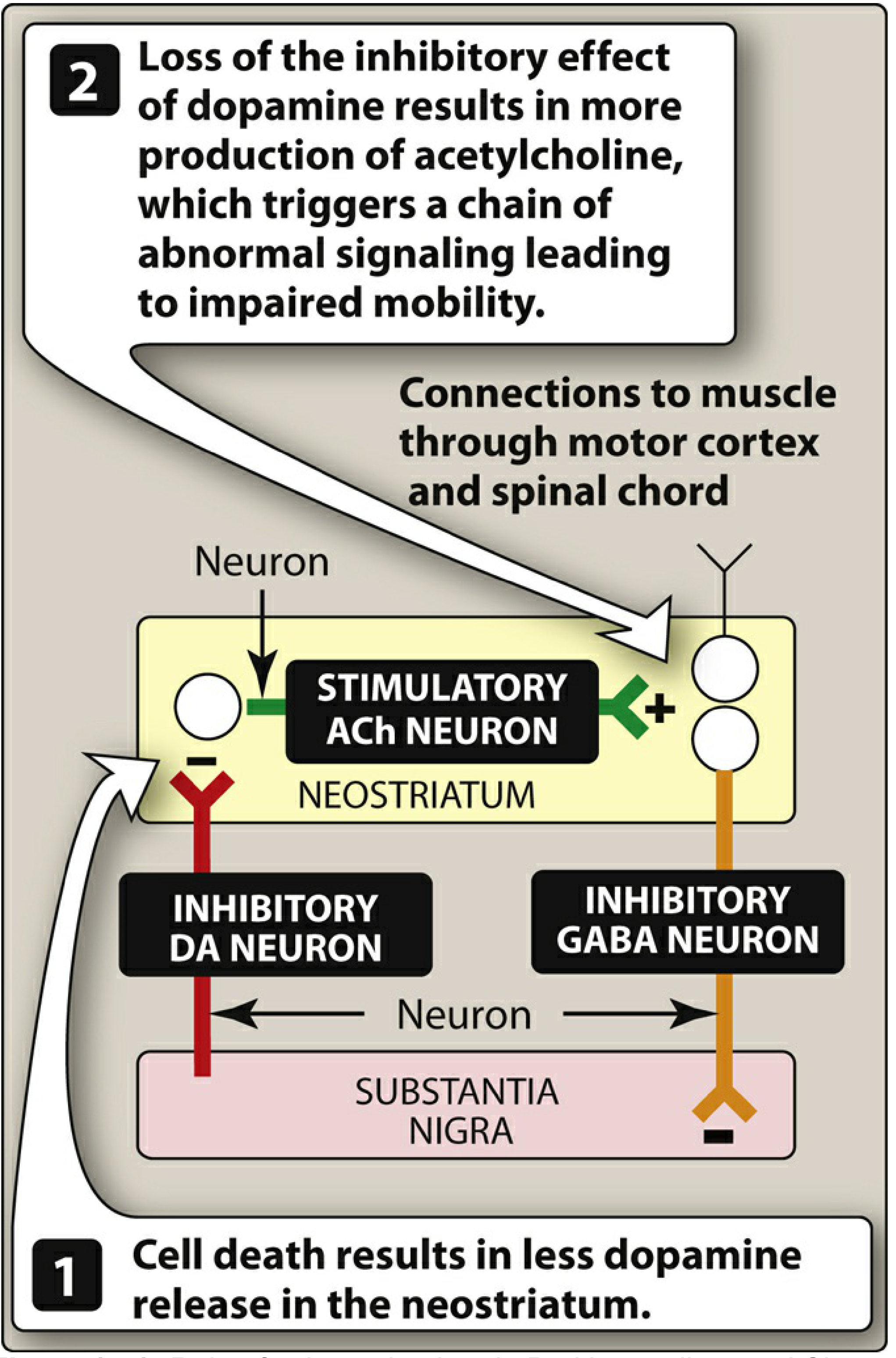
Basal Ganglia Circuit Disruption
- Direct pathway (D1 receptors): dopamine is excitatory - facilitates movement
- Indirect pathway (D2 receptors): dopamine is inhibitory - reduces the brake on movement
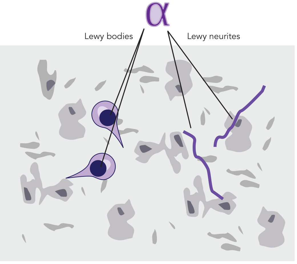
Alpha-Synuclein and Lewy Bodies
Clinical Features
Motor Symptoms (the classic tetrad)
| Feature | Description |
|---|---|
| Resting tremor | "Pill-rolling" tremor at 4-6 Hz, suppressed by voluntary movement |
| Rigidity | Increased tone throughout range of motion; often "cogwheel" type |
| Bradykinesia | Slowness of voluntary movement; most disabling feature |
| Postural instability | Impaired reflex adjustments; late feature, leads to falls |
Other Motor Manifestations
- Masked facies (hypomimia) - reduced facial expression, decreased blink rate
- Micrographia - progressively smaller handwriting
- Hypophonia - soft, monotone voice with hurried muttering quality
- Festinating gait - small, shuffling steps; difficulty initiating (freezing), tendency to lean forward (anteropulsion) or backward (retropulsion)
- En bloc turning - turning without normal torso twist
- Myerson's sign - inability to suppress blinking when glabella is repeatedly tapped (non-specific)
- Dysphagia - recognized by Parkinson himself in 1817; subjective prevalence ~35%, objective up to 82%; involves oral, pharyngeal, and esophageal phases; silent aspiration in 15-33%
Non-Motor Symptoms (often precede motor symptoms)
- Autonomic: orthostatic hypotension, constipation, urinary urgency, sexual dysfunction, seborrhea
- Sleep: REM sleep behavior disorder (RBD) - often an early prodromal marker
- Olfactory: hyposmia/anosmia (may predate motor symptoms by years)
- Neuropsychiatric: depression (most common psychiatric disturbance), anxiety, apathy, psychosis (typically medication-induced visual hallucinations in ~40%, delusions in ~16%)
- Cognitive: mild executive dysfunction is common early; dementia occurs in 15-40% later in the course
Note on psychosis: In PD, hallucinations are usually fleeting and nocturnal, motor symptoms virtually always precede psychosis, and psychosis is generally medication-induced. This distinguishes it from Dementia with Lewy Bodies (DLB), where psychosis is a core feature occurring even without medications. - Bradley and Daroff's Neurology in Clinical Practice
Diagnosis
Supporting features:
- Asymmetric onset
- Good response to levodopa
- Progressive course over 5-15 years
Investigations:
- DaT SPECT (dopamine transporter scan): shows reduced uptake in the striatum; useful when diagnosis is uncertain
- MRI brain: typically normal in idiopathic PD; used to exclude structural causes
Red flags suggesting alternative diagnoses:
- Early postural instability (before 3 years)
- Rapid progression
- Symmetrical onset
- No response to levodopa
- Early falls, early dementia, early autonomic failure, supranuclear gaze palsy, cerebellar signs
Pharmacological Treatment
1. Levodopa + Carbidopa (first-line, most effective)
- Reduces peripheral conversion of levodopa to dopamine (preventing nausea, vomiting, cardiac arrhythmias)
- Increases CNS availability of levodopa
- Allows a 75% reduction in levodopa dose
- Wearing-off phenomenon: as neuronal loss progresses, the drug effect duration shortens between doses
- On-off fluctuations: unpredictable swings between mobile ("on") and immobile ("off") states
- Dyskinesias: involuntary choreiform movements, typically at peak dose
- Nausea, orthostatic hypotension, psychosis
2. COMT Inhibitors (adjunct to levodopa)
- Increase central levodopa uptake
- Raise brain dopamine levels
- Reduce "wearing-off" effects
3. MAO-B Inhibitors
4. Dopamine Receptor Agonists
- Longer duration of action
- Lower risk of dyskinesia
- Useful in patients with levodopa fluctuations
- Initial monotherapy in younger patients delays dyskinesia
5. Anticholinergic Agents
6. Amantadine
Non-Pharmacological and Surgical Treatment
Exercise and Physiotherapy
Deep Brain Stimulation (DBS)
- Advanced PD with motor fluctuations refractory to medical therapy
- Intolerable levodopa-induced dyskinesias
- Preserved cognitive function
Repetitive Transcranial Magnetic Stimulation (rTMS)
Swallowing and Speech Therapy
Complications and Prognosis
- Disease progression is typically insidious, over 5-15 years
- Many patients die of other causes (given late onset) rather than directly from PD
- Leading causes of death in advanced PD: aspiration pneumonia, falls/trauma, immobility complications
- Dementia (PD dementia) occurs in 15-40% of patients late in the course
- PD dementia shares pathological overlap with DLB (both are "synucleinopathies")
Key Differential Diagnoses
| Condition | Distinguishing Features |
|---|---|
| Drug-induced parkinsonism | History of dopamine blockers (antipsychotics, metoclopramide); usually reversible |
| Multiple System Atrophy (MSA) | Early autonomic failure, cerebellar signs, poor levodopa response |
| Progressive Supranuclear Palsy (PSP) | Early postural instability, supranuclear gaze palsy, axial rigidity |
| Corticobasal Degeneration (CBD) | Alien limb, asymmetric apraxia, cortical sensory loss |
| Dementia with Lewy Bodies (DLB) | Dementia precedes or co-occurs with parkinsonism, fluctuating cognition, spontaneous psychosis |
| Essential Tremor | Action/postural tremor, no bradykinesia or rigidity, family history |
Recent Evidence (2024-2026)
- Exercise for motor symptoms (PMID: 39880702, 2025 network meta-analysis): Specific exercise modalities significantly improve motor outcomes - exercise should be considered a core therapeutic component, not optional.
- Virtual reality for balance (PMID: 40142335, 2025 meta-analysis): VR combined with conventional therapy improves balance compared to conventional therapy alone.
- Anxiety management (PMID: 40568748, 2025): Anxiety affects up to 50% of PD patients and remains undertreated; a recent meta-analysis reviewed pharmacological and non-pharmacological approaches.
Pls provide me Harrison based for sure
Parkinson's Disease
Harrison's Principles of Internal Medicine, 22nd Edition (2025)
Authors: C. Warren Olanow, Anthony H.V. Schapira, Christine Klein
Epidemiology
- Estimated ~10.8 million people with PD worldwide; expected to double within 20 years due to population aging
- Mean age of onset: ~60 years (but can occur in the 20s, especially with pathogenic gene mutations)
- Lifetime risk: ~3% for men, ~2% for women
- Frequency increases steeply with age
Pathology
- Degeneration of dopaminergic neurons in the substantia nigra pars compacta (SNc)
- Reduced striatal dopamine
- Lewy pathology - intraneuronal proteinaceous inclusions (Lewy bodies in cell bodies; Lewy neurites in axons) that stain positive for alpha-synuclein (Fig. 446-1)
- Cholinergic neurons of the nucleus basalis of Meynert (NBM)
- Norepinephrine neurons of the locus coeruleus (LC)
- Serotonin neurons of raphe nuclei
- Neurons of the olfactory system, cerebral hemispheres, spinal cord, and peripheral autonomic nervous system
Premotor Stage ("Body-First vs. Brain-First" PD)
- Based on neuronal alpha-synuclein accumulation staging
- Based on alpha-synuclein deposition + distribution of neurodegeneration + pathogenic gene variants
Etiology and Pathogenesis
Genetics
| Gene | Inheritance | Notes |
|---|---|---|
| SNCA (alpha-synuclein) | Autosomal dominant | Point mutations and duplications/triplications; multiplications increase expression and severity |
| LRRK2 (leucine-rich repeat kinase 2) | Autosomal dominant | p.G2019S is the most common mutation; founder effect in Ashkenazi Jewish and North African Berber populations |
| PRKN (Parkin) | Autosomal recessive | Most common AR cause; early-onset PD (median AAO ~32 years) |
| PINK1 | Autosomal recessive | Mitochondrial kinase; early-onset |
| PARK7 (DJ-1) | Autosomal recessive | Rare; early-onset |
| GBA1 (glucocerebrosidase) | Risk factor | Heterozygous mutations are the most common genetic risk factor for PD |
Putative Mechanisms of Cell Death
- Oxidative stress (increased free radicals from dopamine metabolism)
- Mitochondrial dysfunction (Complex I deficiency in SNc)
- Proteasomal/lysosomal dysfunction - failure to clear alpha-synuclein aggregates
- Neuroinflammation - activated microglia in SNc
- Apoptosis as a final common pathway
MPTP Model
Clinical Features
Cardinal Motor Features (TABLE 446-1)
| Cardinal Features | Other Motor Features | Nonmotor Features |
|---|---|---|
| Bradykinesia | Micrographia | Anosmia |
| Rest tremor | Masked facies (hypomimia) | Sensory disturbances (pain, hyposmia) |
| Rigidity | Reduced eye blinking | Mood disorders (depression, anxiety, apathy) |
| Postural instability | Drooling | Sleep disturbances (fragmented sleep, RBD) |
| Hypophonia | Autonomic disturbances | |
| Dysphagia | Orthostatic hypotension | |
| Freezing of gait | GI disturbances | |
| Falling | Genitourinary disturbances | |
| Cognitive impairment/dementia | ||
| Psychosis (hallucinations, delusions) |
Diagnosis - UK Brain Bank Criteria & MDS Criteria
- Supportive criteria (features increasing diagnostic confidence)
- Absolute exclusion criteria
- Red flags (must be counterbalanced by supportive criteria)
Imaging
In atypical parkinsonism (MSA, PSP): striatal dopamine depletion can also be seen, so DaT imaging does NOT reliably distinguish PD from atypical parkinsonism. However, FDG-PET of basal ganglia/thalamic network may help - PD shows decreased GPI activity with increased thalamic activity (opposite in atypical parkinsonism).
Differential Diagnosis
Atypical Parkinsonism (TABLE 446-3 from Harrison's)
| Symptom/Sign | Alternative Diagnosis |
|---|---|
| Early speech and gait impairment, no tremor, no asymmetry, early falls | Atypical parkinsonism (MSA, PSP) |
| Neuroleptic/metoclopramide exposure | Drug-induced parkinsonism |
| Onset before age 40 | Genetic PD, Wilson's disease, DRD |
| Liver disease | Wilson's disease |
| Dementia/hallucinations preceding motor features | Dementia with Lewy Bodies (DLB) |
| Diplopia, impaired vertical (downward) gaze | PSP |
| Poor or no response to adequate levodopa trial | Atypical or secondary parkinsonism |
Wilson's disease must always be ruled out in young-onset parkinsonism, as progression can be prevented with copper chelators.
Treatment
LEVODOPA - The Gold Standard
"Levodopa remains the most effective symptomatic treatment for PD and the gold standard against which new therapies are compared." - Harrison's 22e
- Carbidopa (USA - Sinemet)
- Benserazide (most other countries - Madopar)
- Prevent peripheral levodopa conversion to dopamine, reducing: nausea, vomiting, orthostatic hypotension (mediated via area postrema dopamine receptors outside the BBB)
- Standard oral levodopa/carbidopa (Sinemet)
- Controlled-release (Sinemet CR / Madopar HP)
- Long-acting oral formulation (Rytary)
- Levodopa/carbidopa/entacapone combined (Stalevo)
- Levodopa-carbidopa intestinal gel - continuous intra-intestinal infusion
- Continuous subcutaneous infusions of levodopa
- Inhaled levodopa - absorbed through pulmonary alveoli (approved on-demand therapy for off periods)
Motor Complications of Levodopa (Fig. 446-6)
-
Wearing-off phenomenon: Duration of benefit per dose progressively shortens. Initially, benefit lasts many hours (the "long-duration response"). Eventually, benefit approaches the drug's half-life (60-90 min).
-
On-off phenomenon: Rapid, unpredictable switches between the "on" state (drug working) and "off" state (parkinsonism returns).
-
Dyskinesias: Involuntary choreiform/dystonic movements, typically at peak dose during "on" periods.
- Inhaled levodopa (Inbrija)
- Subcutaneous apomorphine injection (Apokyn)
- Sublingual apomorphine (Kynmobi)
Other Pharmacological Agents
| Drug Class | Examples | Role |
|---|---|---|
| MAO-B inhibitors | Selegiline, rasagiline | Early monotherapy or adjunct; potential neuroprotective effect (ADAGIO trial - rasagiline 1 mg/d showed disease-modifying consistent results) |
| Dopamine agonists | Pramipexole, ropinirole, rotigotine, apomorphine | Monotherapy in early disease (especially younger patients); adjunct in fluctuating disease; longer duration than levodopa; lower dyskinesia risk |
| COMT inhibitors | Entacapone, opicapone, tolcapone | Reduce wearing-off; tolcapone reserved for refractory cases (hepatotoxicity risk) |
| Amantadine | NMDA antagonist | Mild early PD; importantly, reduces levodopa-induced dyskinesias |
| Anticholinergics | Benztropine, trihexyphenidyl | Primarily for tremor in younger patients; avoid in elderly |
Neuroprotection - The Major Unmet Need
"A neuroprotective or disease-modifying therapy that slows or stops disease progression remains the major unmet therapeutic need." - Harrison's 22e
- Agents preventing toxic alpha-synuclein species formation or accumulation
- LRRK2 inhibitors
- GCase (GBA1) enhancers
- GLP-1 agonists (developed for diabetes; anti-inflammatory and pro-mitochondrial effects; results in double-blind trials have been inconsistent)
Surgical Treatment
- Primary targets: subthalamic nucleus (STN) or globus pallidus interna (GPi)
- STN-DBS: reduces all cardinal motor features; often allows levodopa dose reduction
- GPi-DBS: particularly effective for dyskinesias
- Indications: Advanced PD with motor fluctuations refractory to optimized medical therapy; intolerable dyskinesias; preserved cognitive function
- Contraindications/poor candidates: Significant dementia, prominent non-dopaminergic features (freezing, falls), active psychiatric illness, unrealistic expectations
- Focused ultrasound thalamotomy (non-invasive): now available as an alternative for tremor-predominant PD
Emerging / Experimental Therapies (Harrison's 22e)
- Japan (Kyoto University): Phase 1/2 trial since 2018; 7 patients treated
- USA: FDA approved first clinical trial of ESC-derived dopamine neurons; 10 patients in Phase 1
- Europe: Trial of ESC-derived neurons began 2023; 4 patients on low dose with no acute adverse effects
Non-Motor Management
- Depression: Most common psychiatric disturbance; SSRIs are first-line (though may worsen motor symptoms); tricyclics as alternative
- Psychosis: Visual hallucinations in up to 40% - usually medication-induced; clozapine (low dose) or pimavanserin (5-HT2A inverse agonist, does not worsen parkinsonism) are options
- Dementia: 15-40% develop dementia; rivastigmine (ChEI) has some evidence
- Autonomic: Orthostatic hypotension (fludrocortisone, midodrine, droxidopa); constipation; urinary dysfunction
- Sleep/RBD: Melatonin or low-dose clonazepam for RBD
- Anosmia: No treatment; important as a premotor marker
This is not much to write in pg exam
PARKINSON'S DISEASE
Harrison's Principles of Internal Medicine, 22nd Edition — Chapter 446
C. Warren Olanow | Anthony H.V. Schapira | Christine Klein
1. DEFINITION & EPIDEMIOLOGY
- 2nd most common age-related neurodegenerative disease (after Alzheimer's)
- First described by James Parkinson in 1817 as "shaking palsy"
- Prevalence: ~10.8 million worldwide; expected to double in 20 years
- Mean age of onset: ~60 years; can occur in 20s with gene mutations
- Lifetime risk: 3% men, 2% women
- ~15% of cases are familial; the rest are sporadic
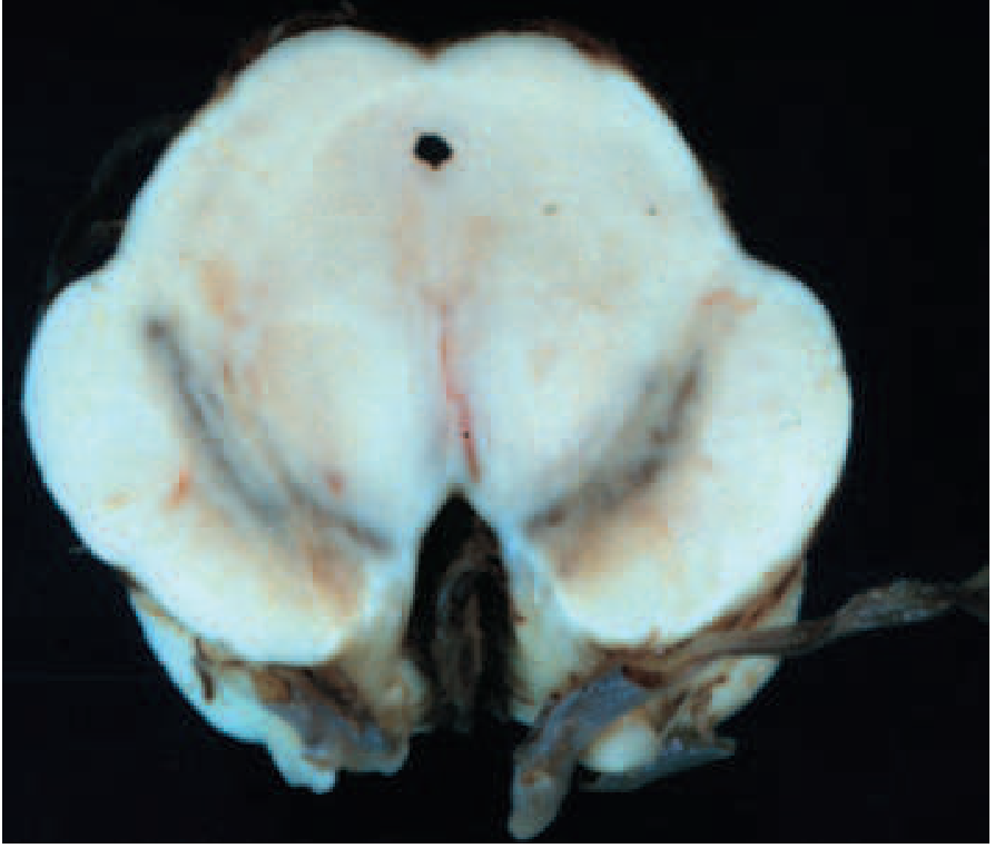
2. PATHOLOGY (FIGURE 446-1 - Harrison's)
Gross pathology
Microscopic pathology
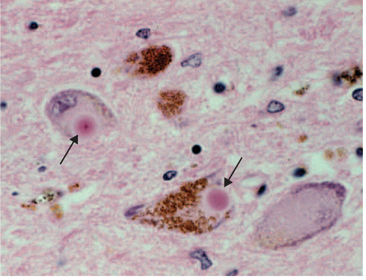
Three hallmark pathological features:
- Degeneration of dopaminergic neurons in the SNc (substantia nigra pars compacta)
- Reduced striatal dopamine
- Lewy pathology — intraneuronal alpha-synuclein aggregates as:
- Lewy bodies (in cell bodies)
- Lewy neurites (in axons)
Nondopaminergic involvement (explains nonmotor features):
- Cholinergic neurons of nucleus basalis of Meynert (NBM) → dementia, cognitive dysfunction
- Norepinephrine neurons of locus coeruleus (LC) → depression, sleep
- Serotonin neurons of raphe nuclei → mood disorders
- Olfactory system → anosmia
- Dorsal motor nucleus of vagus → autonomic dysfunction
- Peripheral autonomic nervous system, GI tract → constipation, gastroparesis
- Spinal cord and cerebral hemispheres
3. ETIOLOGY AND PATHOGENESIS
A. Genetics (TABLE 446-4 — Harrison's)
| Gene | Inheritance | Key Features |
|---|---|---|
| SNCA (alpha-synuclein) | AD | Median AAO 46 yrs; duplications cause classical PD; triplications cause severe, early-onset PD with dementia. Alpha-syn = major component of Lewy bodies |
| LRRK2 | AD | Most common known genetic form. Median AAO 56 yrs; clinically typical PD; slightly slower progression. p.G2019S mutation most common — founder effect in Ashkenazi Jewish and North African Berber populations |
| VPS35 | AD | Median AAO 52 yrs; clinically typical PD; very rare |
| GBA1 | Risk factor | Heterozygous variants = most common genetic risk factor; faster progression + greater cognitive risk |
| PRKN (Parkin) | AR | Most common AR cause; early-onset PD (median AAO 32 yrs, range 9-67 yrs) |
| PINK1 | AR | Mitochondrial kinase; early-onset |
| PARK7 (DJ-1) | AR | Very rare; early-onset |
B. Environmental Factors
- MPTP (methyl-phenyl-tetrahydropyridine) — contaminant of illicit synthetic heroin; caused acute PD syndrome in California addicts in 1980s
- MPTP → oxidized (by MAO-B) to MPP+ → mitochondrial Complex I inhibitor → selective dopamine neuron death
- Importantly: MPTP/MPTP-like compounds have NOT been linked to sporadic PD
- Risk factors (inconsistent evidence): pesticides, rural living, farming, well water, solvents
- Possible protective factors: caffeine, cigarette smoking, NSAIDs, calcium channel blockers
C. Pathogenetic Mechanisms
- Oxidative stress — free radical production from dopamine metabolism
- Mitochondrial dysfunction — Complex I deficiency in SNc
- Proteasomal/lysosomal failure — impaired clearance of alpha-synuclein aggregates
- Neuroinflammation — activated microglia in SNc
- Apoptosis — final common pathway
D. Braak Staging (Alpha-synuclein spread)
- Anosmia, constipation, RBD precede motor symptoms by years to decades
- These are now classified as premotor PD, not just risk factors
- Concept of "body-first" vs "brain-first" PD based on where pathology initiates
4. CLINICAL FEATURES (TABLE 446-1 — Harrison's)
A. Cardinal Motor Features (TRAP)
| Feature | Details |
|---|---|
| Tremor (rest) | "Pill-rolling"; 4-6 Hz; suppressed by voluntary movement; asymmetric onset |
| Rigidity | Cogwheel or lead-pipe; throughout range of motion |
| Akinesia/Bradykinesia | Slowing of movement; the most disabling feature |
| Postural instability | Impaired postural reflexes; late feature; major cause of falls |
B. Other Motor Features
- Micrographia — progressively smaller handwriting
- Masked facies (hypomimia) — reduced facial expression
- Reduced eye blinking
- Drooling (sialorrhoea)
- Hypophonia — soft, monotone voice
- Dysphagia — oral, pharyngeal, esophageal
- Freezing of gait — sudden arrest of movement ("feet glued to the ground"); major cause of falls; may respond to sensory cues (marching, stepping over imaginary line, singing)
- Festinating gait — small, shuffling, rapid steps; anteropulsion; reduced arm swing; en bloc turning
- Retropulsion — backward stepping on pull test
C. Non-Motor Features
| System | Features |
|---|---|
| Autonomic | Orthostatic hypotension, constipation, urinary urgency, sexual dysfunction, seborrhea, sialorrhoea |
| Sleep | REM sleep behaviour disorder (RBD) — often premotor; fragmented sleep; excess daytime sleepiness |
| Sensory | Anosmia/hyposmia (early premotor), pain, restless legs |
| Mood | Depression (~50% of PD patients), anxiety, apathy, panic attacks |
| Cognitive | Mild executive dysfunction early; dementia in up to 80% ultimately — primarily affects executive function and attention; relative sparing of language, memory, calculation |
| Psychosis | Visual hallucinations (typically formed, nonthreatening); often medication-induced |
5. DIAGNOSIS
Clinical Criteria
- Core: Bradykinesia + rigidity
- Plus: rest tremor, asymmetry of onset, good response to levodopa
- Core: Motor parkinsonism (bradykinesia + rigidity/tremor)
- Three additional categories:
- Supportive criteria (increase diagnostic confidence)
- Absolute exclusion criteria
- Red flags (must be counterbalanced by supportive criteria)
- Two certainty levels: Clinically Established PD and Clinically Probable PD
Supportive Criteria for PD (Harrison's)
- Clear beneficial response to dopaminergic therapy
- Rest tremor
- Anosmia
- Cardiac sympathetic denervation on MIBG scintigraphy
Red Flags (suggest atypical parkinsonism):
- Early postural instability or falls (within first 3 years)
- Early dementia (within first 1 year = DLB)
- Absence of rest tremor
- Symmetrical onset
- Poor or no response to adequate levodopa trial
- Early prominent autonomic failure
- Supranuclear gaze palsy (downward gaze palsy → PSP)
- Cerebellar signs
- Pyramidal signs
Investigations
- Shows reduced and asymmetric striatal dopamine transporter uptake
- Most affected: posterior putamen (with relative sparing of caudate)
- Does NOT reliably distinguish PD from atypical parkinsonism (both show reduced DaT)
- PD: decreased GPI activity + increased thalamic activity
- Atypical parkinsonism: reverse pattern
- Alpha-synuclein seed amplification assay (SAA) — detects misfolded alpha-syn in CSF/skin/olfactory epithelium; highly sensitive and specific; increasingly used for early/premotor diagnosis
6. DIFFERENTIAL DIAGNOSIS
Classification of Parkinsonism (TABLE 446-2 — Harrison's)
| Category | Examples |
|---|---|
| PD | Sporadic; Genetic; PD with dementia/DLB |
| Atypical parkinsonism | MSA (MSA-p, MSA-c); PSP (parkinsonian form, Richardson form); Corticobasal syndrome (CBS) |
| Secondary parkinsonism | Drug-induced; Vascular; Tumor; Infection; NPH; Toxins (MPTP, CO, manganese) |
| Other neurodegenerative | Wilson's disease; Huntington's (Westphal variant); Spinocerebellar ataxias; PANK-associated neurodegeneration |
Key Distinguishing Features
| Diagnosis | Key Clue |
|---|---|
| Drug-induced | Neuroleptics, metoclopramide, flunarizine, cinnarizine, amiodarone, lithium, tetrabenazine |
| MSA | Parkinsonism + cerebellar signs + severe autonomic failure; alpha-syn in oligodendrocytes (GCIs); MRI: putaminal rim / hot cross bun sign |
| PSP | Early falls + downward gaze palsy + eyelid apraxia; tau pathology; MRI: hummingbird sign (midbrain atrophy) |
| CBS | Alien limb, apraxia, cortical sensory loss; asymmetric |
| DLB | Dementia develops before or within 12 months of parkinsonism; core: fluctuating cognition + recurrent visual hallucinations + RBD; GBA1 a major risk |
| Wilson's disease | Age <40; liver disease; Kayser-Fleischer rings; MUST exclude — treatable |
| DRD | Age <20; levodopa-responsive; no fluorodopa PET abnormality; no drug-induced dyskinesias; GTP-CH1 mutation |
7. TREATMENT
A. LEVODOPA — The Gold Standard
"Levodopa remains the most effective symptomatic treatment for PD and the gold standard against which new therapies are compared." — Harrison's 22e
- Carbidopa (USA) — brand: Sinemet
- Benserazide (Europe) — brand: Madopar
- Purpose: block peripheral levodopa conversion → prevent nausea, vomiting, orthostatic hypotension (from area postrema dopamine receptors that lie outside the BBB)
| Formulation | Example |
|---|---|
| Standard oral | Sinemet (carbidopa/levodopa) |
| Controlled-release | Sinemet CR / Madopar HP |
| Combined with COMT inhibitor | Stalevo (levodopa + carbidopa + entacapone) |
| Long-acting oral | Rytary |
| Intestinal gel infusion (continuous) | Levodopa-carbidopa intestinal gel |
| Subcutaneous continuous infusion | Available |
| Inhaled (pulmonary alveoli) | Inbrija — on-demand for off periods |
Motor Complications of Chronic Levodopa
-
Wearing-off effect
- Duration of benefit per dose shortens progressively
- Initial "long-duration response" (hours/days) → eventually approaches drug half-life (60-90 min)
- Due to loss of dopaminergic storage capacity as neurons degenerate
-
On-off phenomenon
- Rapid, unpredictable switches: "on" (drug working) ↔ "off" (parkinsonism returns)
- Patients may remain in "off" state for hours despite a levodopa dose
-
Dyskinesias
- Involuntary choreiform/dystonic movements
- Typically at peak dose (peak-dose dyskinesias)
- Inhaled levodopa (Inbrija) — fastest acting
- Subcutaneous apomorphine injection (Apokyn)
- Sublingual apomorphine (Kynmobi)
B. MAO-B INHIBITORS
| Drug | Notes |
|---|---|
| Selegiline | First-line option; DATATOP study: delayed need for levodopa |
| Rasagiline | ADAGIO study: 1 mg/d showed disease-modifying consistent benefit (delayed-start design); 2 mg dose failed (reason unclear) |
C. DOPAMINE AGONISTS
| Drug | Type | Notes |
|---|---|---|
| Pramipexole | Non-ergot | Preferred; also helps depression |
| Ropinirole | Non-ergot | Preferred |
| Rotigotine | Non-ergot (patch) | Transdermal delivery |
| Apomorphine | Non-ergot (SC/SL) | On-demand rescue for off periods |
| Bromocriptine | Ergot | Less used now |
- Longer duration of action
- Lower risk of dyskinesias
- Used as initial monotherapy in younger patients to delay dyskinesias
D. COMT INHIBITORS
| Drug | Notes |
|---|---|
| Entacapone | Preferred; no hepatotoxicity; multiple daily dosing |
| Opicapone | Once daily; no hepatotoxicity; taken on empty stomach |
| Tolcapone | Risk of fulminant hepatic necrosis → reserved for refractory cases only; requires liver function monitoring |
E. AMANTADINE
- NMDA glutamate receptor antagonist (also mild dopaminergic + anticholinergic)
- Uses: mild early PD symptoms; crucially — treatment of levodopa-induced dyskinesias in advanced PD
F. ANTICHOLINERGICS
- Benztropine, trihexyphenidyl
- Primarily for tremor in younger patients
- Avoid in elderly — anticholinergic burden (confusion, memory impairment, urinary retention, constipation)
G. NEUROPROTECTION (Major unmet need — Harrison's 22e)
"A neuroprotective or disease-modifying therapy that slows or stops disease progression remains the major unmet therapeutic need."
- Anti-alpha-synuclein therapies (prevent aggregation/accumulation)
- LRRK2 inhibitors
- GBA1/GCase enhancers
- GLP-1 agonists (anti-inflammatory, pro-mitochondrial) — results in double-blind trials have been inconsistent
H. SURGICAL TREATMENT
- Motor cortex lesions → improved tremor but caused motor deficits → abandoned
- VIM thalamotomy → reduced tremor, no hemiparesis, but no benefit for bradykinesia/rigidity
- Posteroventral pallidotomy (GPi lesion) → improved rigidity, bradykinesia, tremor + markedly reduced dyskinesias; but bilateral lesions → dysphagia, dysarthria, cognitive impairment
- STN lesions → anti-parkinsonian + reduced levodopa need, but risk of hemiballismus
- Electrode implanted into target → connected to subcutaneous stimulator over chest wall
- Disrupts pathological neurophysiological signals without making a brain lesion
- Fully reversible and adjustable (voltage, frequency, pulse duration, electrode configuration)
- Suitable for bilateral procedures safely
| Target | Best for |
|---|---|
| STN (subthalamic nucleus) | All cardinal features; allows levodopa dose reduction |
| GPi (globus pallidus interna) | All cardinal features + especially dyskinesias; less risk of depression vs STN |
| VIM thalamus | Tremor only |
- Severe tremor refractory to medications
- Motor fluctuations / dyskinesias not controlled by drug adjustments
- Good cognitive function
- Good levodopa response (except for tremor)
- Surgical: hemorrhage, infarction, infection
- Hardware: lead displacement, lead break, skin ulceration
- Stimulation: ocular/speech abnormalities, paresthesias, depression, rarely suicidality
8. MANAGEMENT OF NONMOTOR FEATURES (Harrison's 22e)
Harrison's explicitly states: "Nondopaminergic features (especially falls and dementia) are the primary source of disability and main reason for hospitalization and nursing home placement in the levodopa era."
| Problem | Management |
|---|---|
| Depression (~50%) | Pramipexole (also helps motor features); SSRIs; tricyclics; don't withhold in major depression |
| Anxiety | Better dopaminergic control; short-acting benzodiazepines |
| Psychosis | Step-down: stop anticholinergics → stop amantadine → stop DA agonists → reduce levodopa → add atypical antipsychotic |
| Atypical antipsychotics | Clozapine (effective but needs CBC monitoring for agranulocytosis); Quetiapine (used but not proven in RCTs); Pimavanserin (5-HT2A inverse agonist; no motor worsening; mild efficacy; QT prolongation risk) |
| Dementia (PDD) | Rivastigmine (ChEI — approved for PDD); ultimately affects up to 80% |
| REM Sleep Behaviour Disorder | Clonazepam 0.5-1 mg at bedtime; melatonin; polysomnography if needed |
| Constipation | Osmotic laxatives, fibre, hydration; correlates with disease severity |
| Orthostatic hypotension | Fludrocortisone, midodrine, droxidopa, compression stockings |
| Sialorrhoea | Botulinum toxin injections into salivary glands |
| Freezing | Sensory cues (marching in place, stepping over imaginary line, counting); "on" period freezing has no specific treatment |
| Excess daytime sleepiness | Sodium oxybate (Xyrem) being evaluated; patient counselling re: driving |
9. CURRENT MANAGEMENT ALGORITHM (Harrison's 22e)
DIAGNOSIS OF PD
      |
      ├── No functional disability → MAO-B inhibitor (rasagiline/selegiline)
      |                              [potential neuroprotection + mild symptom relief]
      |
      ├── Functional disability present:
      |         |
      |         ├── Age <60 / Cognitively intact → DA AGONIST first
      |         |   (delays dyskinesias; longer duration of action)
      |         |
      |         └── Age >70 / Cognitively impaired → LEVODOPA first
      |             (most effective; agonists poorly tolerated in elderly)
      |
      ├── Motor complications develop (wearing-off/dyskinesias):
      |         → Adjust levodopa dosing/intervals
      |         → Add COMT inhibitor (entacapone/opicapone) for wearing-off
      |         → Add amantadine for dyskinesias
      |         → On-demand therapies for off periods (inhaled levodopa, apomorphine)
      |
      └── Refractory motor complications → Consider DBS (STN or GPi)
10. PROGNOSIS
- Progression typically over 5-15+ years
- Nondopaminergic features (dementia, falls, autonomic failure) cause the most late disability
- Dementia ultimately affects up to 80% of patients
- Leading cause of death: aspiration pneumonia; complications of immobility; falls
11. KEY EXAM FACTS AT A GLANCE
| Point | Fact |
|---|---|
| Pathological hallmark | Lewy bodies (alpha-synuclein) in SNc |
| Most common genetic form | LRRK2 (AD) |
| Most common AR form | PRKN/Parkin |
| Most common genetic risk factor | GBA1 (heterozygous) |
| Drug that causes parkinsonism + watch for | Metoclopramide (a neuroleptic used for GI) |
| Must always rule out in young-onset | Wilson's disease (treatable!) |
| Gold standard treatment | Levodopa |
| Most effective for dyskinesias | Amantadine; GPi-DBS |
| Antipsychotic that doesn't worsen parkinsonism | Pimavanserin (5-HT2A inverse agonist) |
| RBD treatment | Clonazepam 0.5-1 mg at bedtime |
| DBS preferred for dyskinesias | GPi (less depression risk vs STN) |
| Primary disability cause in levodopa era | Falls + dementia (nondopaminergic) |
| Premotor markers | Anosmia, constipation, RBD |
| Alpha-synuclein prion evidence | Lewy bodies found in transplanted fetal neurons from PD patients |
New sepsis guidelines for a long answer question
"sepsis" AND "guidelines" AND "Surviving Sepsis Campaign"
Surviving Sepsis Campaign 2024 2025 updated guidelines sepsis management
https://www.sccm.org/clinical-resources/guidelines/guideline…
https://www.ebmedicine.net/media_library/files/Sepsis-Emerge…
"sepsis" AND "septic shock" AND "management" AND "2026"
SSC 2026 sepsis guidelines full recommendations antibiotics steroids fluids vasopressors new changes from 2021
https://www.infectiousdiseaseadvisor.com/features/surviving-…
SEPSIS AND SEPTIC SHOCK
PG Exam Long Answer — Based on Surviving Sepsis Campaign (SSC) 2026 + Harrison's 22e (Chapter 315)
1. DEFINITIONS (Sepsis-3, 2016 — Still Current)
| Term | Definition |
|---|---|
| Infection | Pathological process caused by invasion of normally sterile tissue by microorganisms |
| Sepsis | Life-threatening organ dysfunction caused by a dysregulated host response to infection. Operationalized as: new SOFA score increase ≥2 from baseline in a patient with suspected/confirmed infection |
| Septic Shock | Subset of sepsis with circulatory, cellular, and metabolic abnormalities profound enough to substantially increase mortality. Operationalized as: vasopressor requirement to maintain MAP ≥65 mmHg + serum lactate >2 mmol/L despite adequate fluid resuscitation. Hospital mortality >40% |
Note: The term "severe sepsis" (Sepsis-2) has been eliminated — it is no longer used. All patients with organ dysfunction from infection are now called "sepsis."
Previous Definitions (Historical Context)
| Old Term | Old Definition |
|---|---|
| SIRS | ≥2 of: Temp <36 or >38°C; HR >90; RR >20 or pCO₂ <32; WBC <4000 or >12,000 or >10% bands |
| Sepsis (old) | Suspected infection + ≥2 SIRS criteria |
| Severe sepsis (old) | Sepsis + organ dysfunction/hypoperfusion |
| Septic shock (old) | Sepsis + SBP <90 or drop ≥40 mmHg despite volume resuscitation |
2. SOFA SCORE (Sequential Organ Failure Assessment)
| System | Score 0 | Score 1 | Score 2 | Score 3 | Score 4 |
|---|---|---|---|---|---|
| Respiration PaO₂/FiO₂ (mmHg) | ≥400 | <400 | <300 | <200 + ventilated | <100 + ventilated |
| Coagulation Platelets (×10³/μL) | ≥150 | <150 | <100 | <50 | <20 |
| Liver Bilirubin (mg/dL) | <1.2 | 1.2–1.9 | 2.0–5.9 | 6.0–11.9 | >12.0 |
| Cardiovascular MAP or vasopressors | MAP ≥70 | MAP <70 | Dopamine <5 or dobutamine | Dopamine 5–15 or epinephrine/norepinephrine ≤0.1 | Dopamine >15 or epinephrine/norepinephrine >0.1 |
| CNS GCS | 15 | 13–14 | 10–12 | 6–9 | <6 |
| Renal Creatinine (mg/dL) or UO | <1.2 | 1.2–1.9 | 2.0–3.4 | 3.5–4.9 or UO <500 mL/d | >5.0 or UO <200 mL/d |
- RR ≥22 breaths/min
- GCS <15 (altered mentation)
- SBP ≤100 mmHg
3. EPIDEMIOLOGY
- ~11 million deaths per year worldwide from sepsis
- ~10.8% of all deaths globally are attributable to sepsis
- ~88% community-onset (within 48h of hospitalization); 12% hospital-onset (after 48h)
- Most common organisms: S. aureus, E. coli, Klebsiella, Streptococcus, P. aeruginosa
- Most common source: Urinary tract (48.9%), respiratory (32.9%), intraabdominal (13.6%), skin/soft tissue (10.3%)
- ~53% of US sepsis cases are culture-positive
- Hospital mortality: sepsis ~25-30%; septic shock >40%
4. PATHOPHYSIOLOGY
A. Dysregulated Host Response
| Phase | Mechanism |
|---|---|
| Pro-inflammatory | PAMPs (pathogen-associated molecular patterns) + DAMPs (damage-associated) → activate PRRs (Pattern Recognition Receptors, e.g., TLRs) → NFκB activation → release of TNF-α, IL-1β, IL-6, IL-12 |
| Vascular | Endothelial injury → loss of barrier function → vasodilation (iNOS-mediated NO overproduction) → relative hypovolemia → capillary leak → oedema |
| Coagulation | Tissue factor upregulation → diffuse microvascular thrombosis → DIC → organ ischaemia |
| Immunosuppression | Lymphocyte apoptosis; T-cell exhaustion; monocyte deactivation — can occur simultaneously with pro-inflammatory phase |
| Mitochondrial dysfunction | Cytopathic hypoxia — cells cannot use O₂ despite adequate delivery |
| Organ dysfunction | Heart (myocardial depression), lungs (ARDS), kidneys (AKI), brain (encephalopathy), liver, coagulation |
B. Haemodynamic Profile in Septic Shock
| Parameter | Finding |
|---|---|
| Cardiac output | Initially HIGH (hyperdynamic/warm shock) |
| SVR | LOW (vasodilatory shock) |
| Skin | Warm, flushed (early); cold, mottled (late) |
| Mixed venous O₂ saturation | Initially elevated (cells cannot extract O₂) |
| Lactate | Elevated (tissue hypoperfusion + cytopathic hypoxia + increased glycolysis) |
5. CLINICAL FEATURES
Motor Presentation
- Fever (>38°C) or hypothermia (<36°C) — hypothermia is ominous
- Tachycardia >90/min; tachypnoea >20/min
- Hypotension — SBP <90 mmHg or MAP <65 mmHg
- Altered mental status — confusion, agitation, drowsiness
- Oliguria — UO <0.5 mL/kg/h
- Warm, flushed skin (early) → mottled, cold peripheries (late, worsening shock)
Organ Dysfunction Manifestations
| Organ | Feature |
|---|---|
| Lung | ARDS — bilateral infiltrates, PaO₂/FiO₂ <300 |
| Kidney | AKI — oliguria, rising creatinine |
| Liver | Jaundice, elevated bilirubin/transaminases |
| Brain | Sepsis-associated encephalopathy — confusion, delirium |
| Coagulation | DIC — prolonged PT/APTT, low platelets, elevated D-dimer, fibrinogen depletion |
| Heart | Myocardial depression — reduced EF, troponin rise |
6. INVESTIGATIONS
Immediate (within minutes)
- Blood cultures — ≥2 sets from different sites BEFORE antibiotics (but do not delay antibiotics beyond 1h for cultures)
- Serum lactate — key biomarker; lactate >2 mmol/L suggests tissue hypoperfusion
- CBC, CRP, procalcitonin — procalcitonin guides antibiotic de-escalation
- LFTs, RFTs, coagulation profile, ABG
- Urinalysis + urine culture
- Chest X-ray (portable)
- Serum glucose, electrolytes
Additional
- Sputum culture, wound cultures, drain fluid — based on suspected source
- Echocardiography (bedside/POCUS) — to assess cardiac function and guide resuscitation
- CT abdomen/pelvis — if intraabdominal source suspected
- Lumbar puncture — if CNS infection suspected
Biomarkers
- Lactate: Single most important biomarker; >4 mmol/L = very high risk (= "cryptic shock" even without hypotension)
- Procalcitonin: Guides antibiotic de-escalation and duration; elevated in bacterial sepsis
- CRP: Less specific but widely used
7. SSC 2026 GUIDELINES — MANAGEMENT
Published: March 23, 2026 | Prescott HC, Antonelli M et al. | Crit Care Med + Intensive Care Med
129 statements; 46 NEW statements; endorsed by 24 professional societies
A. INITIAL RESUSCITATION
Hour-1 Bundle (SSC) — Actions within the FIRST HOUR:
- Measure serum lactate — re-measure if initial >2 mmol/L
- Obtain blood cultures (≥2 sets) BEFORE antibiotics
- Administer broad-spectrum antibiotics (within 1h of septic shock recognition; within 3h of sepsis without shock)
- Administer 30 mL/kg IV crystalloid bolus for sepsis-induced hypoperfusion/hypotension
- Start vasopressors if hypotension persists after or during fluid resuscitation — target MAP ≥65 mmHg
Fluid Resuscitation (SSC 2026 - Updated):
| Recommendation | Strength |
|---|---|
| Use crystalloids as first-line resuscitation fluid | Strong |
| Balanced crystalloids (e.g., Lactated Ringer's, Plasmalyte) preferred over normal saline (0.9% NaCl) — NEW 2026 | Conditional |
| Normal saline associated with hyperchloraemic metabolic acidosis and increased AKI | Evidence |
| Use 30 mL/kg IV crystalloid in the first 3 hours for hypoperfusion | Strong |
| In patients requiring large volumes — albumin can be considered as adjunct | Conditional |
| Against use of starches (HES) and gelatin-based colloids | Strong (against) |
| Active fluid removal (deresuscitation/diuresis) AFTER the acute resuscitation phase | Conditional — NEW 2026 |
| Use dynamic parameters (pulse pressure variation, stroke volume variation, PLR test) to guide further fluid boluses beyond initial 30 mL/kg | Good practice |
Key 2026 change: Balanced crystalloids (Lactated Ringer's or Plasmalyte) are NOW recommended over normal saline — this was not in the 2021 guidelines.
B. VASOPRESSORS (SSC 2026)
| Drug | Recommendation | Strength |
|---|---|---|
| Norepinephrine (noradrenaline) | FIRST-LINE vasopressor | Strong |
| Vasopressin | Add as second agent to reduce norepinephrine dose | Conditional |
| Angiotensin II | Can be considered as second or third agent | Conditional (very low evidence) |
| Epinephrine (adrenaline) | Can be added when MAP target not achieved | Conditional — STRONGER recommendation than 2021 |
| Dopamine | Do NOT use as vasopressor (except in specific cases) | Against |
| Dobutamine | Add if evidence of myocardial dysfunction + persistent hypoperfusion | Conditional |
- Target MAP ≥65 mmHg — STRONG recommendation (preferred over higher targets such as 80-85 mmHg)
- In older adults: Lower MAP targets may be appropriate — NEW 2026 (reduces vasopressor-related harm)
- Peripheral vasopressor administration is acceptable initially rather than delaying while awaiting central venous access — Conditional recommendation
C. ANTIMICROBIAL THERAPY (SSC 2026)
Timing:
| Situation | Recommendation | Strength |
|---|---|---|
| Septic shock | Antibiotics within 1 hour of recognition | Strong |
| Sepsis without shock (high likelihood) | Antibiotics within 3 hours | Strong |
| Possible sepsis (uncertain) | Reassess within 3 hours; if no alternative diagnosis → give antibiotics | Conditional |
| Pre-hospital (en route to hospital) | Antibiotics may be given in transport — NEW 2026 (context-dependent) | Conditional |
7-8% increase in mortality for every 1-hour delay in appropriate antibiotic administration in septic shock (Harrison's 22e).
Antibiotic Selection:
| Situation | Antibiotic Recommendation |
|---|---|
| Gram-negative coverage (non-Pseudomonas risk) | 3rd generation cephalosporin (ceftriaxone or cefotaxime) |
| Pseudomonas risk | Cefepime, piperacillin-tazobactam, or carbapenem (imipenem/meropenem) |
| Highly resistant GNR risk | Two empiric gram-negative antibiotics (combination therapy) |
| MRSA risk (healthcare exposure, hospital-onset) | Add vancomycin or linezolid |
| MSSA risk | Anti-staphylococcal beta-lactam (oxacillin, flucloxacillin) |
| Fungal infection risk (abdominal surgery, TPN, liver failure, multi-site Candida colonization) | Empiric echinocandin (caspofungin, micafungin) |
| Low anaerobic infection risk (e.g., lung/UTI source) | Do NOT add anti-anaerobic antibiotics — NEW 2026 |
| No obvious fungal risk | Against routine empiric antifungal therapy — NEW 2026 |
- Avoid unnecessary anti-anaerobic coverage (e.g., metronidazole, piperacillin-tazobactam) when source is urinary or pulmonary — these disrupt gut microbiome and are associated with higher mortality
Antibiotic Optimization:
- Beta-lactams should be administered before vancomycin
- Prolonged infusion (extended infusion) of beta-lactams after the initial bolus — improves PK/PD target attainment
- De-escalation: Narrow antibiotics as soon as culture + sensitivity results available
- Duration guided by procalcitonin: Use PCT decline to guide stopping antibiotics (reduces duration safely)
- Target 5-7 days for most uncomplicated infections; not more than 7-10 days routinely
Blood Cultures:
- Obtain ≥2 sets before antibiotics when possible
- Do NOT delay antibiotics >1h to obtain cultures in septic shock
D. SOURCE CONTROL
- Identify and control source as soon as possible
- Examples: Drain abscess, resect necrotic bowel, relieve biliary obstruction (ERCP/PTC), debride necrotizing fasciitis
- Remove potentially infected devices (central lines, catheters) promptly
- Source control should occur within 6-12 hours of diagnosis whenever feasible
E. HAEMODYNAMIC MONITORING & RESUSCITATION TARGETS
| Parameter | Target |
|---|---|
| MAP | ≥65 mmHg |
| ScvO₂ (central venous O₂ saturation) | ≥70% |
| Lactate clearance | >10-20% reduction per hour; target normal (<2 mmol/L) |
| Urine output | ≥0.5 mL/kg/h |
| CVP | No longer recommended as primary resuscitation endpoint (unreliable) |
- If lactate >2 mmol/L → remeasure within 2h
- Lactate-guided resuscitation is recommended over CVP-guided resuscitation
- ANDROMEDA-SHOCK trial: Capillary refill time (CRT) ≤3 sec can be an equivalent endpoint to lactate clearance
- Passive leg raise (PLR) test — increase in CO ≥10% predicts fluid responsiveness
- Pulse pressure variation (PPV) ≥13% in ventilated patients
- Stroke volume variation (SVV)
F. CORTICOSTEROIDS (SSC 2026)
| Recommendation | Strength |
|---|---|
| Suggest IV hydrocortisone in adults with septic shock | Conditional |
| Indication: Patients with septic shock not adequately controlled with fluids + vasopressors | Conditional |
| Dose: Hydrocortisone 200 mg/day (either 50 mg IV every 6h, or 200 mg continuous infusion) | Conditional |
| Do NOT use dexamethasone or methylprednisolone routinely | |
| Do NOT use corticosteroids in sepsis WITHOUT shock | |
| Against antipyretic therapy (paracetamol/surface cooling) to improve clinical outcomes — NEW 2026 | Conditional against |
Note on antipyretics: SSC 2026 recommends against using antipyretics/surface cooling to improve outcomes. This does not prohibit their use for comfort or in specific indications (neuro-ICU patients, post-cardiac arrest).
G. MECHANICAL VENTILATION (Sepsis-associated ARDS)
| Recommendation | Strength |
|---|---|
| Low tidal volume ventilation — 6 mL/kg predicted body weight (PBW) | Strong |
| Plateau pressure ≤30 cmH₂O | Strong |
| Driving pressure ≤15 cmH₂O | Strong (new emphasis) |
| Higher PEEP strategy in moderate-severe ARDS (PaO₂/FiO₂ <150) | Conditional |
| Prone positioning ≥12h/day for severe ARDS (PaO₂/FiO₂ <150) | Strong |
| Recruitment manoeuvres — use with caution; against routine aggressive RM | Conditional |
| Neuromuscular blockade (NMB) — can be used for severe ARDS; no routine use for mild-moderate | Conditional |
| Permissive hypercapnia — acceptable to limit plateau pressure | |
| Conservative O₂ strategy — target SpO₂ 92-96%; avoid hyperoxia | Conditional |
| HFNO (High-flow nasal oxygen) — consider before intubation in hypoxaemic respiratory failure | Conditional |
| Avoid NIV (Non-invasive ventilation) in moderate-severe ARDS — increased risk of delayed intubation |
H. SEDATION & ANALGESIA IN SEPSIS
| Recommendation | Strength |
|---|---|
| Minimize sedation — use lightest sedation level | Strong |
| Daily sedation interruption (SAT — spontaneous awakening trials) | Strong |
| Analgesia-first (analgosedation) — treat pain before sedation | Strong |
| Avoid benzodiazepines as primary sedative — use propofol or dexmedetomidine | Strong (against benzodiazepines) |
| ABCDEF bundle (Awakening + Breathing + Coordination + Delirium + Early mobility + Family engagement) | Good practice |
I. RENAL REPLACEMENT THERAPY (RRT)
| Recommendation | Strength |
|---|---|
| Start RRT when there are life-threatening indications (refractory hyperkalaemia, acidosis, fluid overload, uraemia) | Strong |
| Do NOT start RRT based on creatinine/urea thresholds alone — wait for clinical indications | Strong (against) |
| CRRT (Continuous RRT) preferred over intermittent HD in haemodynamically unstable patients | Conditional |
J. GLUCOSE CONTROL (SSC 2026)
| Recommendation | Strength |
|---|---|
| Initiate insulin therapy when blood glucose ≥180 mg/dL (10 mmol/L) | Strong |
| Target glucose 140-180 mg/dL (7.8-10 mmol/L) | Strong |
| Do NOT target normoglycaemia (<110 mg/dL) — risk of hypoglycaemia (NICE-SUGAR trial) | Strong against |
| Monitor glucose every 1-2 hours when on insulin drip |
K. DVT PROPHYLAXIS
| Recommendation | Strength |
|---|---|
| Pharmacological DVT prophylaxis with LMWH or UFH unless contraindicated | Strong |
| LMWH preferred over UFH | Conditional |
| Mechanical (intermittent pneumatic compression) if anticoagulation contraindicated | Conditional |
L. STRESS ULCER PROPHYLAXIS (SSC 2026 — Updated)
| Recommendation | Strength |
|---|---|
| Use PPI for stress ulcer prophylaxis in patients with risk factors for GI bleeding | Conditional |
| Risk factors: coagulopathy, mechanical ventilation, shock, prior GI ulcer history, steroid use | |
| Do NOT use PPIs routinely in all ICU patients — risk of C. difficile and pneumonia |
M. NUTRITION (SSC 2026)
| Recommendation | Strength |
|---|---|
| Early enteral nutrition (within 72h) preferred over parenteral nutrition | Conditional — NEW 2026 strengthened |
| Enteral route preferred whenever gut is functional | |
| Against parenteral nutrition in the early phase if enteral route is possible | |
| Target caloric delivery based on indirect calorimetry where available | Conditional |
| Against probiotics in sepsis/septic shock — NEW 2026 | Conditional against |
N. BLOOD TRANSFUSION (SSC 2026)
| Recommendation | Strength |
|---|---|
| Restrictive transfusion strategy — transfuse when Hb <7 g/dL | Strong |
| Do NOT target Hb >9-10 g/dL | Against liberal strategy |
| Exception: Acute coronary syndrome, active haemorrhage — use higher threshold |
O. POST-SEPSIS CARE (NEW in SSC 2026)
- Psychological rehabilitation — depression, PTSD, anxiety are common post-sepsis
- Physical rehabilitation — early mobilisation in ICU; physiotherapy post-discharge
- Post-ICU follow-up clinic — structured review of physical, cognitive, and psychological sequelae
- PICS (Post-Intensive Care Syndrome) — cognitive impairment, physical disability, mental health issues
- Medication review — de-prescribe unnecessary medications started during ICU stay
- Long-term outcomes: 30-40% of survivors have significant long-term morbidity
8. WHAT'S NEW IN SSC 2026 vs SSC 2021 — EXAM SUMMARY
| Topic | 2021 | 2026 Change |
|---|---|---|
| Crystalloid choice | Crystalloids recommended | Balanced crystalloids preferred over normal saline (NEW) |
| Vasopressors — peripheral | CVC recommended first | Peripheral vasopressors acceptable initially (NEW) |
| MAP target — elderly | MAP ≥65 mmHg | Lower MAP targets may be appropriate in older adults (NEW) |
| Epinephrine | 2nd line, weak rec. | Stronger recommendation for epinephrine as add-on (updated) |
| Anti-anaerobic antibiotics | Broad coverage | Against anti-anaerobes if low-risk source (lung, UTI) (NEW) |
| Empiric antifungals | Conditional for high-risk | Against routine empiric antifungal in undifferentiated sepsis (NEW) |
| Pre-hospital antibiotics | Not addressed | Context-dependent pre-hospital antibiotics addressed (NEW) |
| Antipyretics | Not addressed | Against antipyretics to improve clinical outcomes (NEW) |
| Active fluid removal | Not addressed | Suggest active fluid removal post-resuscitation (NEW) |
| Probiotics | Not addressed | Against probiotics (NEW) |
| Post-discharge care | Not included | Psychological/physical rehabilitation post-sepsis (NEW) |
| Enteral nutrition | Within 72h | Strengthened — early EN within 72h (updated) |
9. SEPSIS CARE BUNDLES (CMS SEP-1)
- Measure lactate
- Blood cultures before antibiotics
- Broad-spectrum antibiotics
- 30 mL/kg IV crystalloid for hypotension or lactate ≥4 mmol/L
10. COMPLICATIONS OF SEPSIS
| Complication | Notes |
|---|---|
| ARDS | ~40% of septic shock patients |
| AKI | Most common organ failure; may require RRT |
| DIC | Haematological emergency |
| Sepsis-associated encephalopathy | Most common neurological complication; mechanism multifactorial |
| Cardiomyopathy | Reversible myocardial depression in 40-50% |
| Liver dysfunction | Centrilobular necrosis, cholestasis |
| SIADH/DI | Electrolyte dysregulation |
| Critical illness polyneuropathy/myopathy | Long-term disability |
| PICS (Post-Intensive Care Syndrome) | Cognitive, psychological, physical sequelae |
11. KEY EXAM FACTS AT A GLANCE
| Fact | Answer |
|---|---|
| Sepsis = | SOFA increase ≥2 + infection |
| Septic shock = | Vasopressors to maintain MAP ≥65 + lactate >2 despite fluids |
| Antibiotic timing in septic shock | Within 1 hour |
| Antibiotic timing in sepsis without shock | Within 3 hours |
| Initial fluid resuscitation volume | 30 mL/kg crystalloid |
| Preferred fluid in SSC 2026 | Balanced crystalloid (Lactated Ringer's / Plasmalyte) over NS |
| First-line vasopressor | Norepinephrine |
| MAP target | ≥65 mmHg |
| Steroid dose | Hydrocortisone 200 mg/day (50 mg q6h or 200 mg infusion) |
| Glucose target | 140-180 mg/dL; start insulin at ≥180 mg/dL |
| TV in ARDS | 6 mL/kg PBW |
| Plateau pressure limit | ≤30 cmH₂O |
| Prone positioning threshold | PaO₂/FiO₂ <150 for ≥12h/day |
| Transfusion threshold | Hb <7 g/dL (restrictive) |
| Lactate in septic shock definition | >2 mmol/L |
| No longer used as endpoint | CVP (not reliable for fluid resuscitation guidance) |
| New 2026 — against use of | Anti-anaerobes in low-risk, antipyretics for outcomes, probiotics |
| New 2026 — now recommended | Balanced crystalloids, peripheral vasopressors, active fluid removal post-resuscitation |
New dyslipdemia guidelines for long answer
"dyslipidemia" AND "guidelines" AND "cardiovascular risk"
dyslipidemia guidelines 2024 2025 2026 ACC AHA ESC EAS updated LDL targets treatment recommendations
https://www.lipid.org/nla/2026-accahamultisociety-dyslipidem…
https://hvt-journal.com/articles/art640
2026 ACC AHA dyslipidemia guideline PREVENT equations risk categories statin intensity ezetimibe PCSK9 inclisiran bempedoic acid Lpa triglycerides full recommendations
https://reference.medscape.com/cc1/p10/dyslipidemia-guidelin…
DYSLIPIDAEMIA
PG Exam Long Answer — Based on 2026 ACC/AHA Guidelines + 2025 ESC/EAS Focused Update + Harrison's Principles of Internal Medicine, 22nd Edition (Chapter 419)
1. DEFINITIONS AND CLASSIFICATION
- Hypercholesterolaemia — elevated LDL-C (primary target)
- Hypertriglyceridaemia — elevated TG
- Low HDL-C
- Mixed hyperlipidaemia — elevated LDL-C + elevated TG
- Elevated Lp(a) — newly recognised independent risk factor
Fredrickson-WHO Classification
| Type | Lipoprotein Elevated | Lipid Elevated | Primary Cause |
|---|---|---|---|
| I | Chylomicrons | TG | LPL deficiency, ApoC-II deficiency |
| IIa | LDL | Cholesterol | FH, Familial defective ApoB |
| IIb | LDL + VLDL | Cholesterol + TG | Combined hyperlipidaemia |
| III | IDL/remnants | Cholesterol + TG | Familial dysbetalipoproteinaemia (ApoE2/E2) |
| IV | VLDL | TG | Familial hypertriglyceridaemia |
| V | VLDL + Chylomicrons | TG | Severe, mixed |
2. LIPID METABOLISM — KEY CONCEPTS (Harrison's Ch. 419)
Lipoproteins and Apolipoproteins
| Lipoprotein | Density | Core Lipid | Key Apolipoprotein | Function |
|---|---|---|---|---|
| Chylomicrons | Lowest | TG | ApoB-48 | Transport dietary (exogenous) lipids from gut to tissues |
| VLDL | Very low | TG | ApoB-100, ApoC-II, ApoE | Transport hepatic TGs to periphery (endogenous pathway) |
| IDL | Intermediate | TG + CE | ApoB-100, ApoE | VLDL remnant; cleared by liver or converted to LDL |
| LDL | Low | Cholesteryl esters | ApoB-100 | Deliver cholesterol to tissues; primary atherogenic lipoprotein |
| HDL | High | Cholesteryl esters | ApoA-I, ApoA-II | Reverse cholesterol transport (RCT) from tissues to liver |
| Lp(a) | Variable | CE + OxPL | ApoB-100 + Apo(a) | Independent ASCVD and aortic stenosis risk; causal |
Key Enzymes and Proteins
- LPL (Lipoprotein lipase) — on capillary endothelium; hydrolyses TGs in chylomicrons and VLDL; requires ApoC-II as cofactor, ApoA-V as facilitator; deficiency → massive hypertriglyceridaemia
- PCSK9 — secreted by liver; binds LDL receptor → targets it for lysosomal degradation → reduces LDL receptors on hepatocyte surface → raises LDL-C. Gain-of-function mutations → FH (ADH type 3); Loss-of-function mutations → protective, very low LDL-C
- CETP (Cholesteryl Ester Transfer Protein) — transfers CE from HDL to TG-rich lipoproteins; inhibition raises HDL
- HMG-CoA reductase — rate-limiting enzyme in cholesterol synthesis; target of statins
- Hepatic lipase (HL) — processes IDL to LDL; involved in HDL catabolism
Exogenous vs Endogenous Pathways
- Exogenous: Dietary fat → absorbed in duodenum/jejunum → chylomicrons (ApoB-48) → lymph → blood → LPL removes TG → chylomicron remnants → taken up by liver via ApoE/LDLR
- Endogenous: Liver secretes VLDL (ApoB-100) → LPL removes TG → IDL → either liver clearance (via ApoE) or further processed to LDL → taken up by LDL receptor on liver/peripheral cells
3. PRIMARY (GENETIC) DYSLIPIDEMIAS
A. Familial Hypercholesterolaemia (FH)
| Feature | Heterozygous FH | Homozygous FH |
|---|---|---|
| Frequency | ~1 in 250 | ~1 in 300,000-1,000,000 |
| LDL-C | 190-400 mg/dL | 400-1000 mg/dL |
| Gene mutations | LDLR (90%), APOB, PCSK9 (gain-of-function) | Both alleles of above genes |
| Cardiovascular onset | 3rd-5th decade | Childhood (aortic stenosis, MI by teen years) |
| Clinical signs | Xanthomas, xanthelasma, corneal arcus | Severe + planar/cutaneous xanthomas |
| Treatment response | Good response to statins | Poor; needs PCSK9 inhibitors, LDL apheresis, evinacumab |
- LDLR mutations (most common) — reduced LDL receptor expression/function
- APOB mutations — reduced ApoB-100 binding to LDL receptor (familial defective ApoB, FDB)
- PCSK9 gain-of-function — increased PCSK9 activity → receptor degradation (ADH type 3)
B. Familial Dysbetalipoproteinaemia (Type III Hyperlipoproteinaemia)
- ApoE2/E2 homozygosity → impaired chylomicron and VLDL remnant clearance
- Both cholesterol AND TG elevated (mixed dyslipidaemia)
- Palmar xanthomas (pathognomonic — orange-yellow creases on palms)
- Responds well to fibrates and statins
C. Familial Hypertriglyceridaemia
- VLDL overproduction or reduced LPL activity
- TG 200-500 mg/dL typically; risk of pancreatitis if >1000 mg/dL
D. Familial Combined Hyperlipidaemia (FCHL)
- Most common familial dyslipidaemia — 1-2% of population
- Overproduction of ApoB-containing lipoproteins (VLDL + LDL)
- Phenotype variable (IIa, IIb, or IV)
- Associated with metabolic syndrome; high ASCVD risk
4. SECONDARY CAUSES OF DYSLIPIDAEMIA
| Condition | Lipid Effect |
|---|---|
| Hypothyroidism | ↑ LDL-C (reduced LDL receptor expression) |
| Type 2 Diabetes / Insulin resistance | ↑ TG, ↓ HDL, small dense LDL particles |
| Nephrotic syndrome | ↑ LDL-C, ↑ TG (reduced albumin → liver upregulates lipoprotein synthesis) |
| Chronic kidney disease | ↑ TG, ↓ HDL |
| Obesity | ↑ TG, ↓ HDL, ↑ LDL |
| Alcohol excess | ↑ TG, ↑ HDL (variable) |
| Medications | |
| - Corticosteroids | ↑ LDL, ↑ TG |
| - Thiazide diuretics | ↑ LDL, ↑ TG |
| - Beta-blockers | ↑ TG, ↓ HDL |
| - Isotretinoin | ↑ TG, ↓ HDL |
| - Protease inhibitors (HIV) | ↑ TG, ↑ LDL |
| - Antipsychotics (clozapine, olanzapine) | ↑ TG |
| - Oestrogen | ↑ TG, ↑ HDL |
| Hypothyroidism | ↑ LDL |
| Cholestasis/liver disease | ↑ TC, ↑ LDL |
5. CLINICAL FEATURES
Physical Signs of Dyslipidaemia
| Sign | Significance |
|---|---|
| Xanthelasma | Yellow plaques on eyelids; FH (but also found in normal cholesterol) |
| Tendon xanthomas (Achilles, extensor tendons of hands) | FH (heterozygous and homozygous) |
| Tuberous xanthomas (elbows, knees) | FH, Type III |
| Eruptive xanthomas (buttocks, trunk) | Severe hypertriglyceridaemia (TG >1000 mg/dL) |
| Palmar (planar) xanthomas | Pathognomonic for Type III (familial dysbetalipoproteinaemia) |
| Corneal arcus | If <45 years → strongly suggests FH; non-specific >45 years |
| Lipaemia retinalis | Whitish/creamy retinal vessels; TG >2000 mg/dL |
| Acute pancreatitis | Severe hypertriglyceridaemia (TG >1000 mg/dL) |
| Hepatosplenomegaly | Type I (chylomicronaemia syndrome) |
6. SCREENING AND INVESTIGATIONS
When to Screen (2026 ACC/AHA)
- All adults ≥20 years: Fasting lipid panel at least once
- Children: Screening between ages 9-11 (especially if FH suspected or family history of premature ASCVD)
- Screen earlier if: family history of FH, premature ASCVD, high-risk medical conditions
Lipid Panel Components
| Parameter | Normal | Borderline High | High/Low |
|---|---|---|---|
| Total cholesterol (TC) | <200 mg/dL | 200-239 | ≥240 |
| LDL-C | <100 mg/dL (optimal) | 130-159 | ≥160 (high); ≥190 (very high) |
| HDL-C | ≥60 mg/dL (protective) | 40-59 | <40 mg/dL men / <50 mg/dL women (low) |
| Triglycerides (TG) | <150 mg/dL | 150-199 | 200-499 (high); ≥500 (very high) |
| Non-HDL-C | <130 mg/dL (optimal) | — | ≥160 (treat similarly to LDL) |
Additional Tests (2026 ACC/AHA — New Emphasis)
| Test | Role |
|---|---|
| ApoB | Better reflects atherogenic particle number than LDL-C; particularly useful in metabolic syndrome/diabetes |
| Lp(a) | Universal screening at least ONCE in all adults — Class I recommendation (NEW 2026) |
| hs-CRP | Additional risk marker; >2 mg/L supports statin initiation in borderline cases |
| CAC score (CT calcium scoring) | Powerful risk stratifier; CAC=0 supports deferring statin; CAC ≥100 supports treatment; CAC ≥300-999 → treat to LDL <55; CAC ≥1000 → high-priority treatment |
7. CARDIOVASCULAR RISK ASSESSMENT
2026 ACC/AHA — PREVENT Equations (MAJOR CHANGE from 2018)
The Pooled Cohort Equations (PCE) are replaced by the new PREVENT-ASCVD equations (Predicting Risk of cardiovascular disease EVENTs)
| Feature | Old PCE | New PREVENT (2026) |
|---|---|---|
| Race-specific | Yes (separate Black/White equations) | Race-free (socioeconomic factors instead) |
| Risk period | 10-year only | Both 10-year AND 30-year lifetime risk |
| Age range | 40-79 years | 30-79 years (extended to younger patients) |
| Accuracy | Overestimated risk by 40-50% in contemporary cohorts | More accurate for modern populations |
| New variables | No | Includes HbA1c, eGFR, blood pressure treatment status |
Risk Categories (2026 ACC/AHA)
| Risk Category | Definition |
|---|---|
| Very High Risk | Clinical ASCVD + ≥2 high-risk conditions (age ≥65, DM, HTN, CKD, recent ACS <12 months, prior CABG/PCI, multivessel CAD, prior stroke/TIA, PAD, Lp(a) ≥50 mg/dL on maximally tolerated therapy) |
| High Risk (Secondary) | Clinical ASCVD, not very high risk |
| High Risk (Primary) | PREVENT 10-year risk ≥10%; or LDL-C ≥190 mg/dL; or DM age 40-75 |
| Intermediate Risk | PREVENT 10-year risk 5-<10% |
| Borderline Risk | PREVENT 10-year risk 3-<5% |
| Low Risk | PREVENT 10-year risk <3% |
2025 ESC/EAS — SCORE2/SCORE2-OP
| Risk Category | SCORE2 / SCORE2-OP | LDL-C Target |
|---|---|---|
| Low | <2% | <3.0 mmol/L (<116 mg/dL) |
| Moderate | 2-10% | <2.6 mmol/L (<100 mg/dL) |
| High | 10-20% | <1.8 mmol/L (<70 mg/dL) + ≥50% reduction |
| Very High | ≥20%, or established ASCVD, DM with organ damage, severe CKD | <1.4 mmol/L (<55 mg/dL) + ≥50% reduction |
| Extreme Risk | Very high risk + recurrent ASCVD event while on maximally tolerated statin | <1.0 mmol/L (<40 mg/dL) — optional |
8. LDL-C TREATMENT TARGETS
2026 ACC/AHA LDL-C Goals (MAJOR CHANGE — LDL targets restored after 2018 removed them)
"LDL goals are back — lower is better, lower for longer." — 2026 ACC/AHA
| Patient Group | LDL-C Goal | Additional |
|---|---|---|
| ASCVD at Very High Risk | <55 mg/dL + ≥50% reduction | Non-HDL-C <85 mg/dL |
| ASCVD not at Very High Risk | <70 mg/dL | Non-HDL-C <100 mg/dL |
| Primary prevention — High risk (PREVENT ≥10%) | <70 mg/dL + ≥50% reduction | Non-HDL-C <100 mg/dL |
| Primary prevention — Intermediate risk (5-<10%) | <100 mg/dL, 30-49% reduction | Non-HDL-C <130 mg/dL |
| Primary prevention — Borderline risk (3-<5%) | <100 mg/dL | Consider based on risk enhancers |
| FH (primary prevention) | <100 mg/dL; <70 mg/dL if with ASCVD risk factors or subclinical atherosclerosis | |
| LDL-C ≥190 mg/dL (no ASCVD) | <100 mg/dL; <70 mg/dL if FH + risk factors | |
| CAC ≥300-999 AU | <55 mg/dL | |
| CAC ≥1000 AU | <55 mg/dL + ≥50% reduction | Highest priority |
2025 ESC/EAS LDL-C Goals
| Risk | Target |
|---|---|
| Low | <116 mg/dL (<3.0 mmol/L) |
| Moderate | <100 mg/dL (<2.6 mmol/L) |
| High | <70 mg/dL (<1.8 mmol/L) + ≥50% reduction from baseline |
| Very High | <55 mg/dL (<1.4 mmol/L) + ≥50% reduction |
| Extreme | <40 mg/dL (<1.0 mmol/L) — optional/aspirational |
9. LIFESTYLE MODIFICATIONS (FIRST LINE FOR ALL PATIENTS)
| Intervention | Effect on Lipids |
|---|---|
| Diet — reduce saturated fats (<7% of calories), eliminate trans fats | ↓ LDL-C 10-20% |
| Increase soluble fibre (oats, psyllium, legumes) | ↓ LDL-C 5-10% |
| Plant sterols/stanols (2 g/day) | ↓ LDL-C 5-15% |
| Weight loss (if obese) | ↓ LDL-C, ↓ TG, ↑ HDL |
| Aerobic exercise (150 min/week moderate intensity) | ↑ HDL, ↓ TG; modest LDL effect |
| Reduce alcohol | ↓ TG significantly |
| Smoking cessation | ↑ HDL |
| Mediterranean diet | ↓ ASCVD events (PREDIMED trial) |
| Treat hypothyroidism | ↓ LDL-C |
10. PHARMACOLOGICAL TREATMENT
A. STATINS (HMG-CoA Reductase Inhibitors) — FIRST LINE
- Doubling the statin dose produces only ~6% further reduction ("rule of 6%")
- Most LDL-lowering occurs at the starting dose
| Intensity | Statin | Dose | Expected LDL↓ |
|---|---|---|---|
| High intensity (≥50% LDL reduction) | Atorvastatin | 40-80 mg daily | ~50-60% |
| Rosuvastatin | 20-40 mg daily | ~50-55% | |
| Moderate intensity (30-49% LDL reduction) | Atorvastatin | 10-20 mg | ~40% |
| Rosuvastatin | 5-10 mg | ~38% | |
| Simvastatin | 20-40 mg | ~35% | |
| Pravastatin | 40-80 mg | ~35% | |
| Lovastatin | 40 mg | ~30% | |
| Fluvastatin | 40-80 mg | ~25-30% | |
| Low intensity (<30%) | Simvastatin 10 mg, Pravastatin 10-20 mg | <30% |
- Statin-associated muscle symptoms (SAMS): Myalgia (most common), myopathy (rare), rhabdomyolysis (rare but dangerous — check CK if myopathy suspected)
- Risk increased by: hypothyroidism, CYP3A4 inhibitors (e.g., clarithromycin, cyclosporin), high dose, elderly, renal/hepatic impairment, Asian patients
- Hepatotoxicity: Mild transaminase elevation common; clinically significant hepatitis rare — routine LFT monitoring no longer recommended by most guidelines
- New-onset type 2 diabetes: Small increased risk (especially high-intensity statins) — cardiovascular benefits far outweigh this risk
- Statin intolerance: If SAMS, try a different statin at lower dose before abandoning statin therapy
B. EZETIMIBE — SECOND STEP
C. PCSK9 INHIBITORS — THIRD STEP (Powerful; High Cost)
| Drug | Type | Dose | LDL-C Reduction |
|---|---|---|---|
| Evolocumab (Repatha) | Monoclonal antibody (anti-PCSK9) | 140 mg SC every 2 weeks OR 420 mg monthly | ~60% additional |
| Alirocumab (Praluent) | Monoclonal antibody (anti-PCSK9) | 75-150 mg SC every 2 weeks | ~60% additional |
| Inclisiran (Leqvio) | siRNA (silences PCSK9 mRNA) | 284 mg SC at baseline, 3 months, then every 6 months | ~50% additional |
- FOURIER (evolocumab) — 27% RRR in MACE in secondary prevention
- ODYSSEY OUTCOMES (alirocumab) — 15% RRR in MACE post-ACS
- ORION trials (inclisiran) — sustained LDL-C reduction; non-inferior to antibodies
- Very high-risk ASCVD on maximally tolerated statin + ezetimibe if LDL still >55 mg/dL
- FH with inadequate response to statins ± ezetimibe
- With elevated Lp(a) ≥50 mg/dL (PCSK9 mAb specifically recommended — Class IIa, 2026)
D. BEMPEDOIC ACID — NEW IN 2026 GUIDELINES
- Class I: In high-risk patients with statin-attributed muscle symptoms → add bempedoic acid and/or ezetimibe to achieve LDL <70 mg/dL
- Class IIa: In very high-risk ASCVD on maximally tolerated statin → add bempedoic acid (±ezetimibe ±PCSK9 mAb) to reach LDL <55 mg/dL
- Class IIa: In severe hypercholesterolaemia on maximally tolerated statin → add bempedoic acid
E. FIBRATES (Fibric Acid Derivatives)
| Drug | Dose |
|---|---|
| Fenofibrate | 145 mg daily (preferred over gemfibrozil with statins) |
| Gemfibrozil | 600 mg BID — avoid with statins (increased myopathy risk via CYP2C8 inhibition) |
| Bezafibrate | 400 mg daily |
F. OMEGA-3 FATTY ACIDS
| Agent | Dose | Effect | Trial |
|---|---|---|---|
| Icosapent ethyl (IPE) (Vascepa) — pure EPA | 4 g/day | TG ↓ 25-30%; LDL-C neutral; REDUCES MACE | REDUCE-IT: 25% RRR in MACE in high-TG patients on statin |
| Omega-3 CA (Epanova) — EPA+DHA | 4 g/day | TG ↓ 25-30% | STRENGTH trial: No MACE benefit (failed) |
| OTC Fish oil | Variable | Modest TG reduction; may ↑ LDL (DHA effect) | Not recommended for CV risk reduction |
KEY POINT: Only icosapent ethyl (pure EPA, no DHA) has demonstrated CV benefit in the statin era (REDUCE-IT). The DHA-containing omega-3 preparations have NOT shown benefit.
G. NIACIN (Nicotinic Acid)
- Mechanism: Reduces hepatic VLDL secretion, ↑ HDL, ↓ TG, ↓ LDL
- Currently NOT recommended for routine use — HPS2-THRIVE and AIM-HIGH trials showed no added CV benefit over statin + significant flushing, hepatotoxicity, and new-onset DM
- May be considered in rare cases where alternative therapies unavailable
H. BILE ACID SEQUESTRANTS (Resins)
| Drug | Dose |
|---|---|
| Cholestyramine | 4-16 g BID |
| Colestipol | 5-20 g BID |
| Colesevelam | 3.75 g daily (better tolerated) |
- Mechanism: Bind bile acids in gut → prevent reabsorption → liver uses cholesterol for more bile acids → ↑ LDL receptor expression → ↓ LDL-C
- LDL-C ↓ 15-30%; may ↑ TG
- Avoid in hypertriglyceridaemia
- Can impair absorption of fat-soluble vitamins, other medications (take other medications 1h before or 4h after resin)
- Colesevelam also reduces blood glucose (approved for T2DM)
I. SPECIAL AGENTS FOR RARE CONDITIONS
| Drug | Mechanism | Indication | Effect |
|---|---|---|---|
| Evinacumab (Evkeeza) | Anti-ANGPTL3 antibody | Homozygous FH ≥5 years | LDL-C ↓ ~50% even in receptor-negative HoFH |
| Lomitapide | MTP inhibitor | HoFH (adults) | LDL-C ↓ ~40-50% |
| Mipomersen | ApoB antisense oligonucleotide | HoFH | LDL-C ↓ ~25% (hepatic steatosis side effect) |
| Pelacarsen | Lp(a)-targeted antisense | Elevated Lp(a) — trials ongoing | Lp(a) ↓ ~80% |
| Olpasiran/Muvalaplin | siRNA/oral Lp(a) inhibitors | Lp(a)-mediated ASCVD | Lp(a) ↓ >90% — trials ongoing |
11. TREATMENT ALGORITHM
ASSESS CARDIOVASCULAR RISK (PREVENT-ASCVD / SCORE2)
              |
    ┌─────────┴──────────┐
    │                    │
CLINICAL ASCVD       PRIMARY PREVENTION
    │                    │
    ├── Very High Risk    ├── PREVENT ≥10% (High Risk)
    │   LDL target <55   │   LDL target <70 mg/dL
    │                    │   Start HIGH-intensity statin
    └── Not Very High    │
        LDL target <70   ├── PREVENT 5-<10% (Intermediate Risk)
                         │   LDL target <100 mg/dL
STEP 1: HIGH-INTENSITY  │   MODERATE-to-HIGH intensity statin
STATIN                  │
(Atorvastatin 40-80mg   ├── PREVENT 3-<5% (Borderline Risk)
or Rosuvastatin 20-40mg)│   Use risk enhancers/CAC to decide
        |               │
     If LDL goal        └── PREVENT <3% (Low Risk)
     not achieved           Lifestyle only; consider statin
        |                   only if LDL very high (>190)
STEP 2: ADD EZETIMIBE 10mg
        |
     If LDL goal still
     not achieved
        |
STEP 3: ADD PCSK9 INHIBITOR
   Evolocumab / Alirocumab (biweekly SC)
   OR Inclisiran (every 6 months SC)
        |
     If statin-intolerant or still not at goal:
        |
STEP 4: ADD/SUBSTITUTE BEMPEDOIC ACID
   (±ezetimibe; no muscle risk; option in statin intolerance)
        |
     SPECIAL CASES:
   - TG ≥500 mg/dL: FIBRATE (fenofibrate preferred) to prevent pancreatitis
   - TG ≥135 mg/dL on statin + high CV risk: ICOSAPENT ETHYL 4 g/day
   - HoFH: EVINACUMAB + LDL apheresis
   - Elevated Lp(a) ≥50 mg/dL: PCSK9 mAb (reduces Lp(a) ~25-30%)
12. LIPOPROTEIN(a) — Lp(a) — IMPORTANT 2026 UPDATES
What is Lp(a)?
- LDL particle with an additional protein — apolipoprotein(a) [apo(a)] covalently attached to ApoB-100
- Levels are genetically determined (90% heritable) by LPA gene on chromosome 6q26-27 — largely unaffected by diet/lifestyle
- Independent causal risk factor for ASCVD and calcific aortic valve disease
- High Lp(a) confers risk even with normal LDL-C
2026 ACC/AHA — NEW Class I Recommendation
Universal Lp(a) measurement at least ONCE in every adult — brings US practice in line with ESC/EAS
- Intensify statin therapy
- Lower LDL-C targets
- PCSK9 inhibitors reduce Lp(a) by ~25-30% — specifically recommended when Lp(a) ≥50 mg/dL + ASCVD if on maximally tolerated statin (Class IIa, 2026 ACC/AHA)
- Novel agents in trials: Pelacarsen (antisense), Olpasiran/Muvalaplin (siRNA/oral) — Lp(a) >90% reduction
13. TREATMENT OF HYPERTRIGLYCERIDAEMIA
| TG Level | Risk | Management |
|---|---|---|
| 150-199 mg/dL (borderline high) | Low ASCVD risk | Lifestyle: reduce refined carbs, alcohol, sugar |
| 200-499 mg/dL (high) | Elevated ASCVD risk | Treat underlying cause; statin (primary driver); icosapent ethyl if CV risk high |
| ≥500 mg/dL (very high) | Risk of pancreatitis | Fibrate FIRST (fenofibrate); reduce fat + carbs + alcohol; treat secondary causes |
| ≥1000 mg/dL (severe) | High pancreatitis risk | Hospitalisation; IV insulin ± plasma exchange in severe pancreatitis |
14. SPECIAL POPULATIONS
Diabetes
- All patients with DM + ASCVD → high-intensity statin → LDL <55 mg/dL (ADA + 2026 ACC/AHA)
- Age 40-75, DM without ASCVD → moderate-intensity statin (at minimum)
- Statins associated with mild ↑ risk of new-onset T2DM — cardiovascular benefits outweigh this risk
- Small dense LDL particles in T2DM are more atherogenic (more easily glycated, more oxidised)
Chronic Kidney Disease (CKD)
- High cardiovascular risk; statins recommended
- Adjust doses: rosuvastatin dose reduction in severe CKD; avoid simvastatin 80 mg
- Dialysis patients: statin initiation de novo not recommended (4D, AURORA trials — no benefit); continue statin if already taking one
Elderly (>75 years)
- Secondary prevention: continue statin — clear benefit
- Primary prevention: individualise decision; 2026 guidelines allow lower MAP targets reflecting benefit-risk balance
- Start low, go slow; watch for drug interactions (polypharmacy)
Pregnancy
- Stop statins during pregnancy and breastfeeding (teratogenic — Category X)
- Bile acid sequestrants (cholestyramine) are safe in pregnancy
- PCSK9 mAbs — safety not established
HIV Patients (2025 ESC/EAS — New Section)
- HAART drugs (especially protease inhibitors) cause dyslipidaemia
- Statin interactions: simvastatin contraindicated with most PIs; rosuvastatin preferred; pravastatin safe
- HIV itself is a major independent ASCVD risk factor — treat aggressively
15. WHAT'S NEW IN 2026 ACC/AHA vs 2018
| Topic | 2018 ACC/AHA | 2026 ACC/AHA |
|---|---|---|
| LDL-C targets | No specific numerical goals | RESTORED — <55, <70, <100 based on risk |
| Risk calculator | Pooled Cohort Equations (PCE) | PREVENT equations (race-free, 30-year risk) |
| Lp(a) screening | Risk enhancer consideration | Class I — Universal Lp(a) screening in ALL adults |
| Inclisiran | Not available | Included as PCSK9 option for very high-risk |
| Bempedoic acid | Not available | Multiple Class I & IIa recommendations |
| Subclinical atherosclerosis | CAC mentioned | More detailed CAC-based treatment targets |
| Primordial prevention | Limited | New section on preventing onset of dyslipidaemia in young |
| Children | Limited | Expanded guidance — screen 9-11 yrs; treat FH early |
| Post-ASCVD event care | Standard | More granular very high-risk definition |
16. KEY EXAM FACTS AT A GLANCE
| Fact | Answer |
|---|---|
| First-line pharmacotherapy | High-intensity statin |
| Most effective statin + dose | Atorvastatin 80 mg or Rosuvastatin 40 mg |
| Doubling statin dose gives additional LDL reduction of | ~6% only ("rule of 6") |
| Ezetimibe mechanism | NPC1L1 inhibitor — blocks intestinal cholesterol absorption |
| PCSK9 mAb effect on LDL | ~60% reduction |
| Inclisiran advantage | Given every 6 months (twice yearly SC injection) |
| Bempedoic acid — unique advantage | No muscle side effects (prodrug inactive in muscle); upstream of HMG-CoA reductase (ACL inhibitor) |
| Only omega-3 with proven CV benefit | Icosapent ethyl (pure EPA) 4 g/day (REDUCE-IT) |
| Avoid fibrate + statin combination | Gemfibrozil (use fenofibrate instead) |
| HoFH treatment | Evinacumab (ANGPTL3 inhibitor) + LDL apheresis |
| New 2026 — universal screening recommendation | Lp(a) at least once in all adults |
| Risk tool replacing Pooled Cohort Equations | PREVENT-ASCVD equations (race-free) |
| ESC extreme risk LDL target | <40 mg/dL (<1.0 mmol/L) |
| ACC very high-risk LDL target | <55 mg/dL + ≥50% reduction |
| Type III dyslipidaemia diagnosis | ApoE2/E2 + palmar xanthomas (pathognomonic) |
| Corneal arcus <45 years | Strongly suggests FH |
| Eruptive xanthomas indicate | TG >1000 mg/dL |
| Statin contraindicated in | Pregnancy (Category X) |
New stroke guidelines
"stroke" AND "guidelines" AND "AHA" AND "management"
AHA ASA stroke guidelines 2024 2025 2026 updated ischemic stroke management tPA thrombectomy
https://www.emdocs.net/2026-guideline-update-early-managemen…
https://www.stroke-manual.com/wp-content/uploads/2026/01/202…
2026 AHA ASA stroke guideline full recommendations tenecteplase contraindications thrombectomy ASPECTS secondary prevention anticoagulation new vs 2018
https://dig.pharmacy.uic.edu/faqs/2026-2/april-2026-faqs/upd…
STROKE (CEREBROVASCULAR ACCIDENT)
PG Exam Long Answer — Based on 2026 AHA/ASA Acute Ischemic Stroke Guidelines (PMID: 41582814) + Harrison's Principles of Internal Medicine, 22nd Edition (Chapters 437-439)
1. DEFINITION AND CLASSIFICATION
| Type | Proportion | Mechanism |
|---|---|---|
| Ischemic stroke | ~87% | Thrombosis / embolism / hypoperfusion |
| Intracerebral haemorrhage (ICH) | ~10% | Hypertension, amyloid angiopathy, AVM |
| Subarachnoid haemorrhage (SAH) | ~3% | Aneurysm rupture, AVM |
2. PATHOPHYSIOLOGY OF ISCHAEMIC STROKE (Harrison's)
Ischaemic Cascade
| CBF (mL/100g/min) | Effect |
|---|---|
| Normal: 50-60 | Normal function |
| <20-25 | Electrical failure — neurons stop firing (neurological deficit appears) |
| <16-18 | Ischaemic penumbra — neurons silent but potentially viable |
| <10 | Irreversible infarction — cell death within 4-10 minutes |
The Ischaemic Penumbra
- Core = dead tissue; penumbra = at-risk but salvageable tissue surrounding the core
- Reperfusion of penumbra within the therapeutic window = basis of thrombolysis and thrombectomy
- The penumbra is detected on perfusion imaging (CT perfusion or MRI DWI/PWI mismatch)
Cellular Injury Mechanisms
- Energy failure → failure of Na-K ATPase → cell swelling
- Glutamate excitotoxicity → NMDA receptor activation → Ca²⁺ influx → cell death
- Free radical generation → lipid peroxidation
- Nitric oxide (iNOS) → cytotoxicity
- Apoptosis (caspase activation)
- Reperfusion injury → paradoxically more damage on restoring blood flow
3. AETIOLOGY OF ISCHAEMIC STROKE (TOAST Classification)
| Category | % | Causes |
|---|---|---|
| Large artery atherosclerosis | 20% | Carotid stenosis, intracranial atherosclerosis |
| Cardioembolism | 20% | AF (most common), MI with LV thrombus, prosthetic valves, endocarditis, cardiomyopathy, PFO |
| Small vessel disease (lacunar) | 20% | Hypertension, diabetes → lipohyalinosis of small penetrating arteries |
| Other determined aetiology | 5% | Dissection, hypercoagulable states, vasculitis, antiphospholipid syndrome, oral contraceptives, sickle cell, CADASIL |
| Cryptogenic (undetermined) | 35% | No cause found after full workup; often due to occult AF (now called ESUS — Embolic Stroke of Undetermined Source) |
Cardioembolism — Details (Harrison's)
- Non-rheumatic AF = most common cause of cerebral embolism overall
- Average annual stroke risk with AF = ~5%
- Risk stratified by CHA₂DS₂-VASc score (see below)
- Emboli lodge most often in: ICA, MCA, PCA, or their branches
- Other sources: MI (antero-apical wall), prosthetic valves, rheumatic MS, ischaemic cardiomyopathy, PFO (paradoxical embolism — present in ~15% of general population)
4. RISK FACTORS
Non-Modifiable
- Age (risk doubles each decade after 55)
- Male sex (younger), female sex (higher lifetime risk due to longevity)
- Race/ethnicity (Black, Hispanic higher risk)
- Family history
- Prior stroke/TIA
Modifiable (ABCDEF mnemonic)
- Atrial fibrillation — single biggest modifiable risk
- Blood pressure — hypertension is the most important overall risk factor
- Cholesterol — dyslipidaemia (↑ LDL, ↓ HDL)
- Diabetes mellitus
- Exercise (lack of)
- Fibromuscular dysplasia, Smoking, Obesity, Alcohol excess, Oral contraceptives, Homocysteinaemia
5. CLINICAL FEATURES
Vascular Territory Syndromes (Harrison's)
Anterior Circulation (Internal Carotid Artery territory)
| Vessel | Clinical Features |
|---|---|
| MCA — full occlusion | Contralateral hemiplegia + hemisensory loss + homonymous hemianopia + gaze deviation toward ipsilateral side |
| MCA — dominant hemisphere | + Global aphasia (Broca's if superior division; Wernicke's if inferior division) |
| MCA — non-dominant hemisphere | + Anosognosia, constructional apraxia, hemispatial neglect |
| ACA | Contralateral leg > arm weakness; abulia; gait apraxia; urinary incontinence |
| ICA (internal carotid) | Variable; may mimic full MCA; ipsilateral monocular blindness (amaurosis fugax) if ophthalmic artery involved |
Posterior Circulation (Vertebrobasilar territory)
| Vessel | Clinical Features |
|---|---|
| PCA | Contralateral homonymous hemianopia (most common); thalamic pain; alexia without agraphia (dominant side) |
| Basilar artery | "Locked-in" syndrome (bilateral pontine infarct); conjugate gaze palsy; quadriplegia; dysarthria; coma |
| PICA (posterior inferior cerebellar artery) | Lateral medullary syndrome (Wallenberg's): ipsilateral facial hemianalgesia, Horner's syndrome, dysphagia, vertigo, nausea; contralateral hemianalgesia (body); ipsilateral limb ataxia; hiccups |
| AICA | Ipsilateral facial weakness, deafness, tinnitus, Horner's; contralateral hemianaesthesia |
| Vertebral artery | Similar to PICA territory; may have medullary infarct |
Lacunar (Small Vessel) Syndromes (Harrison's)
- Pure motor hemiparesis — posterior limb internal capsule or pons; face + arm + leg all involved
- Pure sensory stroke — ventral thalamus
- Ataxic hemiparesis — ventral pons or internal capsule
- Dysarthria-clumsy hand syndrome — ventral pons or genu of internal capsule
- Small vessels: prone to lipohyalinosis (hypertension, diabetes)
- Risk factors: Hypertension and age (principal risk factors)
6. INVESTIGATIONS
Immediate (Target: door-to-imaging <25 minutes; door-to-needle <60 minutes)
- Non-contrast CT brain — FIRST investigation; excludes haemorrhage (thrombolysis decision); detects early ischaemic signs (loss of grey-white differentiation, hyperdense MCA sign)
- CT Angiography (CTA) — identifies large vessel occlusion (LVO) — essential for thrombectomy decision; done simultaneously with NCCT
- CT Perfusion (CTP) — identifies core infarct vs salvageable penumbra; particularly important for extended time window decisions (4.5-24h)
- Blood glucose (finger stick) — exclude hypoglycaemia mimicking stroke; treat immediately if abnormal
- ECG — detect AF
- 12-lead ECG, cardiac monitoring (24-48h) — detect paroxysmal AF
Urgent Labs
- CBC, coagulation (PT, APTT, INR), platelets
- RFTs, electrolytes
- Cardiac biomarkers (troponin)
- Blood glucose
MRI (when available / if CT equivocal)
- DWI (Diffusion-Weighted Imaging) — most sensitive for acute ischaemia within minutes
- FLAIR — older infarct; wake-up stroke timing (DWI+/FLAIR- = within 4.5h)
- SWI/GRE — haemorrhage, microbleeds
Workup for Aetiology (after stabilisation)
- Echocardiography (TTE/TOE) — cardiac source, PFO, vegetation
- Carotid Doppler/CTA — carotid stenosis
- Prolonged cardiac monitoring (Holter, implantable loop recorder) — occult AF
- MRA/CTA of intracranial vessels
- Lipid profile, HbA1c, homocysteine
- Hypercoagulable screen (protein C, protein S, antithrombin III, antiphospholipid antibodies) — young stroke, no other cause
7. ASSESSMENT TOOLS
NIHSS (NIH Stroke Scale)
- 15-item neurological exam; scores 0-42
- 0-4: Minor stroke
- 5-15: Moderate stroke
- 16-20: Moderate-severe
- >20: Severe stroke
- Used to guide treatment decisions (EVT requires NIHSS ≥6)
ASPECTS (Alberta Stroke Programme Early CT Score)
- Assesses early ischaemic changes on NCCT of MCA territory
- Score 0-10 (10 = normal; 0 = all 10 regions infarcted)
- ASPECTS ≥6: Typically considered for thrombolysis
- ASPECTS 3-10: EVT eligibility (within 6h — 2026 guideline)
- ASPECTS 3-5: EVT may still be considered 6-24h (selected patients — 2026 guideline)
CHA₂DS₂-VASc Score (for AF stroke risk)
| Risk Factor | Score |
|---|---|
| C — Congestive heart failure | 1 |
| H — Hypertension | 1 |
| A₂ — Age ≥75 | 2 |
| D — Diabetes | 1 |
| S₂ — Prior Stroke/TIA | 2 |
| V — Vascular disease (MI, PAD) | 1 |
| A — Age 65-74 | 1 |
| Sc — Sex category (Female) | 1 |
- Score 0 (male) or 1 (female): No anticoagulation (annual stroke risk <1%)
- Score 1 (male): Consider anticoagulation
- Score ≥2: Anticoagulate (annual stroke risk increases significantly)
ABCD² Score (TIA risk stratification — 2-day stroke risk)
| A — Age ≥60 | 1 |
|---|---|
| B — BP ≥140/90 | 1 |
| C — Clinical features | Unilateral weakness=2; Speech disturbance=1 |
| D — Duration | ≥60 min=2; 10-59 min=1 |
| D — Diabetes | 1 |
- Score ≥4: High risk → urgent hospital admission and workup
8. MANAGEMENT OF ACUTE ISCHAEMIC STROKE
2026 AHA/ASA GUIDELINE — ACUTE MANAGEMENT
A. PREHOSPITAL AND SYSTEMS OF CARE
| Recommendation | Strength |
|---|---|
| Mobile Stroke Units (MSUs) — endorsed for systems that have them to reduce onset-to-treatment time | COR 2a (NEW 2026) |
| EMS triage — bypass closest hospital for PSC/CSC/TSC with EVT capability if LVO suspected | COR 1 |
| Telemedicine/Telestroke for remote hospital consultation | COR 1 |
| Track key time metrics: door-to-needle, door-to-puncture, successful reperfusion | COR 1 |
- ASC (Acute Stroke-Ready) → PSC (Primary Stroke Centre) → TSC (Thrombectomy-Capable Stroke Centre) → CSC (Comprehensive Stroke Centre)
B. INITIAL MEDICAL SUPPORT
| Parameter | Recommendation |
|---|---|
| ABCs | Airway, breathing, circulation first; supplemental O₂ only if SpO₂ <94% |
| Blood glucose | Check immediately; treat hypoglycaemia (<60 mg/dL) urgently |
| Fever | Treat fever (>38°C) — detrimental to ischaemic brain |
| Blood pressure — pre-thrombolysis | Reduce to <185/110 mmHg before IVT |
| Blood pressure — no reperfusion therapy | Reduce if >220/120 mmHg only (lower BP may worsen penumbra) |
| Blood pressure — after IVT | Maintain <180/105 mmHg for 24h |
| BP reduction after successful EVT — NEW 2026 | AVOID intensive SBP target <140 mmHg in first 72h after successful recanalization (mTICI 2b, 2c, 3) — this is harmful (COR 3: Harm) |
| Hyperglycaemia management — NEW 2026 | Target 140-180 mg/dL (less aggressive than prior guidelines — COR 2b) |
| DVT prophylaxis | Subcutaneous heparin or LMWH (safe; concomitant use acceptable); pneumatic compression stockings (proven benefit, safe alternative) |
C. INTRAVENOUS THROMBOLYSIS (IVT) — 2026 AHA/ASA MAJOR UPDATES
Drug Choice — MAJOR 2026 CHANGE
| Drug | Dose | Route | 2026 Status |
|---|---|---|---|
| Tenecteplase (TNK) | 0.25 mg/kg IV (max 25 mg) | Single IV bolus | COR 1 — co-equal with alteplase (NEW) |
| Alteplase (tPA) | 0.9 mg/kg IV (max 90 mg); 10% as bolus, rest over 60 min | IV infusion | COR 1 — remains standard |
KEY 2026 CHANGE: Tenecteplase is now a Class I, equal recommendation to alteplase (previously only a COR 2a "reasonable alternative"). Tenecteplase advantage: single IV bolus (simpler; no infusion pump needed; practical in pre-hospital and EVT bridge settings)
Standard Time Window (Well-Established)
- Within 4.5 hours of symptom onset or last known well
- Benefits documented extensively from NINDS, ECASS III, IST-3 trials
Extended Time Window — NEW 2026 (COR 2a)
- (a) Wake up with stroke symptoms within 9 hours from the midpoint of sleep, OR
- (b) 4.5-9 hours from last known well
Penumbra detected by CT perfusion or MRI DWI/FLAIR mismatch (FLAIR-negative, DWI-positive = within 4.5h of onset)
IVT Eligibility — Standard Inclusion Criteria
- AIS with measurable neurological deficit
- Age ≥18 years (also consider in ≥80 years — benefit established)
- Within 4.5h of onset (or extended window as above)
- BP controlled to <185/110 mmHg before administration
IVT Absolute Contraindications (2026 — Modified from 2018)
- Active intracranial haemorrhage on CT
- Prior intracranial haemorrhage
- Ischaemic stroke within 3 months
- Suspected or confirmed aortic dissection
- Active internal bleeding (excluding menstruation)
- Significant head trauma within 3 months
- Infective endocarditis (risk of mycotic aneurysm haemorrhage)
- Intra-axial intracranial neoplasm
- BP not controllable to <185/110 mmHg
2026 UPDATE on contraindications: Multiple prior "relative contraindications" have been reclassified or removed based on new evidence. The 2026 guideline streamlines the contraindication list, enabling more patients to receive IVT.
IVT in Specific Situations (Selected 2026 Recommendations)
| Situation | Recommendation |
|---|---|
| Minor disabling stroke (low NIHSS) | IVT reasonable (COR 2a) |
| Mild non-disabling stroke | IVT may not provide net benefit; individualise (COR 2b) |
| Elderly (≥80 years) | Benefit established — do not withhold based on age alone |
| Pre-treated with anticoagulants — DOAC within 48h | Avoid IVT OR use reversal agent + reassess; individualise |
| Warfarin — INR ≤1.7 | IVT can be given |
| Recent large infarct (ASPECTS ≤5) | Higher haemorrhagic risk; individualise |
Symptomatic Intracranial Haemorrhage (sICH) after IVT
- Occurs in ~6% of patients (6.4% in NINDS trial)
- Management: Stop tPA infusion; check coagulation; cryoprecipitate (for fibrinogen) ± platelets; urgent neurosurgical consultation
D. ENDOVASCULAR THROMBECTOMY (EVT / Mechanical Thrombectomy) — 2026 UPDATES
Standard Window: Within 6 hours
- AIS from anterior circulation proximal LVO (ICA or M1 segment of MCA)
- NIHSS ≥6
- Prestroke mRS 0-1 (functionally independent)
- ASPECTS 3-10
- Within 6 hours from symptom onset
Extended Window: 6-24 hours — 2026 GUIDELINE
- AIS from anterior circulation proximal LVO (ICA or M1)
- Age <80 years
- NIHSS ≥6
- Prestroke mRS 0-1
- ASPECTS 3-5 (moderate infarct core acceptable in extended window)
- No significant mass effect on imaging
Additional EVT Considerations (2026)
| Situation | Recommendation |
|---|---|
| M2 segment MCA occlusion | EVT reasonable (COR 2a) |
| Basilar artery occlusion | EVT recommended (COR 1) for eligible patients |
| Posterior circulation (vertebrobasilar) LVO | EVT reasonable (COR 2a) |
| ASPECTS 0-2 (large established infarct) | EVT generally not recommended; individualise |
| mTICI score | Target mTICI 2b-3 (successful recanalization); mTICI 2b, 2c, 3 = successful |
IVT Bridge before EVT
- For eligible patients undergoing EVT who are also IVT-eligible: Give IVT first (bridge therapy), then proceed to EVT — do not delay EVT for IVT to complete
Door-to-Reperfusion Time Goals
- Door-to-needle (IVT): ≤60 minutes (target ≤45 minutes in high-performing centres)
- Door-to-puncture (EVT): ≤90 minutes
- Door-to-recanalization: ≤120 minutes
E. ANTITHROMBOTIC THERAPY (ACUTE PHASE)
| Situation | Recommendation |
|---|---|
| Aspirin 162-325 mg within 24-48h | COR 1 — for all AIS not receiving IVT/EVT; give 24h after IVT |
| Aspirin + Clopidogrel (DAPT) for TIA/minor stroke | COR 1 — within 24h; continue for 21 days then switch to monotherapy (based on CHANCE and POINT trials) |
| Ticagrelor + Aspirin | Alternative to aspirin+clopidogrel for TIA/minor stroke (THALES trial — similar benefit) |
| Heparin/anticoagulation acutely | NOT recommended routinely for acute ischaemic stroke (risk of haemorrhagic transformation outweighs benefit) |
| LMWH | Only for DVT prophylaxis (subcutaneous low dose) |
CYP2C19 polymorphism: Clopidogrel poor metabolisers (loss-of-function allele) have reduced antiplatelet effect — consider ticagrelor in these patients (CHANCE-2 trial)
F. HYPERGLYCAEMIA MANAGEMENT (NEW 2026)
| 2018 Guideline | 2026 Update |
|---|---|
| Target glucose 140-180 mg/dL | Target 140-180 mg/dL maintained |
| Aggressive glucose control not recommended | LESS aggressive glycaemic management endorsed: avoid hypoglycaemia (COR 2b) |
| Avoid hypoglycaemia | Hypoglycaemia equally harmful to hyperglycaemia in AIS |
G. DYSPHAGIA SCREENING (2026 — Strengthened Recommendation)
- Screen for dysphagia BEFORE oral feeding, medications, or fluids in all AIS patients — COR 1
- Use validated bedside swallowing screen (e.g., 3-oz water test, GUSS)
- If fails: NPO, NG tube feeding; formal SLT evaluation
- Aspiration pneumonia is a leading cause of post-stroke death
H. PAEDIATRIC STROKE (NEW — First Time in 2026 Guidelines)
| Recommendation | Detail |
|---|---|
| IVT with alteplase | Can be considered in paediatric AIS (28 days to 18 years) with disabling deficits, within 4.5h of onset; alteplase 0.9 mg/kg (max 90 mg); SAFE (TIPS trial), efficacy uncertain |
| EVT | Can be considered in paediatric LVO; limited evidence |
| Note | Tenecteplase safety/efficacy NOT established in paediatrics — use alteplase |
9. IN-HOSPITAL COMPLICATIONS MANAGEMENT
| Complication | Management |
|---|---|
| Malignant MCA oedema (herniation risk within 24-96h of large MCA infarct) | Osmotherapy (mannitol 20% or hypertonic saline); consider decompressive hemicraniectomy within 48h if age <60 and deteriorating (DESTINY, HAMLET, DECIMAL trials — significant mortality benefit) |
| Cerebellar infarct with hydrocephalus/mass effect | Urgent neurosurgical review; suboccipital decompressive craniectomy ± EVD |
| Haemorrhagic transformation | Withhold anticoagulation; supportive |
| Seizures | Treat if clinical; prophylactic AEDs NOT recommended routinely |
| DVT/PE | Pneumatic compression (proven benefit); LMWH (safe concomitant use); early mobilisation |
| Aspiration pneumonia | Swallowing screen; early tube feeding; head of bed ≥30°; oral hygiene |
| Urinary tract infection | Avoid indwelling catheter if possible; early removal |
| Post-stroke depression | Screen and treat; SSRIs (may also aid motor recovery) |
| Pressure ulcers | Regular turning; skin care |
10. STROKE UNIT CARE (COR 1)
- Dedicated stroke unit care reduces mortality and disability vs general ward care (absolute benefit ~5% reduction in death, ~5% reduction in dependency)
- Components: Dedicated nurses + physicians + therapists; standardised order sets; monitoring
- Telemedicine/Telestroke: Extends expert stroke care to remote/rural hospitals (COR 1)
11. REHABILITATION (COR 1)
- Begin as soon as patient is medically stable (early mobilisation within 24-48h if stable)
- Multidisciplinary team: Physical therapy + occupational therapy + speech/language therapy
- Constraint-induced movement therapy (CIMT): Immobilise unaffected limb to force use of affected limb — improves hemiparesis even years after stroke
- Goal: Return patient to home; prevent complications (contractures, pneumonia, DVT)
- SSRIs may help prevent post-stroke depression; controversial for motor recovery
- Newer approaches: Robotic therapy, rTMS, tDCS — under investigation
12. SECONDARY PREVENTION OF STROKE
A. Antiplatelet Therapy (Non-Cardioembolic Stroke)
| Agent | Evidence | Notes |
|---|---|---|
| Aspirin 75-325 mg/day | COR 1 — monotherapy | Irreversibly inhibits COX → ↓ TXA₂ |
| Clopidogrel 75 mg/day | COR 1 — monotherapy alternative | ADP receptor blocker; slightly better than aspirin (CAPRIE) |
| Aspirin + extended-release dipyridamole | COR 1 — equivalent to clopidogrel | (PROFESS — non-inferior to clopidogrel) |
| DAPT (ASA + clopidogrel) | COR 1 — first 21 days only (TIA or minor stroke) | CHANCE trial (Chinese); POINT trial (international); then switch to monotherapy |
| Ticagrelor + Aspirin | COR 1 — alternative to DAPT for 21 days | THALES trial; particularly in CYP2C19 poor metabolisers |
| Long-term DAPT | NOT recommended | MATCH, SPS3 — increased bleeding, no benefit |
B. Anticoagulation (Cardioembolic Stroke — AF)
| Drug | Mechanism | Trial | Key Finding |
|---|---|---|---|
| Apixaban (Eliquis) | Factor Xa inhibitor | ARISTOTLE | Superior to warfarin — ↓ stroke + ↓ major bleeding + ↓ mortality |
| Rivaroxaban (Xarelto) | Factor Xa inhibitor | ROCKET-AF | Non-inferior; ↓ ICH |
| Dabigatran (Pradaxa) | Direct thrombin inhibitor | RE-LY | 150mg: superior to warfarin; 110mg: non-inferior with ↓ major bleeding |
| Edoxaban | Factor Xa inhibitor | ENGAGE AF | Non-inferior to warfarin |
| Warfarin | Vitamin K antagonist | Multiple | 67% reduction in AF stroke; INR 2-3; requires monitoring |
When to start anticoagulation after AIS (Harrison's): Generally wait 2-14 days depending on infarct size (to avoid haemorrhagic transformation). Small infarct → start early (~2-3 days); large infarct or haemorrhagic transformation → wait up to 14 days.
- Dabigatran → Idarucizumab (Praxbind)
- Apixaban/Rivaroxaban → Andexanet alfa (Ondexxya) — FDA approved
- Left atrial appendage occlusion (LAAO/Watchman device): Non-inferior to warfarin for stroke prevention; for patients who cannot take anticoagulants (COR 2a; 2023 ACC/AHA AF guidelines)
C. PFO-Related Stroke (Cryptogenic Stroke in Young Patients)
- If cryptogenic stroke with PFO + atrial septal aneurysm (ASA) + significant right-to-left shunt → PFO closure + antiplatelet therapy superior to antiplatelet alone (CLOSE, RESPECT, REDUCE trials)
- Age <60, no other cause found: Consider PFO closure after multidisciplinary discussion
D. Carotid Artery Disease
| Severity | Symptomatic | Asymptomatic |
|---|---|---|
| ≥70% stenosis | CEA recommended within 2 weeks of TIA/minor stroke (high urgency); CAS as alternative if surgical risk high | CEA may be considered (average surgical risk); NNT higher |
| 50-69% | CEA beneficial (less than ≥70%); consider in men, early post-TIA | Less benefit; individualise |
| <50% | CEA not beneficial | Medical therapy only |
CEA must be done within 2 weeks for maximum benefit in symptomatic high-grade stenosis — this is a time-critical intervention.
E. Risk Factor Modification
| Risk Factor | Target / Intervention |
|---|---|
| Hypertension | Target BP <130/80 mmHg; restart antihypertensives in hospital before discharge |
| Dyslipidaemia | High-intensity statin (atorvastatin 40-80 mg) for all ischaemic stroke/TIA; LDL <70 mg/dL (or <55 mg/dL if very high risk) |
| Diabetes | HbA1c <7%; metformin first-line; SGLT-2 inhibitors + GLP-1 agonists reduce ASCVD events |
| Smoking | Cessation mandatory; varenicline + counselling |
| Obesity | Weight loss; lifestyle modification |
| Atrial fibrillation | Rate control + DOAC/warfarin (CHA₂DS₂-VASc ≥2 in men, ≥3 in women) |
| Physical inactivity | ≥150 min/week moderate aerobic exercise |
13. TIA MANAGEMENT (URGENT)
- TIA = neurological emergency — 10% risk of stroke within 2 days; 5% in first 48h
- ABCD² score ≥4 → urgent hospital admission
- Management ("Time is Brain"):
- Urgent brain imaging (MRI DWI preferred; NCCT if MRI unavailable)
- Urgent carotid imaging (duplex/CTA) if anterior circulation TIA
- Start DAPT immediately (aspirin + clopidogrel for 21 days) — COR 1
- Cardiac monitoring (48h minimum) for AF detection
- Statin and antihypertensive (start in hospital)
- Urgent carotid endarterectomy if ≥50% symptomatic carotid stenosis (within 2 weeks)
- Anticoagulate if AF detected
14. HAEMORRHAGIC STROKE
Intracerebral Haemorrhage (ICH)
- Hypertension (most common) — basal ganglia, thalamus, pons, cerebellum
- Cerebral amyloid angiopathy (CAA) — lobar haemorrhage in elderly; recurrent
- Anticoagulation, thrombolytics
- AVM, cavernous malformation, tumour, vasculitis
- Sudden onset headache + focal deficit + declining consciousness
- Vomiting common (↑ICP)
- Haematoma may expand in first 24h → clinical deterioration
- Reverse anticoagulation urgently (vitamin K + PCC/FFP; idarucizumab for dabigatran)
- Blood pressure: Target SBP <140 mmHg acutely (INTERACT2, ATACH-2 trials)
- Neurosurgical consultation: Evacuation for accessible lobar haematoma / cerebellar haematoma >3cm with deterioration / hydrocephalus
- Stop antiplatelet/anticoagulants acutely
Subarachnoid Haemorrhage (SAH)
- NCCT brain (95% sensitive within 12h; sensitivity decreases after)
- Lumbar puncture if CT negative and high clinical suspicion — xanthochromia (12h-2 weeks post SAH)
- CTA/DSA for aneurysm detection
- Secure aneurysm: Endovascular coiling preferred over surgical clipping in most cases (ISAT trial)
- Nimodipine 60 mg every 4 hours for 21 days — reduces vasospasm-related morbidity (NOT blood pressure management)
- Monitor for and treat vasospasm (TCD monitoring; triple-H therapy or endovascular rescue)
- EVD for hydrocephalus
15. WHAT'S NEW IN 2026 AHA/ASA vs 2018 GUIDELINES
| Topic | 2018/2019 | 2026 CHANGE |
|---|---|---|
| Tenecteplase | COR 2a (alternative to alteplase) | COR 1 — co-equal with alteplase |
| Tenecteplase dose | 0.4 mg/kg (minor stroke/LVO-EVT) | 0.25 mg/kg (max 25 mg) — unified dose for all eligible patients |
| Extended IVT window | 4.5h standard | 4.5-9h with perfusion imaging (COR 2a) — wake-up/unknown onset |
| EVT — ASPECTS | ASPECTS ≥6 typical | ASPECTS 3-5 included in 6-24h window (COR 1) |
| EVT time window | Up to 24h (DAWN/DEFUSE-3 criteria) | Refined criteria maintained; expanded ASPECTS range |
| BP after EVT | Not clearly defined | Intensive SBP <140 harmful in 72h after successful recanalization (COR 3: Harm) |
| Hyperglycaemia | Control to 140-180 mg/dL | Less aggressive management; avoid hypoglycaemia (COR 2b) |
| Dysphagia screening | Recommended | Strengthened — mandatory before ANY oral intake (COR 1) |
| Paediatric stroke | Not specifically addressed | First dedicated paediatric AIS recommendations — alteplase safe in children 28 days-18 years |
| Mobile Stroke Units | Not addressed | Endorsed (COR 2a) — reduce treatment delays |
| IVT contraindications | Long list | Streamlined — several prior relative contraindications removed |
16. KEY EXAM FACTS AT A GLANCE
| Fact | Answer |
|---|---|
| Most common type of stroke | Ischaemic (87%) |
| Most common cause of cardioembolic stroke | Non-rheumatic (non-valvular) atrial fibrillation |
| First investigation in acute stroke | Non-contrast CT brain (to exclude haemorrhage) |
| Most sensitive MRI sequence for acute ischaemia | DWI (Diffusion-Weighted Imaging) |
| tPA standard time window | 4.5 hours from onset |
| New 2026 extended IVT window | 4.5-9 hours with perfusion imaging (penumbra confirmed) |
| 2026 major drug change | Tenecteplase = Class I (single IV bolus; 0.25 mg/kg max 25mg) |
| EVT standard window | 6 hours (NIHSS ≥6, ASPECTS 3-10, prestroke mRS 0-1) |
| EVT extended window | 6-24 hours (age <80, ASPECTS 3-5 acceptable) |
| BP threshold before IVT | <185/110 mmHg |
| BP target after IVT | <180/105 mmHg for 24h |
| BP HARMFUL after EVT (COR 3) | SBP <140 mmHg in first 72h after successful recanalization |
| DAPT duration after TIA/minor stroke | 21 days (then monotherapy) |
| DAPT best avoided long-term because | MATCH, SPS3 — increased bleeding, no benefit over monotherapy |
| Best anticoagulant for AF stroke prevention | DOACs (apixaban, rivaroxaban, dabigatran) preferred over warfarin |
| DOAC reversal for apixaban/rivaroxaban | Andexanet alfa |
| DOAC reversal for dabigatran | Idarucizumab |
| CHA₂DS₂-VASc ≥2 in males → | Anticoagulate |
| Symptomatic carotid stenosis ≥70% | CEA within 2 weeks |
| "Worst headache of life" | SAH — LP if CT negative (xanthochromia) |
| Nimodipine in SAH | Prevents vasospasm — 60 mg q4h for 21 days |
| SAH aneurysm — preferred treatment | Endovascular coiling (ISAT trial) |
| Lacunar stroke — most important risk factors | Hypertension and age |
| Lateral medullary (Wallenberg) syndrome — vessel | PICA (posterior inferior cerebellar artery) |
| Pure motor hemiparesis — lacunar location | Posterior limb of internal capsule or pons |
| New 2026 — first time included | Paediatric AIS recommendations |