
Benign and malignant diseases of the breast, HPE of breast tissue, TNM staging, benign breast lesion
Reading File
Now I'll run parallel searches across all the key topics requested.
Finding Sources
Finding Sources
Finding Sources
Finding Sources
Finding Sources
Reading File
Excellent - I have the TNM staging. Now let me read the key breast sections from Robbins Pathology (HPE) and Sabiston/Schwartz (clinical).
Reading File
Reading File
Reading File
Excellent data so far. Let me now read more of the Robbins pathology section (HPE details) and the Schwartz staging continuation, plus search for benign lesions specifically.
Reading File
Reading File
Finding Sources
Reading File
Reading File
Now let me get the detailed HPE (benign epithelial lesions and invasive carcinoma morphology) from Robbins:
Reading File
I now have comprehensive data from multiple textbooks. Let me compile the full answer.
Loading Image
Loading Image
I now have all the information needed for a comprehensive answer. Let me compile it.
Diseases of the Breast: Benign & Malignant, HPE, TNM Staging
Sources: Robbins & Kumar Basic Pathology (10e), Robbins Cotran & Kumar Pathologic Basis of Disease, Schwartz's Principles of Surgery (11e), Sabiston Textbook of Surgery
1. Normal Breast Histology - Foundation for Understanding Disease
The functional unit is the lobule, supported by specialized intralobular stroma. Two cell layers line the lobules:
- Inner luminal epithelial cells - produce milk during lactation
- Basally located myoepithelial cells - contractile (aid milk ejection); support the basement membrane
Ducts convey milk to the nipple. Breast size is determined mainly by interlobular stroma (increases at puberty, involutes with age). Each constituent is a source of both benign and malignant lesions, as shown below:
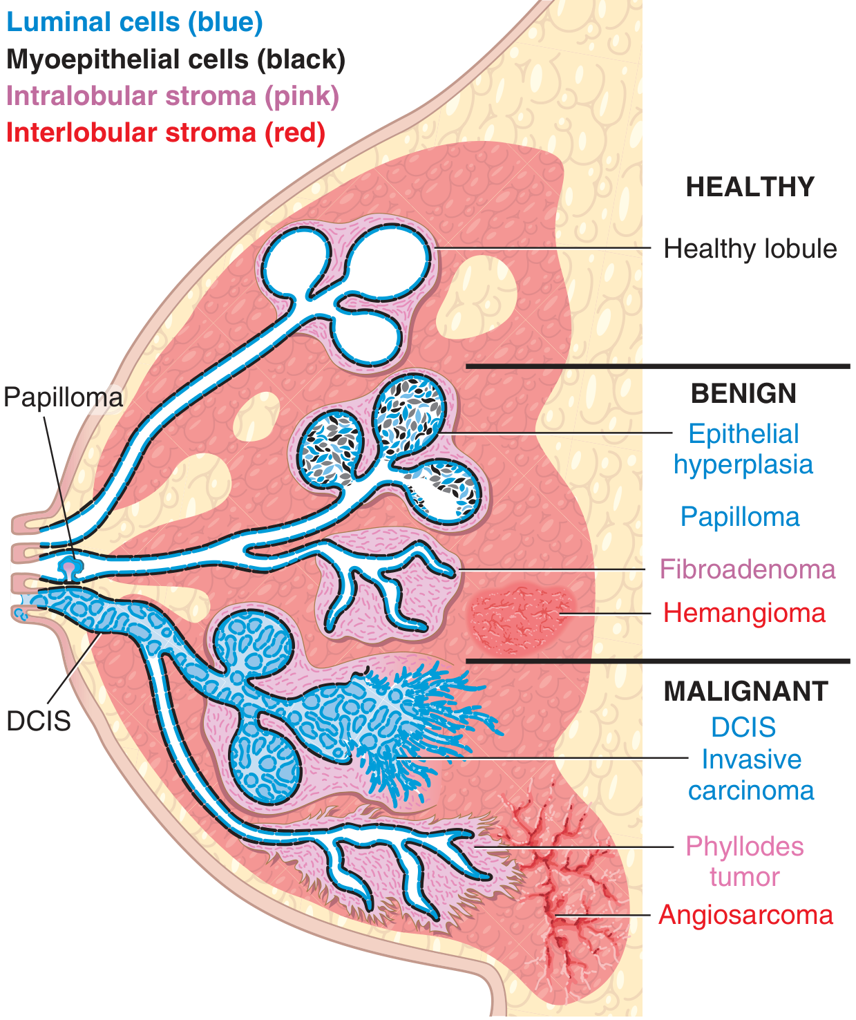
Fig. 17.23 - Origins of breast disorders. Benign epithelial lesions include intraductal papillomas and epithelial hyperplasia. Malignant lesions include DCIS and invasive carcinoma. Intralobular stroma (pink) gives rise to fibroadenomas and phyllodes tumors; interlobular stroma (red) gives rise to hemangiomas and angiosarcomas.
2. Clinical Presentations of Breast Disease
| Symptom | Key Points |
|---|---|
| Pain (mastalgia) | Common; cyclic edema/swelling; localized pain from ruptured cyst or fat necrosis; ~5% of painful masses = cancer |
| Inflammatory changes | Rare; usually lactational abscess (Staph aureus); always exclude inflammatory carcinoma |
| Nipple discharge | Unilateral, spontaneous, bloody = most worrisome for malignancy; most common benign cause = intraductal papilloma |
| Diffuse nodularity | Usually physiologic "lumpiness" |
| Palpable mass | >95% are benign; round/oval with circumscribed borders; malignant masses tend to have irregular borders |
- Most lesions (>90%) are benign
- ~45% of cancers have symptoms; remainder detected by screening mammography
- Sensitivity/specificity of mammography increases with age; likelihood of malignancy rises from ~10% at age 40 to >25% after age 50
3. Benign Breast Lesions
A. Inflammatory Processes
- Acute mastitis / Lactational abscess: Staphylococcus aureus (most common) or streptococci; occurs within the first month of breastfeeding; treated with antibiotics + continued milk expression; rarely requires I&D
- Non-lactational abscess: Mixed anaerobic organisms; less common
B. Benign Epithelial Lesions (Classified by Cancer Risk)
(Robbins & Kumar Basic Pathology, Table 17.6)
| Category | Lesions | Relative Risk of Breast Cancer |
|---|---|---|
| Non-proliferative changes | Simple cysts (often with apocrine metaplasia), fibrosis, adenosis | No increased risk |
| Proliferative lesions without atypia | Epithelial hyperplasia, sclerosing adenosis, complex sclerosing lesion (radial scar), papilloma | 1.5-2x increased risk |
| Proliferative disease with atypia | Atypical ductal hyperplasia (ADH), Atypical lobular hyperplasia (ALH) | 4-5x increased risk |
HPE Details:
- Nonproliferative changes: Single layers of epithelial cells; cysts lined by luminal cells with apocrine metaplasia; secretions may calcify (mammographic calcifications); ruptured cysts cause chronic inflammation and palpable nodularity ("fibrocystic changes"); adenosis = increased acini per lobule
- ADH: Uniform cells forming sharply marginated spaces or rigid bridges - closely resembles DCIS but limited in extent
- ALH: Monomorphic cells with bland, round nuclei - closely resembles LCIS but limited in extent
C. Stromal Neoplasms (Intralobular Stroma)
Fibroadenoma:
- Most common benign tumor of the breast
- Composed of neoplastic stromal cells + reactive proliferation of epithelial cells
- As fibroblasts proliferate, they push/distort epithelial cells into elongated, slit-like structures
- HPE: Circumscribed mass, low cellularity, biphasic (epithelial + stromal) - expansile growth with pushing borders
- Does NOT increase risk of breast cancer development
- Management: If diagnosed by core biopsy and ≤3 cm, observation is appropriate
Phyllodes Tumor:
- Stromal cells outgrow epithelial cells; bulbous nodules of proliferating stroma covered by epithelium
- Characteristic "phyllodes" (leaf-like) growth pattern on HPE
- High-grade forms: epithelium may be scant or absent (sarcomatous appearance)
- May recur following excision; rarely malignant
- Share driver mutations with fibroadenomas - they are part of a related neoplasm spectrum
Benign Interlobular Stromal Lesions:
- Myofibroblastoma: Only breast tumor equally common in males
- Lipoma: Palpable; fat-containing on mammography
- Fibromatosis: Clonal fibroblast/myofibroblast proliferation; locally aggressive but does NOT metastasize; associated with FAP/Gardner syndrome; some post-traumatic
4. Malignant Breast Diseases
A. Carcinoma In Situ
Ductal Carcinoma In Situ (DCIS):
- Malignant cells confined within the duct-lobular system; no invasion through basement membrane
- HPE: Ductlike spaces distort the lobule; calcified secretory material common
- Comedo type DCIS: High-grade proliferation with large central zones of necrosis + calcifications ("comedonecrosis"); linear/branching calcifications on mammogram
- Paget disease of the nipple: Tumor cells within the squamous epithelium of the nipple (large cells with clear halo = Paget cells); presents as unilateral crusting exudate; almost always associated with underlying invasive carcinoma or high-grade DCIS
- Treatment: Surgical excision + radiation → >95% survival at 20 years
- Progression: ~1% per year to invasive cancer in the same quadrant; same grade/ER/HER2 as associated DCIS
Lobular Carcinoma In Situ (LCIS):
- Monomorphic cells fill the breast lobule
- Almost always an incidental finding (rarely associated with calcifications or stromal reactions)
- Risk factor for invasive carcinoma in either breast (~1% per year; one-third ultimately develop invasive cancer)
- Unlike DCIS, surgical removal of the identified lesion may not lower risk
- Management: Close clinical/radiologic follow-up ± antiestrogen risk reduction (tamoxifen)
B. Invasive (Infiltrating) Carcinomas
Invasive Ductal Carcinoma (IDC) / "No Special Type" (NST):
- Most common invasive breast cancer (~70-80%)
- HPE: Tubule formation; desmoplastic stromal reaction (reactive fibrosis); irregular spicular margins on mammography (invasion of adjacent radiolucent tissue)
- Spreads to axillary lymph nodes (most important prognostic factor)
Invasive Lobular Carcinoma (ILC):
- Sheets of tightly cohesive cells (or "Indian file" single-cell pattern)
- May appear as well-circumscribed masses on mammography mimicking benign lesions
- Loss of E-cadherin (CDH1 mutation) - key molecular feature
Special Histologic Types:
| Type | HPE Features | Prognosis |
|---|---|---|
| Medullary carcinoma | Poorly differentiated cells; syncytial growth; marked lymphocytic infiltrate | Relatively favorable |
| Mucinous (colloid) carcinoma | Clusters of tumor cells floating in pools of mucin | Favorable |
| Tubular carcinoma | Well-formed open tubules; low grade | Excellent |
| Metaplastic carcinoma | Mixed carcinoma + sarcoma-like elements | Aggressive |
| Inflammatory carcinoma | Dermal lymphatic invasion; erythema + peau d'orange; T4d | Very poor |
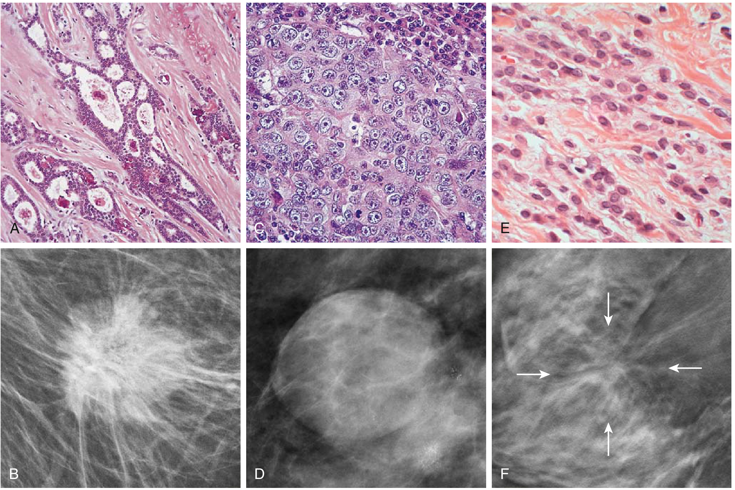
Fig. 17.30 - (A) IDC: tubule formation with desmoplastic stroma; (B) Dense spicular mass on mammogram; (C) Medullary carcinoma: sheets of tightly cohesive cells; (D) Well-circumscribed mass on mammogram; (E) ILC: single-file invasion; (F) ILC mammographic pattern with diffuse infiltration
C. Malignant Stromal Tumors
- Angiosarcoma: Most common interlobular stromal malignancy (<0.05% of breast malignancies); sporadic (young females, mean age 35, poor prognosis) or secondary (post-radiation or chronic lymphedema = Stewart-Treves syndrome); molecular hallmark = high-level MYC amplification
- Primary breast lymphoma: Usually diffuse large B-cell lymphoma; breast implant-associated anaplastic large cell lymphoma (BIA-ALCL) is T-cell type
5. Histopathological Examination (HPE) of Breast Tissue
What HPE Assesses:
- Architecture: Ductal vs lobular pattern; in situ vs invasive; margins
- Nuclear grade: Low (1), intermediate (2), high (3)
- Tubule formation: Used for Nottingham grade (% tubule formation score)
- Mitotic rate: Mitoses per 10 HPF
- Nottingham Grading System (Elston-Ellis): Tubule formation + nuclear pleomorphism + mitotic count → Grade 1 (well), 2 (moderate), 3 (poorly differentiated)
- Special stains: ER, PR (hormone receptors), HER2/neu (IHC ± FISH)
- Ki-67 proliferative index
- Lymphovascular invasion (LVI)
- Sentinel lymph node biopsy analysis: Isolated tumor cells (ITCs ≤0.2 mm), micrometastases (0.2-2 mm), macrometastases (>2 mm)
Molecular Subtypes (HPE-based biomarkers):
| Subtype | ER | PR | HER2 | Characteristics |
|---|---|---|---|---|
| Luminal A | + | + | - | Low grade, best prognosis |
| Luminal B | + | ± | ± | Higher grade, worse prognosis than A |
| HER2-enriched | - | - | + | Aggressive; responds to trastuzumab |
| Triple-negative (TNBC) | - | - | - | Aggressive; BRCA1 association; no targeted therapy |
Predictive Genomic Tests:
- Oncotype DX (21-gene assay): Node-negative, ER-positive; generates recurrence score to guide chemotherapy decision
- Other: Nottingham Prognostic Index, PREDICT
6. TNM Staging for Breast Cancer (AJCC 8th Edition)
(Source: Schwartz's Principles of Surgery, 11e)
T - Primary Tumor
| Category | Definition |
|---|---|
| TX | Primary tumor cannot be assessed |
| T0 | No evidence of primary tumor |
| Tis (DCIS) | Ductal carcinoma in situ |
| Tis (Paget) | Paget disease of nipple NOT associated with invasive carcinoma or DCIS in underlying parenchyma |
| T1 | Tumor ≤20 mm |
| T1mi | ≤1 mm (microinvasion) |
| T1a | >1 mm but ≤5 mm |
| T1b | >5 mm but ≤10 mm |
| T1c | >10 mm but ≤20 mm |
| T2 | >20 mm but ≤50 mm |
| T3 | >50 mm |
| T4 | Any size with direct extension to chest wall and/or skin |
| T4a | Extension to chest wall (pectoralis muscle invasion alone does NOT qualify) |
| T4b | Ulceration and/or satellite nodules and/or skin edema (peau d'orange) not meeting inflammatory criteria |
| T4c | T4a + T4b |
| T4d | Inflammatory carcinoma |
Size should be measured to the nearest mm. If slightly less/greater than a cutoff, round to the nearest mm.
N - Regional Lymph Nodes (Pathologic pN)
| Category | Definition |
|---|---|
| pNX | Cannot be assessed |
| pN0 | No regional LN metastasis / ITCs only |
| pN0(i+) | ITCs only (clusters ≤0.2 mm) |
| pN0(mol+) | Positive RT-PCR; no ITCs detected |
| pN1 | Micrometastases OR metastases in 1-3 axillary LNs; clinically negative internal mammary with micro/macrometastases by SLNB |
| pN1mi | Micrometastases: >0.2 mm but ≤2.0 mm (~200 cells) |
| pN1a | Metastases in 1-3 axillary LNs (at least one >2.0 mm) |
| pN1b | Metastases in ipsilateral internal mammary sentinel nodes (excl. ITCs) |
| pN1c | pN1a + pN1b |
| pN2 | Metastases in 4-9 axillary LNs; or positive ipsilateral internal mammary by imaging (no axillary metastases) |
| pN2a | Metastases in 4-9 axillary LNs (at least one >2.0 mm) |
| pN2b | Clinically detected internal mammary + pathologically negative axillary nodes |
| pN3 | ≥10 axillary LNs; or infraclavicular (Level III) LNs; or ipsilateral internal mammary by imaging with ≥1 positive Level I/II axillary LN; or supraclavicular LNs |
| pN3a | ≥10 axillary LNs (at least one >2.0 mm) OR infraclavicular nodes |
| pN3b | pN1a/pN2a + cN2b (positive internal mammary by imaging); or pN2a + pN1b |
| pN3c | Ipsilateral supraclavicular LNs |
M - Distant Metastasis
| Category | Definition |
|---|---|
| M0 | No clinical/radiographic evidence of distant metastases |
| cM0(i+) | No clinical/radiographic evidence, but deposits ≤0.2 mm detected molecularly or microscopically |
| M1 | Distant metastases by clinical/radiographic means; or histologically proven >0.2 mm |
Anatomic Stage Groups
| Stage | T | N | M | Notes |
|---|---|---|---|---|
| 0 | Tis | N0 | M0 | In situ only |
| IA | T1 | N0 | M0 | |
| IB | T0-T1 | N1mi | M0 | Micrometastases only |
| IIA | T0-T1 / T2 | N1 / N0 | M0 | |
| IIB | T2 / T3 | N1 / N0 | M0 | |
| IIIA | T0-T3 / T3 | N2 / N1 | M0 | |
| IIIB | T4 | N0-N2 | M0 | Chest wall/skin involvement |
| IIIC | Any T | N3 | M0 | |
| IV | Any T | Any N | M1 | Distant mets |
7. Prognostic & Predictive Factors for Invasive Breast Cancer
(Schwartz's Principles of Surgery, Table 17-12)
| Tumor Factors | Host Factors |
|---|---|
| Nodal status (most important) | Age |
| Tumor size | Menopausal status |
| Histologic/nuclear grade | Family history |
| Lymphatic/vascular invasion | Previous breast cancer |
| Pathologic stage | Immunosuppression |
| Hormone receptor status (ER, PR) | Nutrition |
| DNA content (ploidy, S-phase fraction) | Prior chemotherapy/radiation |
| HER2/neu expression | |
| Extent of intraductal component |
Key Concepts Summary
- All breast cancers arise from the terminal duct lobular unit (TDLU) - the "ductal" and "lobular" designations are histomorphologic, not anatomic
- Myoepithelial cells are the key marker of in situ vs. invasive disease - intact myoepithelial layer = in situ; absent = invasive (IHC: p63, smooth muscle actin, calponin)
- E-cadherin loss distinguishes lobular (negative) from ductal (positive) carcinoma
- Nottingham grade (tubule formation + nuclear pleomorphism + mitotic count) is the standard grading system used in HPE reports
- DCIS risk for invasion: ~1%/year; treatment is excision + radiation; >95% survival at 20 years
- LCIS is a bilateral risk marker (not a direct precursor), not treated with excision alone
- Inflammatory carcinoma (T4d) is a clinical diagnosis (erythema, peau d'orange, rapid onset), NOT defined by the presence of tumor in dermal lymphatics on biopsy alone
This is a shared conversation. Sign in to Orris to start your own chat.