
In cbc what is most important
A CBC (Complete Blood Count) measures several parameters, but the most clinically important ones are: hemoglobin (for anemia/polycythemia), white blood cell count with differential (for infection, inflammation, or hematologic malignancy), and platelets (for bleeding or clotting risk). The WBC differential - specifically the neutrophil, lymphocyte, and blast percentages - often provides the most actionable diagnostic information.
Searching sources...
Reading File
Finding Sources
Searching the Web
CBC complete blood count most important parameters clinical significance
Finding Sources
Finding Sources
Finding Sources
Finding Sources
Reading File
Reading File
Reading File
I now have plenty of textbook content to give a thorough answer. Here is a comprehensive breakdown:
Most Important Parameters in a CBC
A CBC (Complete Blood Count) measures three main cell lines. Each has its own critical parameters. Here's what matters most and why:
1. Red Blood Cell (RBC) Parameters - Hemoglobin is #1
Hemoglobin (Hb) is arguably the single most important CBC value. It directly reflects oxygen-carrying capacity and is the primary number used to diagnose and grade anemia or polycythemia.
Normal ranges (Goldman-Cecil Medicine):
- Female: 12-16 g/dL
- Male: 13.5-17.5 g/dL
MCV (Mean Corpuscular Volume) is the next most important RBC parameter because it classifies the type of anemia:
- Microcytic (MCV <80 fL) - think iron deficiency, thalassemia
- Normocytic (MCV 80-100 fL) - think chronic disease, acute blood loss, hemolysis
- Macrocytic (MCV >100 fL) - think B12/folate deficiency, liver disease, hypothyroidism
As stated in the Textbook of Family Medicine: "The first step is to classify the anemia, based on MCV... In early anemia, MCV may change before Hb and Hct."
Other RBC indices:
| Parameter | What it tells you |
|---|---|
| Hematocrit (Hct) | % volume of blood occupied by RBCs |
| MCH (30-34 pg) | Average Hb content per cell |
| MCHC (30-36 g/dL) | Average Hb concentration per cell |
| RDW (12-15%) | Variation in RBC size (anisocytosis) - useful for distinguishing iron deficiency from thalassemia trait |
| Reticulocyte count | Bone marrow response to anemia |
2. WBC Count + Differential - Critical for Infection & Malignancy
The total WBC count flags infection, inflammation, or marrow problems. But the 5-part differential is where real diagnostic value lies:
| Component | Normal | Clinical significance |
|---|---|---|
| Neutrophils | 50-70% | Bacterial infection, stress, steroids; if low = neutropenia (infection risk) |
| Lymphocytes | 20-40% | Viral infection; if very high = CLL or viral lymphocytosis |
| Monocytes | 2-8% | Chronic infections (TB, EBV), monocytic leukemia |
| Eosinophils | 1-4% | Allergy, parasites, eosinophilic disorders |
| Basophils | 0-1% | CML, hypersensitivity |
Blasts on the differential are abnormal at any percentage and immediately raise concern for acute leukemia.
Indications for manual peripheral smear review (per Textbook of Family Medicine) include: hemolysis, RBC inclusions, myelodysplasia, megaloblastic changes, thrombocytosis, thrombocytopenia, leukocytosis, and immature/abnormal cells.
3. Platelets - Essential for Bleeding Risk
Normal: ~150,000-400,000/μL
Key thresholds (from Textbook of Family Medicine):
- <50,000-70,000/μL - clinical bleeding begins to appear
- <10,000-20,000/μL - risk of major spontaneous bleeding (intracranial, GI)
Causes of low platelets (thrombocytopenia): decreased production (aplastic anemia, leukemia, B12/folate deficiency), increased destruction (ITP, heparin-induced, DIC, sepsis, HELLP syndrome), or splenic sequestration.
Summary: What to Look at First in a CBC
| Priority | Parameter | Why |
|---|---|---|
| 1st | Hemoglobin | Most direct clinical impact (anemia/polycythemia) |
| 2nd | WBC + differential | Infection, malignancy, immunosuppression |
| 3rd | Platelets | Bleeding risk |
| 4th | MCV | Classifies anemia type, directs next tests |
| 5th | RDW | Distinguishes causes of microcytic/normocytic anemia |
In practice, any "panic value" (Hb <7, WBC >30,000 or <2,000, platelets <20,000) demands immediate clinical attention regardless of context.
Sources: Goldman-Cecil Medicine (CBC section, Table 144-2/144-3); Textbook of Family Medicine, 9e (CBC chapter)
How do I interpret an abnormal WBC differential?
A WBC differential breaks down the 5 white cell types. Abnormalities fall into two patterns: too many (leukocytosis/specific cell excess) or too few (leukopenia/specific cell deficiency). Neutrophilia usually means bacterial infection; lymphocytosis usually means viral infection; eosinophilia means allergy or parasites; and blasts on a differential always mean something serious.
Searching sources...
Finding Sources
Finding Sources
Finding Sources
Finding Sources
Reading File
Reading File
Reading File
Reading File
Reading File
Reading File
Reading File
Finding Sources
Finding Sources
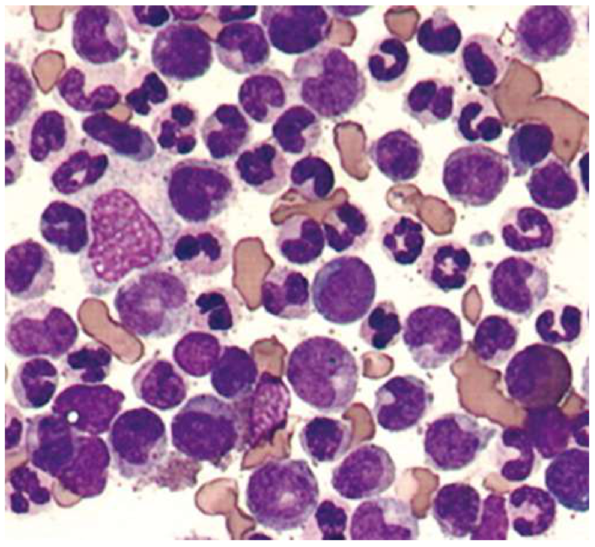
Excellent - I now have comprehensive textbook data. Let me fetch the leukemoid reaction image from Goldman-Cecil for a visual aid.
Loading Image
Here is a thorough, textbook-grounded guide to interpreting an abnormal WBC differential:
Interpreting an Abnormal WBC Differential
Normal WBC range: 4,500-10,000/μL (mean 7,500/μL). Because neutrophils make up ~60% of the WBC, most leukocytosis and leukopenia reflect changes in the neutrophil count.
Step 1: Use Absolute Counts, Not Just Percentages
Always convert percentages to absolute counts before interpreting:
Absolute count = Total WBC × (% cell type / 100)
Percentages alone can mislead. For example, a "low" lymphocyte % may simply reflect neutrophilia, while the absolute lymphocyte count is normal.
Step 2: Interpret Each Cell Line
Neutrophils (Normal ANC: 1,800-7,700/μL)
NEUTROPHILIA (ANC elevated)
Per Goldman-Cecil Medicine, neutrophilia is almost always reactive first. Think:
| Category | Examples |
|---|---|
| Acute infection | Bacterial pneumonia, abscess, sepsis, C. difficile |
| Chronic inflammation | RA, IBD, granulomatous disease, chronic hepatitis |
| Stress/physiologic | Surgery, exercise, MI, catecholamine release |
| Drugs | Corticosteroids, beta-agonists, lithium, G-CSF |
| Smoking | 25-50% of chronic smokers |
| Post-splenectomy | Permanent mild neutrophilia |
| Malignancy | CML, polycythemia vera, cytokine-secreting tumors |
Key flag - "left shift": circulating immature neutrophils (bands, metamyelocytes) indicating the marrow is being pushed hard by infection or inflammation. A WBC >50,000/μL with pronounced left shift = leukemoid reaction (must distinguish from CML using BCR-ABL molecular testing).
Additional smear findings in severe infection: toxic granulation, Döhle bodies, cytoplasmic vacuoles.
NEUTROPENIA (ANC low) - severity matters most:
| Grade | ANC | Infection Risk |
|---|---|---|
| Mild | 1,000-1,500/mm³ | Low |
| Moderate | 500-1,000/mm³ | Moderate |
| Severe | <500/mm³ | High - empiric antibiotics if febrile |
Causes: chemotherapy, aplastic anemia, autoimmune (lupus), drug-induced agranulocytosis, viral infections, congenital neutropenia syndromes, B12/folate deficiency. Note: some ethnic groups (especially African ancestry) have a lower normal ANC baseline - this is a benign genetic variant, not pathology.
Lymphocytes (Normal: 1,000-4,800/μL; lymphocytosis if >5,000/μL)
LYMPHOCYTOSIS
| Cause | Details |
|---|---|
| Viral infections | EBV (mononucleosis), CMV, hepatitis, HIV |
| Atypical bacterial | Pertussis, Bartonella (cat-scratch), TB, syphilis |
| Hypersensitivity | Drug reactions, serum sickness |
| CLL | Most common primary cause in adults >40 y |
| Monoclonal B-cell lymphocytosis | Found in ~5% of adults >40; progresses to CLL at 1-2%/year |
LYMPHOPENIA (<1,000/μL): Think HIV/AIDS, corticosteroids, cytotoxic chemotherapy, autoimmune disease, or severe malnutrition.
Monocytes (Normal: 200-800/μL; monocytosis if >500/μL)
| Cause | Examples |
|---|---|
| Chronic infections | TB, syphilis, SBE, some viruses |
| Autoimmune/granulomatous | SLE, RA, sarcoidosis |
| Hematologic malignancy | CMML, AML (monocytic subtypes), Hodgkin/NHL |
| Recovery | Post-chemotherapy marrow recovery |
Eosinophils (Normal: 100-400/μL; eosinophilia if >400/μL)
The classic mnemonic is NAACP: Neoplasm, Allergy/Asthma, Addison's disease, Collagen vascular disease, Parasites.
| Mild (<1,500/μL) | Drug reactions, allergic rhinitis, asthma, eczema |
|---|---|
| Moderate (1,500-5,000/μL) | Parasitic infections (helminths), inflammatory conditions |
| Severe/Hypereosinophilia (>5,000/μL) | Eosinophilic granulomatosis with polyangiitis (Churg-Strauss), hypereosinophilic syndrome, parasitic infestation |
Basophils (Normal: 0-100/μL; basophilia if >100/μL)
Rare finding. When present, think CML (almost pathognomonic when combined with neutrophilia and left shift), other myeloproliferative neoplasms, or hypersensitivity reactions.
Step 3: Red Flags - When to Escalate Immediately
| Finding | Concern |
|---|---|
| Blasts on differential (any %) | Acute leukemia - urgent hematology referral |
| WBC >50,000 with left shift | Leukemoid reaction vs. CML - check BCR-ABL |
| Pancytopenia (WBC + Hb + platelets all low) | Aplastic anemia, bone marrow failure, leukemia |
| Hypersegmented neutrophils (>5 lobes) | B12/folate deficiency (megaloblastic anemia) |
| Unexplained monocytosis + anemia | CMML (chronic myelomonocytic leukemia) |
| ANC <500 + fever | Febrile neutropenia - medical emergency |
Step 4: When to Do a Peripheral Smear
Manual smear review is indicated for: suspected hemolysis, leukocytosis, leukopenia, thrombocytopenia, immature/abnormal cells flagged by the analyzer, suspected myelodysplasia or megaloblastic changes.
Here is what a leukemoid reaction looks like on smear - note the mix of myeloid cells at all stages of maturity:
Quick Summary Table
| Cell | Elevated = Think | Low = Think |
|---|---|---|
| Neutrophils | Bacterial infection, inflammation, steroids, CML | Chemotherapy, aplastic anemia, autoimmune, drugs |
| Lymphocytes | Viral infection, CLL, EBV/CMV | HIV, steroids, chemotherapy |
| Monocytes | TB, SBE, CMML, IBD, sarcoid | Rare (aplastic anemia) |
| Eosinophils | Allergy, parasites, asthma, vasculitis | Steroids (suppress eosinophils) |
| Basophils | CML, myeloproliferative disorders | Non-specific |
Sources: Goldman-Cecil Medicine (Chapter 153, Leukocytosis and Leukopenia); Tintinalli's Emergency Medicine (Neutropenia section)
This is a shared conversation. Sign in to Orris to start your own chat.