
Ecg left ventricular hypertrophy criteria
Reading File
ECG criteria for LVH include voltage-based criteria (Sokolow-Lyon, Cornell, etc.) combined with secondary ST-T changes. The most commonly used systems are Sokolow-Lyon, Cornell, and Romhilt-Estes point score.
Searching sources...
Finding Sources
Searching the Web
ECG left ventricular hypertrophy criteria Sokolow Cornell Romhilt-Estes 2024
Finding Sources
Reading File
Reading File
Reading File
Loading Image
Here is a thorough, well-sourced breakdown of ECG criteria for Left Ventricular Hypertrophy (LVH):
ECG Criteria for Left Ventricular Hypertrophy (LVH)
Pathophysiology: LVH increases the amplitude of electrical forces directed leftward and posteriorly, producing tall left precordial R waves and deep right precordial S waves. Repolarization abnormalities (ST depression + T-wave inversion) may follow, in leads with prominent R waves - this is called the "LV strain" pattern.
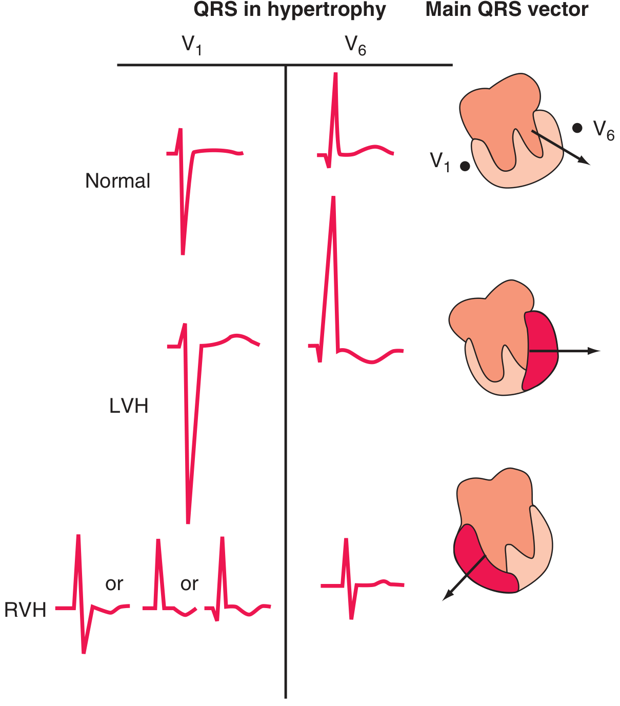
1. Sokolow-Lyon Criteria (1949)
Most widely used, simple voltage criteria
| Finding | Threshold |
|---|---|
| SV1 + RV5 (or RV6) | ≥ 35 mm |
| R wave in aVL alone | ≥ 11 mm |
- Sensitivity: ~20-40% | Specificity: ~85-90%
- Tends to under-diagnose in obese patients
2. Cornell Criteria (1985) - Sex-specific
Better performance in women
| Sex | Formula | Threshold |
|---|---|---|
| Men | SV3 + RaVL | > 28 mm |
| Women | SV3 + RaVL | > 20 mm |
Cornell Product (adds QRS duration for improved accuracy):
-
Men: (SV3 + RaVL) × QRS duration > 2,440 mm·ms
-
Women: same formula with female voltage threshold
-
Sensitivity: ~15-30% | Specificity: ~95-99%
3. Romhilt-Estes Point Score System (1968)
Definite LVH = ≥ 5 points; Probable LVH = 4 points
| Criterion | Points |
|---|---|
| Voltage: R or S in any limb lead ≥ 20 mm, OR S in V1/V2 ≥ 30 mm, OR R in V5/V6 ≥ 30 mm | 3 |
| ST-T changes (LV strain pattern) - without digoxin | 3 |
| ST-T changes (LV strain pattern) - with digoxin | 1 |
| Left atrial abnormality: P terminal in V1 ≥ 1 mm amplitude AND ≥ 0.04 s duration | 3 |
| Left axis deviation ≥ -30° | 2 |
| QRS duration ≥ 90 ms | 1 |
| Intrinsicoid deflection (Q-R interval) ≥ 50 ms in V5 or V6 | 1 |
- Sensitivity: ~50-66% | Specificity: ~65-85% (more sensitive but less specific than voltage-only criteria)
4. Peguero-Lo Presti Criteria (2017)
Newer criterion, better in obese patients
| Formula | Threshold |
|---|---|
| Deepest S in any precordial lead (SD) + S in V4 | ≥ 28 mm (both sexes) |
- Better performance than Sokolow-Lyon in obese patients and women
5. Simple Voltage Criteria (Other)
| Criterion | Threshold |
|---|---|
| R in aVL | ≥ 11 mm (Sokolow limb lead modification) |
| R in aVL | > 20 mm (women) or > 28 mm (men) - Harrison's |
| RaVL + SV3 (Lewis index variant) | See Cornell |
| Tallest R + deepest S in any precordial lead | ≥ 45 mm (simple rule) |
6. Secondary (Non-Voltage) ECG Features of LVH
These increase diagnostic confidence when voltage criteria are borderline:
- LV strain pattern: ST depression + T-wave inversion in lateral leads (I, aVL, V5-V6) - opposite to main QRS deflection
- Left atrial abnormality (P mitrale): Broad, notched P waves in limb leads; biphasic P in V1 with deep terminal component
- Left axis deviation (axis -30° to -90°)
- QRS duration prolongation (may progress to LBBB)
- Increased QRS duration ≥ 90 ms
- Slow R-wave progression in precordial leads
Pediatric LVH Voltage Criteria (Age-dependent)
| Age | RV6 (mm) | SV1 (mm) | SV1 + RV6 (mm) |
|---|---|---|---|
| 0-7 days | >12 | >23 | >28 |
| 7 days - 1 year | >23 | >15-18 | >35 |
| 1-3 years | >21-23 | >21 | >38 |
| 3-5 years | >24-25 | >22 | >42 |
| >5 years | >25-27 | >26 | >47 |
Additional pediatric LVH features: Q wave >4 mm in V5/V6, or inverted T wave in V6.
- Tintinalli's Emergency Medicine, Table 130-9
Diagnostic Accuracy Summary
| Criterion | Sensitivity | Specificity |
|---|---|---|
| Sokolow-Lyon | ~13-40% | ~85-90% |
| Cornell voltage | ~15-30% | ~95-99% |
| Cornell product | ~20-35% | ~90-97% |
| Romhilt-Estes ≥5 | ~30-66% | ~65-85% |
| Peguero-Lo Presti | ~31% | ~88% |
Clinical Pearls
-
False negatives are common in obesity, COPD/emphysema, and right bundle branch block - all reduce voltage
-
False positives are common in young or athletic individuals (high normal voltage)
-
Echocardiography (or cardiac MRI) is the gold standard for LVH diagnosis - ECG has poor sensitivity overall
-
LVH on ECG is an independent predictor of cardiovascular morbidity and mortality, including sudden cardiac death
-
The presence of left atrial abnormality on ECG increases the likelihood of true LVH when voltage criteria are borderline
-
LVH may progress to LBBB over time
-
Harrison's Principles of Internal Medicine 22E (2025), p. 1914
-

24375.jpg
What is this xray ,is it cardiomegaly or pneumonia,or left pleural effusion,or what and diaphragm is also not visible
Your credits are exhausted. You can purchase a top-up or subscribe to a plan to continue using Orris. If you're already subscribed, your daily credits will reset tomorrow.
This is a shared conversation. Sign in to Orris to start your own chat.