THORAX — Complete Exam-Ready Notes (23 Topics)
1. TYPICAL INTERCOSTAL NERVE (4 Marks)
Simple Explanation
Each rib space has its own nerve, like a "wire" running along the space to supply the muscles and skin. The nerve from spaces 3–6 behaves in a standard/"typical" way, so it's called the typical intercostal nerve. Nerves of spaces 1, 2, 7–11 behave differently (atypical) because they also supply the upper limb or abdominal wall.
University Answer
Origin: Anterior (ventral) ramus of the corresponding thoracic spinal nerve (T3–T6 are typical).
Course (in the intercostal space, below the vein and artery = VAN from above downward):
- Runs in the subcostal groove on the lower border of the rib above, between the pleura/innermost intercostal muscle and the internal intercostal muscle.
- Lies below the intercostal vein and artery (VAN – Vein, Artery, Nerve, from above down).
- Gives a collateral branch near the angle of the rib, running along the upper border of the rib below.
- Continues forward and pierces the internal intercostal muscle and anterior intercostal membrane near the sternum as the anterior cutaneous branch.
Branches:
| Branch | Distribution |
|---|
| Collateral branch | Supplies intercostal muscles, runs above lower rib |
| Lateral cutaneous branch | Pierces muscles in mid-axillary line, divides into anterior & posterior branches – supplies skin of thorax/abdomen |
| Anterior cutaneous branch | Terminal branch near sternum, supplies skin near midline |
| Muscular branches | To intercostal muscles, levatores costarum, serratus posterior |
Applied Anatomy:
- Used for intercostal nerve block in thoracotomy pain relief and rib fracture pain.
- Herpes zoster affects a single dermatome supplied by one intercostal nerve ("shingles" band-like rash).
- T7 nerve supplies the xiphisternum area, T10 supplies the umbilicus (important dermatome landmark).
Diagram to Draw
Cross-section of a typical intercostal space showing (from above down): intercostal vein, artery, nerve (VAN), lying in the subcostal groove between internal intercostal and innermost intercostal muscles. Label: rib above, rib below, three muscle layers, pleura, collateral branch.
Must write: VAN order, subcostal groove position, T3–T6 = typical, collateral branch, clinical nerve block.
2. THORACIC DUCT (4 Marks)
Simple Explanation
This is the biggest lymph vessel in the body — a drainage pipe that collects lymph (fluid + fat) from the whole body below the diaphragm and the left half above it, and empties it into a vein in the neck.
University Answer
Definition: Largest lymphatic channel in the body, draining lymph from lower limbs, abdomen, left half of thorax, left upper limb, and left half of head and neck.
Origin: From the upper end of the cisterna chyli (a lymph sac lying in front of L1–L2 vertebrae, behind the aorta).
Course:
- Enters thorax through the aortic opening of diaphragm (T12), lying between the aorta (left) and azygos vein (right).
- Ascends in the posterior mediastinum, behind the esophagus.
- At T5 vertebral level, crosses from right to left behind the esophagus.
- Ascends along the left side of esophagus into the superior mediastinum, then into the root of the neck.
- Arches laterally behind the carotid sheath, in front of the vertebral artery, thyrocervical trunk, and phrenic nerve.
- Termination: at the junction of the left subclavian and left internal jugular veins (left venous angle / Pirogoff's angle).
Relations in thorax:
| Structure | Relation |
|---|
| Thoracic aorta | On the left (lower thorax) |
| Azygos vein | On the right |
| Esophagus | Anterior |
| Vertebral column | Posterior |
Tributaries: Intercostal lymph trunks, left bronchomediastinal trunk, left jugular and left subclavian lymph trunks (near termination).
Applied Anatomy:
- Injury during neck/thoracic surgery → chylothorax (chyle leak into pleural cavity).
- Blockage → chylous ascites or lymphedema.
Diagram to Draw
Outline of posterior mediastinum showing thoracic duct starting from cisterna chyli (T12), crossing midline at T5, ending at left venous angle. Label aorta, esophagus, azygos vein, T5 crossing point, termination into left subclavian + IJV junction.
Must write: Cisterna chyli origin, T5 crossing, termination at left venous angle, relation to aorta/azygos/esophagus.
3. TYPICAL INTERCOSTAL SPACES (3 Marks)
Simple Explanation
The gap between two ribs is the intercostal space. Spaces 3rd to 6th are "typical" because their muscles, vessels and nerves follow the standard/textbook pattern.
University Answer
Definition: The space between two adjacent ribs, occupied by the three intercostal muscles and neurovascular bundle. 3rd–6th spaces are typical.
Contents (from outside in):
- Skin, superficial fascia
- External intercostal muscle — fibers run downward and forward (like hands in pocket)
- Internal intercostal muscle — fibers run downward and backward
- Innermost intercostal muscle (incomplete layer)
- Neurovascular bundle (VAN – vein, artery, nerve) in subcostal groove
- Endothoracic fascia
- Parietal pleura
Intercostal muscles table:
| Muscle | Direction of fibers | Extent |
|---|
| External intercostal | Downward & forward | Tubercle of rib to costochondral junction |
| Internal intercostal | Downward & backward | Angle of rib to sternum |
| Innermost intercostal | Same as internal | Only in middle 1/3 of space |
Neurovascular bundle order: Vein (top), Artery (middle), Nerve (bottom) — in the costal groove of the upper rib.
Applied Anatomy: Needle for pleural tap/thoracocentesis is inserted just above the rib (lower border of intercostal space) to avoid the neurovascular bundle which lies in the groove of the rib above.
Diagram to Draw
Cross section of one typical intercostal space between two ribs, three muscle layers labelled, VAN bundle in subcostal groove, safe site of needle insertion marked just above lower rib.
Must write: 3 muscle layers with fibre direction, VAN order, safe triangle for thoracocentesis.
4. CONSTRICTIONS OF OESOPHAGUS (2 Marks)
Simple Explanation
The oesophagus (food pipe) is not a uniform tube — it is narrowed at four points where things (like swallowed foreign bodies) commonly get stuck.
University Answer
Definition: Four normal anatomical narrowings along the 25 cm length of the esophagus.
| Constriction | Level | Distance from incisor teeth | Caused by |
|---|
| 1. Cricopharyngeal (commonest & narrowest) | C6 | 15 cm | Cricopharyngeus muscle |
| 2. Aortic (arch of aorta crossing) | T4 | 22.5 cm | Arch of aorta |
| 3. Bronchial (left bronchus crossing) | T4/T5 | 27 cm | Left main bronchus |
| 4. Diaphragmatic | T10 | 40 cm | Esophageal opening of diaphragm |
Applied Anatomy (high-yield):
- Sites of impaction of swallowed foreign bodies and food boluses.
- Common sites for corrosive stricture after acid/alkali ingestion.
- Guide for endoscope/nasogastric tube insertion depth.
- Cricopharyngeal constriction is the narrowest and most common site of impaction.
Diagram to Draw
Simple vertical line diagram of esophagus with 4 narrow points marked, with vertebral levels (C6, T4, T4/5, T10) and distances from incisors labelled beside each constriction.
Must write: All 4 levels + distances + cricopharyngeal = narrowest.
5. CARDIAC PLEXUS (4 Marks)
Simple Explanation
The heart's rhythm and blood vessels are controlled by a network of nerves (sympathetic + parasympathetic) sitting near the base of the heart and great vessels — this network is the cardiac plexus.
University Answer
Definition: A nerve plexus formed by sympathetic and parasympathetic (vagal) fibers, situated near the arch of aorta and roots of great vessels, supplying the heart.
Divided into two parts (continuous with each other):
| Part | Location |
|---|
| Superficial cardiac plexus | Below arch of aorta, in front of right pulmonary artery, between arch of aorta and pulmonary trunk |
| Deep cardiac plexus | Behind arch of aorta, in front of tracheal bifurcation |
Contributions:
- Sympathetic: Superior, middle, inferior cervical sympathetic ganglia + upper thoracic ganglia (cardiac nerves)
- Parasympathetic: Vagus nerve — cardiac branches from cervical, thoracic (recurrent laryngeal) parts
Branches distributed via:
- Coronary plexuses (right and left) — accompany coronary arteries
- Fibers reach SA node, AV node, myocardium, coronary vessels
Functions:
| Fiber type | Effect on heart |
|---|
| Sympathetic | Increases heart rate, force of contraction, coronary vasodilation during exercise |
| Parasympathetic (vagal) | Decreases heart rate (bradycardia), constricts coronary vessels |
Applied Anatomy:
- Referred cardiac pain (angina) travels via sympathetic afferents to T1–T4 dermatomes → pain in left arm, jaw.
- Vasovagal syncope — exaggerated vagal (parasympathetic) stimulation slowing the heart.
Diagram to Draw
Base of heart with arch of aorta, pulmonary trunk, and tracheal bifurcation. Show superficial cardiac plexus (below aortic arch, right of ligamentum arteriosum) and deep cardiac plexus (behind aortic arch) with vagal and sympathetic fibers converging, and coronary plexuses following coronary arteries.
Must write: Superficial vs deep plexus location, sympathetic + parasympathetic contributions, coronary plexus continuation, referred pain applied anatomy.
6. LEFT VENTRICLE OF HEART (4 Marks)
Simple Explanation
The left ventricle is the strongest pumping chamber of the heart — it pushes oxygenated blood to the entire body, so it has the thickest muscular wall.
University Answer
Location: Forms the left border, apex, and most of the diaphragmatic (inferior) surface of the heart. Lies posterior and to the left of right ventricle.
Wall thickness: ~3× thicker than right ventricle (has to generate systemic pressure).
Cavity divided into:
- Inflow part (posterior) — receives blood from left atrium through mitral (bicuspid) valve
- Outflow part (aortic vestibule) (anterosuperior) — leads to aorta through aortic valve
Internal features:
| Feature | Description |
|---|
| Trabeculae carneae | Muscular ridges (finer & more numerous than right ventricle) |
| Papillary muscles | 2 large ones – anterior and posterior |
| Chordae tendineae | Connect papillary muscles to cusps of mitral valve |
| Mitral valve | Bicuspid – anterior & posterior cusps |
| Aortic valve | 3 semilunar cusps – right, left, posterior |
| Absence of moderator band | Unlike right ventricle |
Openings:
- Mitral orifice (postero-inferior) — admits 2 fingers
- Aortic orifice (antero-superior) — admits 2 fingers, guarded by 3 semilunar cusps with sinuses of Valsalva behind (right and left aortic sinuses give origin to coronary arteries)
Applied Anatomy:
- Left ventricular hypertrophy — seen in systemic hypertension, aortic stenosis.
- Mitral stenosis/regurgitation — auscultated at apex (mitral area, 5th left intercostal space, midclavicular line).
- Left ventricle forms the apex beat location.
Diagram to Draw
Longitudinal section of heart showing left atrium, mitral valve with 2 papillary muscles and chordae tendineae, left ventricular cavity with trabeculae carneae, aortic vestibule, aortic valve with 3 cusps and coronary ostia in right/left sinuses.
Must write: Thick wall reason, mitral valve (bicuspid), 2 papillary muscles, aortic vestibule, coronary artery origin from aortic sinuses.
7. BLOOD SUPPLY OF HEART (4 Marks)
Simple Explanation
The heart is a muscle and needs its own blood supply — this comes from two coronary arteries (right and left) that branch off the aorta right after it leaves the heart, plus veins that drain into the coronary sinus.
University Answer
Arterial supply — two coronary arteries, both first branches of ascending aorta from aortic sinuses:
| Artery | Origin | Main branches | Supplies |
|---|
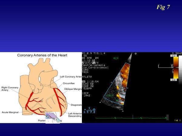
| Right Coronary Artery (RCA) | Right aortic sinus | SA nodal artery, marginal artery, posterior interventricular (in right dominance) | Right atrium, right ventricle, SA & AV node (usually), inferior wall of LV |
| Left Coronary Artery (LCA) | Left aortic sinus | Divides into Anterior interventricular (LAD) + Circumflex | Left atrium, most of left ventricle, anterior interventricular septum |
Coronary dominance: ~70% right dominant (RCA gives posterior interventricular artery).
Venous drainage — mainly via Coronary Sinus (in posterior AV groove, opens into right atrium):
| Vein | Drains into |
|---|
| Great cardiac vein | Coronary sinus (left end) |
| Middle cardiac vein | Coronary sinus |
| Small cardiac vein | Coronary sinus |
| Oblique vein of left atrium | Coronary sinus |
| Anterior cardiac veins | Directly into right atrium |
| Venae cordis minimae (Thebesian veins) | Directly into cardiac chambers |
Applied Anatomy:
- Coronary artery occlusion → myocardial infarction; site of infarct depends on artery blocked (LAD = anterior wall MI, RCA = inferior wall MI).
- Coronary artery bypass grafting (CABG) uses saphenous vein/internal thoracic artery grafts.
- Angina pectoris — pain due to relative ischemia.
Diagram to Draw
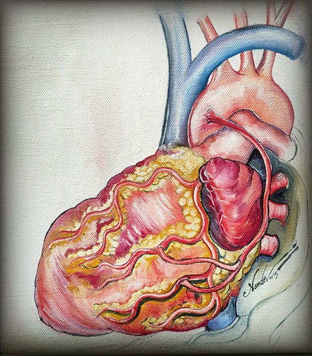
Anterior and diaphragmatic (inferior) view of heart showing RCA in right AV groove with marginal branch, LCA dividing into LAD (anterior interventricular groove) and circumflex artery (left AV groove), coronary sinus in posterior AV groove receiving great/middle/small cardiac veins.
Must write: Origin from aortic sinuses, RCA vs LCA branches, coronary dominance concept, coronary sinus as main venous drainage, MI applied anatomy.
8. ARCH OF AORTA (3 Marks)
Simple Explanation
After leaving the heart, the aorta curves over the top of the heart like a walking-stick handle — this curved part is the arch of aorta, and it gives off three branches that supply the head, neck, and arms.
University Answer
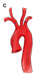
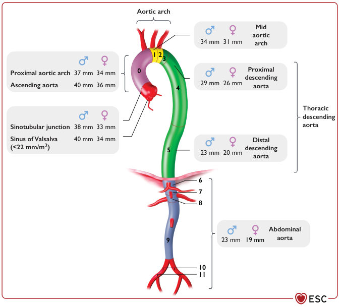
Extent: Continuation of ascending aorta; begins behind the 2nd right sternocostal joint, arches backward and to the left, ends at the lower border of T4 vertebra (becoming descending thoracic aorta).
Level: Its highest point reaches the middle of the manubrium sterni.
Three branches (right to left):
- Brachiocephalic (innominate) trunk — divides into right common carotid + right subclavian artery
- Left common carotid artery
- Left subclavian artery
Relations:
| Position | Structure |
|---|
| Anterosuperior | Left phrenic and left vagus nerve, left superior intercostal vein, cardiac plexus branches |
| Postero-inferior | Trachea, esophagus, left recurrent laryngeal nerve, ligamentum arteriosum, deep cardiac plexus |
| Right | Superior vena cava, trachea |
| Left | Left lung and pleura |
| Below | Bifurcation of pulmonary trunk, left bronchus |
Applied Anatomy:
- Left recurrent laryngeal nerve hooks around the arch (below ligamentum arteriosum) → aneurysm of arch can cause hoarseness of voice (compression of this nerve).
- Site of coarctation of aorta and aortic aneurysms.
- Ligamentum arteriosum (remnant of ductus arteriosus) attaches here.
Diagram to Draw
Arch of aorta arising from ascending aorta, arching over left pulmonary artery/left bronchus, three branches labelled (brachiocephalic, left common carotid, left subclavian), ligamentum arteriosum and left recurrent laryngeal nerve looping under the arch, ending at T4.
Must write: T4 termination level, 3 branches in order, recurrent laryngeal nerve relation, ligamentum arteriosum.
9. RIGHT ATRIUM (3 Marks)
Simple Explanation
The right atrium is the receiving chamber on the right side of the heart — it collects "used" (deoxygenated) blood from the whole body through two big veins and sends it to the right ventricle.
University Answer
Location: Forms the right border of the heart; lies anterior and to the right of left atrium.
Divided into two parts by the crista terminalis:
| Part | Origin | Wall character |
|---|
| Sinus venarum | Develops from sinus venosus | Smooth wall |
| Atrium proper | Develops from primitive atrium | Rough wall due to musculi pectinati |
Openings into right atrium:
- Superior vena cava — opens above, no valve
- Inferior vena cava — opens below, guarded by rudimentary valve of IVC (Eustachian valve)
- Coronary sinus — opens between IVC opening and AV orifice, guarded by valve of coronary sinus (Thebesian valve)
- Right atrioventricular (tricuspid) orifice — leads to right ventricle
- Foramina of venae cordis minimae (Thebesian veins)
Important internal landmarks:
- Crista terminalis — muscular ridge separating sinus venarum from atrium proper; corresponds externally to sulcus terminalis
- Fossa ovalis — depression on interatrial septum, remnant of foramen ovale (fetal)
- Auricle — anterior pouch-like extension, lined by pectinate muscles
- SA node — lies in upper part of crista terminalis, at junction of SVC and right atrium (pacemaker of heart)
Applied Anatomy:
- Patent foramen ovale/ASD — failure of closure of fossa ovalis region.
- SA node location important in cardiac catheterization/pacemaker placement.
Diagram to Draw
Interior of right atrium (coronal section) showing SVC and IVC openings, crista terminalis, musculi pectinati, fossa ovalis, coronary sinus opening with valve, tricuspid orifice, and SA node marked at SVC-RA junction.
Must write: Crista terminalis divides sinus venarum/atrium proper, 3 openings (SVC, IVC, coronary sinus), fossa ovalis, SA node location.
10. CO-ARCTATION OF AORTA (3 Marks)
Simple Explanation
This is a birth defect where the aorta has an abnormal narrowing, usually near where the ductus arteriosus was attached, which forces blood to find detour routes to reach the lower body.
University Answer
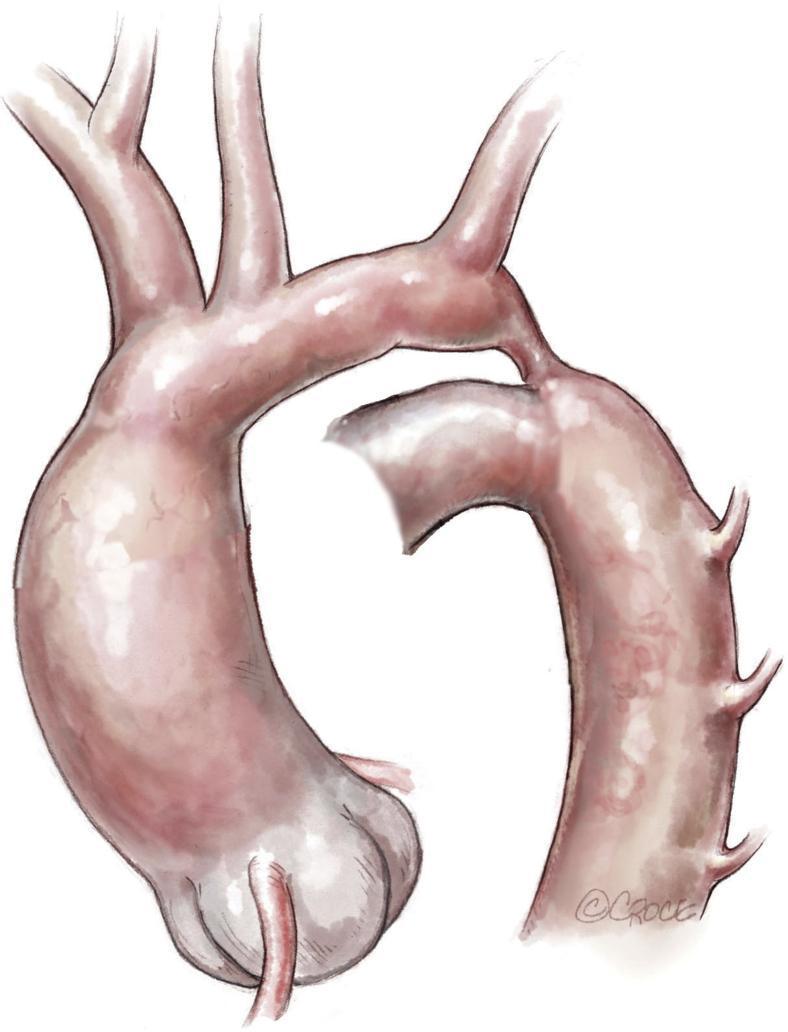
Definition: Congenital localized narrowing of the aorta, most commonly just distal to the origin of the left subclavian artery, near the site of attachment of the ligamentum arteriosum (former ductus arteriosus).
Types:
| Type | Location relative to ductus arteriosus | Presentation |
|---|
| Preductal (infantile) | Proximal to ductus, ductus remains patent | Presents early in infancy, ductus-dependent lower body perfusion |
| Postductal (adult) | Distal to ductus | Often asymptomatic till adulthood |
Pathophysiology/Collateral circulation (postductal type):
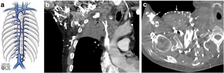
Blood bypasses the narrowing via anastomoses between branches of subclavian artery (above block) and intercostal/epigastric arteries (below block):
- Internal thoracic artery → anterior intercostal arteries → posterior intercostal arteries → descending aorta
- Superior epigastric → inferior epigastric artery
- Scapular anastomosis (suprascapular, transverse cervical ↔ subscapular/circumflex scapular)
Clinical features:
- Hypertension in upper limbs, weak/delayed femoral pulses ("radio-femoral delay")
- Notching of ribs on chest X-ray (due to dilated, tortuous posterior intercostal arteries eroding rib undersurface)
- Systolic murmur
Applied Anatomy: Classic viva/exam favorite — rib notching sign and radio-femoral delay are must-know clinical correlations.
Diagram to Draw
Aortic arch with a narrowed segment just beyond left subclavian artery origin (near ligamentum arteriosum), collateral vessels drawn as dilated tortuous connections between subclavian branches and intercostal arteries bypassing the narrowing.
Must write: Site (near ligamentum arteriosum), pre-/postductal types, collateral pathway via intercostal arteries, rib notching + radio-femoral delay.
11. DESCENDING THORACIC AORTA (3 Marks)
Simple Explanation
After the arch, the aorta continues down through the chest, lying just to the left of the spine, giving off branches to the ribs, esophagus, bronchi and other chest structures.
University Answer
Extent: Continuation of arch of aorta from lower border of T4 to T12, where it passes through the aortic opening of diaphragm to become abdominal aorta.
Position: Begins to the left of vertebral column, gradually moves anterior to it, lying in the midline by T12.
Relations:
| Position | Structure |
|---|
| Anterior | Root of left lung, pericardium, esophagus (crosses from right to left in lower part) |
| Posterior | Vertebral column, hemiazygos veins |
| Right | Esophagus (upper part), thoracic duct, azygos vein |
| Left | Left pleura and lung |
Branches:
| Type | Branches |
|---|
| Visceral | Pericardial, bronchial, esophageal, mediastinal branches |
| Parietal | Posterior intercostal arteries (9 pairs, 3rd–11th spaces), subcostal artery, superior phrenic arteries |
Applied Anatomy:
- Site of aortic aneurysm and traumatic aortic rupture (classically at ligamentum arteriosum/isthmus, at the level where descending aorta is fixed).
- Posterior intercostal arteries are important in coarctation collateral circulation.
Diagram to Draw
Vertical view of posterior mediastinum showing descending thoracic aorta from T4 to T12, giving off posterior intercostal arteries bilaterally, bronchial and esophageal branches anteriorly, ending at aortic opening (T12).
Must write: T4 to T12 extent, 9 pairs posterior intercostal arteries, visceral vs parietal branches, relation to esophagus and azygos.
12. RIGHT CORONARY ARTERY (3 Marks)
Simple Explanation
This is one of the two arteries feeding the heart muscle — it mainly feeds the right side of the heart and, in most people, also feeds the heart's natural pacemakers.
University Answer
Origin: From the right (anterior) aortic sinus of ascending aorta.
Course: Passes forward between pulmonary trunk and right auricle, descends in the right atrioventricular (coronary) groove, curves around the right (acute) margin of the heart, continues in the posterior AV groove to reach the crux of the heart (junction of all four grooves).
Branches:
| Branch | Supplies |
|---|
| SA nodal artery (~60% of people) | Sinoatrial node |
| Right marginal artery | Right border of heart |
| Posterior interventricular (descending) artery — in right-dominant hearts (~70%) | Posterior 1/3 of interventricular septum, part of both ventricles |
| AV nodal artery (branch near crux) | Atrioventricular node |
| Branches to right atrium and right ventricle | — |
Area supplied: Right atrium, most of right ventricle, part of left ventricle (posterior/inferior wall), posterior 1/3 of interventricular septum, SA node (60%) and AV node (~90%) in most individuals.
Applied Anatomy:
- RCA occlusion → inferior wall MI, and often causes bradyarrhythmias/heart blocks due to SA/AV nodal involvement.
- Right dominance (RCA giving posterior interventricular artery) is the most common coronary pattern.
Diagram to Draw
Anterior and inferior views of heart: RCA emerging from right aortic sinus, running in right AV groove, curving around acute margin, ending near crux giving posterior interventricular artery; mark SA nodal and marginal branches.
Must write: Origin (right aortic sinus), course in right AV groove, branches (SA nodal, marginal, posterior interventricular), right dominance concept, inferior MI correlation.
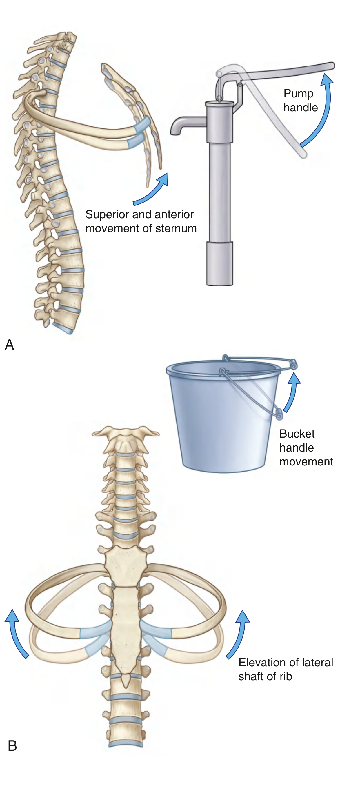
13. PUMP HANDLE AND BUCKET HANDLE MOVEMENT (4 Marks)
Simple Explanation
When you breathe in, your ribcage doesn't just sit still — the upper ribs and sternum swing forward-upward like an old water-pump handle, and the lower ribs flare outward-upward like the handle of a bucket being lifted. Both movements increase chest volume for breathing in.
University Answer
Context: These describe the mechanical movements of ribs and sternum during inspiration, increasing the anteroposterior and transverse diameters of the thorax.
1. Pump-handle movement:
- Occurs mainly at upper ribs (1st–6th) and sternum.
- Since the posterior end of ribs (at costovertebral/costotransverse joints) is fixed and higher than the anterior (sternal) end, the ribs rotate around an axis passing through the neck of the rib.
- This raises the sternal end of the ribs and pushes the sternum upward and forward, like the handle of an old-fashioned water pump.
- Increases the antero-posterior diameter of thorax.
2. Bucket-handle movement:
- Occurs mainly at lower ribs (7th–10th).
- The middle part of these ribs is lower than both its vertebral and sternal ends.
- During inspiration, the rib rotates so that the shaft moves laterally and upward, like the handle of a bucket being lifted from the side.
- Increases the transverse diameter of thorax.
Muscles responsible: External intercostals, and accessory muscles (scalenes, sternocleidomastoid) elevate ribs in forced inspiration; diaphragm contraction also assists overall expansion.
Applied Anatomy:
- Explains why in quiet breathing upper thorax movement is minimal, but becomes visible in forced/laboured breathing.
- Loss of these movements (e.g., in ankylosing spondylitis with costovertebral joint fusion) → reduced chest expansion, patient relies more on diaphragmatic breathing.
Diagram to Draw
Two side-by-side sketches: (A) lateral view of upper ribs/sternum showing axis of rotation through rib neck, arrow showing sternum moving up and forward (pump-handle). (B) Anterior/cross-sectional view of a lower rib showing its shaft moving up and out (bucket-handle), with arrows indicating increased transverse diameter.
Must write: Pump-handle = upper ribs, ↑AP diameter; Bucket-handle = lower ribs, ↑transverse diameter; axis of rotation concept.
14. OPENINGS OF DIAPHRAGM WITH VERTEBRAL LEVELS & CONTENTS (4 Marks)
Simple Explanation
The diaphragm is a dome-shaped sheet separating the chest and abdomen, but it can't be a solid wall — it has holes for the food pipe, the big blood vessels, and nerves to pass through, each at a specific spinal level.
University Answer
Three major openings:
| Opening | Vertebral Level | Contents |
|---|
| Vena caval opening (in central tendon) | T8 | Inferior vena cava, few twigs of right phrenic nerve |
| Esophageal opening (in muscular part, right crus sling) | T10 | Esophagus, anterior & posterior vagal trunks, esophageal branches of left gastric vessels |
| Aortic opening (behind diaphragm, between crura, not truly "in" the muscle) | T12 | Aorta, thoracic duct, sometimes azygos vein |
Mnemonic: I-8-10-Eaten-12-Aortic → "Vena Cava ate (8) ten (10) eggs at aortic (12)" — i.e., number of the letters/level increases: VC8, Esophagus10, Aorta12 (8,10,12 — even numbers going up by 2).
Minor/accessory openings:
| Opening | Content |
|---|
| Two crura (openings within/beside) | Greater, lesser, least splanchnic nerves; sympathetic trunk (behind medial arcuate ligament) |
| Foramen of vena azygos / behind medial crus | Azygos vein, hemiazygos vein |
| Between sternal & costal parts | Superior epigastric vessels |
| Behind lateral arcuate ligament | Subcostal nerve and vessels |
Applied Anatomy:
- Hiatus hernia — stomach herniates through a wide esophageal opening into thorax.
- Diaphragmatic hernia (congenital) — through posterolateral defect (foramen of Bochdalek) or retrosternal (foramen of Morgagni).
Diagram to Draw
Superior view of diaphragm showing central tendon with IVC opening (T8), esophageal hiatus with vagal trunks (T10), aortic opening between crura (T12), and sympathetic trunk/splanchnic nerves piercing the crura. Label vertebral levels alongside each opening.
Must write: All 3 major openings with exact vertebral levels and full content list, mnemonic 8-10-12.
15. BRONCHOPULMONARY SEGMENTS OF RIGHT LUNG (4 Marks)
Simple Explanation
Each lung is divided into small, independent functional units, each supplied by its own segmental bronchus and artery — like separate rooms in a house each with its own door (bronchus) and water pipe (artery). If disease is limited to one segment, that segment alone can be surgically removed.
University Answer
Definition: A bronchopulmonary segment is the largest subdivision of a lung lobe, supplied by a segmental (tertiary) bronchus, together with a corresponding branch of the pulmonary artery, and having its own connective tissue septum. It is a surgically resectable unit (segmentectomy).
Right lung has 10 bronchopulmonary segments (3 lobes):
| Lobe | Segments |
|---|
| Superior (Upper) lobe (3) | Apical, Posterior, Anterior |
| Middle lobe (2) | Lateral, Medial |
| Inferior (Lower) lobe (5) | Superior (apical basal), Medial basal, Anterior basal, Lateral basal, Posterior basal |
Key features:
- Each segment is pyramidal, with apex toward the hilum and base toward the lung surface.
- Segmental bronchus + segmental artery run centrally (together) in the segment.
- Segmental (intersegmental) veins run in the septa between segments — used by surgeon as a plane of separation.
- No collateral air spread between adjacent segments (bronchi are the only air supply).
Applied Anatomy:
- Basis of segmental resection in localized disease (TB, bronchiectasis, tumor) — sparing rest of the lung.
- Right lung, being wider and shorter bronchus more vertical, is the common site of aspirated foreign bodies.
- Segmental anatomy is used in interpreting CT chest/bronchoscopy localization.
Diagram to Draw
Lateral/medial view of right lung with 3 lobes outlined; label all 10 segments in their approximate positions (apical/posterior/anterior in upper lobe; lateral/medial in middle lobe; superior/medial-basal/anterior-basal/lateral-basal/posterior-basal in lower lobe). Show a bronchial tree diagram alongside: trachea → right main bronchus → 3 lobar bronchi → 10 segmental bronchi.
Must write: Definition emphasizing segmental bronchus + artery + own connective tissue septum, exact count (10) and names by lobe, surgical significance.
16. PLEURA WITH ITS APPLIED ANATOMY (4 Marks)
Simple Explanation
The pleura is like a plastic bag with two layers wrapped around each lung — one layer sticks to the lung, the other lines the chest wall, and a thin film of fluid between them lets the lung slide smoothly during breathing.
University Answer
Definition: A serous membrane (mesothelium + connective tissue) forming a closed sac around each lung, with two layers.
Layers:
| Layer | Description |
|---|
| Visceral (pulmonary) pleura | Closely invests the lung, extends into fissures, insensitive to pain |
| Parietal pleura | Lines inner surface of thoracic wall; sensitive to pain (supplied by intercostal & phrenic nerves) |
Parietal pleura is divided into 4 parts:
- Costal pleura — lines ribs/intercostal spaces
- Diaphragmatic pleura — covers upper surface of diaphragm
- Mediastinal pleura — covers lateral surface of mediastinum, forms pulmonary ligament and hilum
- Cervical pleura (cupola/dome) — extends above rib 1 into the root of neck, covered by suprapleural membrane (Sibson's fascia)
Pleural cavity: Potential space between visceral and parietal layers, containing a thin film of serous fluid, normally at negative pressure.
Nerve supply: Visceral pleura — autonomic (insensitive to pain); Parietal pleura — costal & diaphragmatic peripheral part by intercostal nerves, central diaphragmatic & mediastinal part by phrenic nerve.
Applied Anatomy (high-yield):
- Pleural effusion — fluid accumulation in pleural cavity.
- Pneumothorax — air in pleural cavity, lung collapse.
- Pleurisy — inflammation, causes pain referred via intercostal nerves (chest wall) or phrenic nerve (shoulder tip pain, since C3-C5 also supply shoulder skin).
- Thoracocentesis done through costodiaphragmatic recess in a safe zone above the rib.
- Cervical pleura vulnerable to injury in supraclavicular procedures (risk of pneumothorax); also relevant to apical lung tumors (Pancoast tumor) invading brachial plexus.
Diagram to Draw
Coronal section through thorax showing both lungs covered by visceral pleura, parietal pleura lining chest wall/diaphragm/mediastinum, cervical dome above rib 1 with suprapleural membrane, and pleural cavity space between the two layers.
Must write: Visceral vs parietal, 4 parts of parietal pleura, nerve supply difference, pain referral pattern, clinical conditions (effusion/pneumothorax/pleurisy).
17. DIAPHRAGM MUSCLE (4 Marks)
Simple Explanation
The diaphragm is the main breathing muscle — a dome-shaped sheet separating the thorax from the abdomen. When it contracts, it flattens and pulls air into the lungs.
University Answer
Definition: A dome-shaped musculotendinous partition separating thoracic and abdominal cavities; the principal muscle of inspiration.
Origin (peripheral, from three parts):
| Part | Origin |
|---|
| Sternal part | Back of xiphoid process (2 slips) |
| Costal part | Inner surfaces of lower 6 ribs and their costal cartilages (interdigitating with transversus abdominis) |
| Vertebral part (crura + arcuate ligaments) | Right crus from bodies of L1–L3; left crus from bodies of L1–L2; medial arcuate ligament (over psoas major); lateral arcuate ligament (over quadratus lumborum); median arcuate ligament (connecting two crura, over aorta) |
Insertion: All fibers converge and insert into a central tendon (trefoil/clover-shaped, no bony attachment).
Nerve supply: Phrenic nerve (C3, C4, C5) — motor supply entirely; also sensory to central part of diaphragm (peripheral part gets sensory from lower intercostal nerves too).
Mnemonic: "C3, 4, 5 keep the diaphragm alive"
Actions:
- Main muscle of inspiration — contraction flattens the dome, increasing vertical diameter of thoracic cavity.
- Raises intra-abdominal pressure — aids in micturition, defecation, parturition, vomiting (all "straining" actions).
Blood supply: Superior and inferior phrenic arteries, musculophrenic and pericardiacophrenic arteries.
Applied Anatomy:
- Phrenic nerve injury → diaphragmatic paralysis, paradoxical movement, elevated hemidiaphragm on X-ray.
- Hiccup — spasmodic diaphragmatic contraction.
- Referred pain from diaphragm (central part, via phrenic nerve C3–C5) felt at the shoulder tip (same dermatome as supraclavicular nerves).
Diagram to Draw
Superior view of diaphragm showing sternal, costal, and vertebral (crura + arcuate ligaments) origins, central tendon, three major openings, and phrenic nerve branches spreading over it.
Must write: 3 parts of origin, central tendon insertion (no bony attachment), phrenic nerve C3,4,5 motor + sensory to central part, main inspiratory action, referred shoulder pain.
18. DIAPHRAGM AND ITS MAJOR OPENINGS (4 Marks)
(Builds on Q14 & Q17 — combined answer format as typically asked)
Simple Explanation
Same diaphragm as above, but this question wants you to focus specifically on the muscle plus its big three holes together as one connected answer.
University Answer
Give a short account of the diaphragm (origin — sternal, costal, vertebral parts; insertion into central tendon; nerve supply — phrenic C3,4,5; action — main inspiratory muscle) [as in Q17], followed by the three major openings with levels and contents in a table [as in Q14]:
| Opening | Level | Contents |
|---|
| Vena caval opening | T8 | IVC, right phrenic nerve twigs |
| Esophageal opening | T10 | Esophagus, vagal trunks, esophageal vessels |
| Aortic opening | T12 | Aorta, thoracic duct, azygos vein (variable) |
High-yield extra point for this combined question: Note that the esophageal opening is formed by muscular fibers of the right crus (acts as a physiological sphincter helping prevent gastroesophageal reflux), while the aortic opening lies behind the diaphragm (osseo-muscular, between the crura and vertebral column) so the aorta is not compressed during diaphragmatic contraction — this is why aortic flow is undisturbed by breathing.
Applied Anatomy: Combine hiatus hernia (esophageal opening) + rib notching relevance (aortic opening in coarctation) + IVC opening's importance in liver trauma/IVC filter placement.
Diagram to Draw
Same as Q14/Q17 combined: full diaphragm outline (superior view) with origins labelled around the periphery, central tendon, and all three openings with vertebral levels marked centrally.
Must write: Full diaphragm structure + 3 openings table + right crus sphincter action for esophagus.
19. CARDIODIAPHRAGMATIC RECESS (2 Marks)
Simple Explanation
This is a small extra pocket of pleural space near the heart, at the point where the pleura reflects from the chest wall down onto the diaphragm close to the heart — normally the lung doesn't fully fill it during quiet breathing.
University Answer
Definition: A part of the costodiaphragmatic recess located anteriorly, near the region where the pericardium bulges and where costal pleura meets diaphragmatic pleura close to the heart (related to the cardiac notch of left lung and area of superficial cardiac dullness).
Location: On the left side, in relation to the deep cardiac notch of the left lung — anterior costomediastinal/costodiaphragmatic area adjacent to the pericardial sac (also referred to as an accentuated recess where the anterior border of the left lung does not fully occupy the pleural reflection because of the cardiac notch).
Significance:
- Because the left lung's anterior margin is deflected laterally at the cardiac notch (to accommodate the heart), an area of pleura here is not filled by lung even in full inspiration — a potential recess.
- Clinically corresponds to the area of "superficial cardiac dullness" on percussion, which is smaller than the actual area of the heart because part of the heart is covered here only by pleura reflections and thin lung margin.
Applied Anatomy: Important landmark in cardiac percussion and in pericardiocentesis planning (avoiding pleural recesses to prevent pneumothorax while accessing pericardium).
Diagram to Draw
Anterior view of thorax showing outline of heart, pericardium, and left lung with cardiac notch; shade the small triangular cardiodiaphragmatic recess area between lung margin and pericardium/diaphragm reflection.
Must write: Located near cardiac notch of left lung, related to costodiaphragmatic recess, correlates with superficial cardiac dullness.
20. PLEURAL RECESS (3 Marks)
Simple Explanation
Because the lungs don't perfectly fill every corner of the pleural sac even on deep breathing, some extra "spare space" pockets are left — these are the pleural recesses, and fluid tends to collect here first in disease.
University Answer
Definition: Areas of the pleural cavity where two parts of parietal pleura are in contact (or nearly so) because the lung margins do not extend into them, even during deepest inspiration.
Two important recesses:
| Recess | Location | Formed between |
|---|
| Costodiaphragmatic recess | Between costal and diaphragmatic pleura, around periphery of diaphragm (deepest posteriorly, ~ up to rib 10-12 in mid-axillary line during expiration vs. lung border at rib 8) | Costal pleura + diaphragmatic pleura |
| Costomediastinal recess | Anteriorly, behind the sternum, more prominent on the left (due to cardiac notch) | Costal pleura + mediastinal pleura |
Costodiaphragmatic recess — the deepest and clinically most important:
- During quiet respiration, inferior border of the lung lies 2 ribs higher than the pleural reflection at each point, creating this recess.
- Deepest point: in the mid-axillary line, extending down to the level of rib 10 (pleura extends to rib 12).
Applied Anatomy:
- Site of choice for pleural tap (thoracocentesis) and where pleural effusion first accumulates (gravity-dependent).
- Costomediastinal recess (left side, near cardiac notch) is relevant to auscultation and cardiac notch anatomy (links with Q19).
- On erect chest X-ray, small effusions are best seen filling the costophrenic angle (radiological correlate of costodiaphragmatic recess).
Diagram to Draw
Lateral view of thoracic cavity, showing lung's inferior border ending 2 rib-levels above the pleural reflection line, with the gap between them shaded as the costodiaphragmatic recess; also show the smaller costomediastinal recess anteriorly near the heart.
Must write: Definition (lung doesn't fill it), 2 recesses (costodiaphragmatic & costomediastinal), costodiaphragmatic = deepest/site of fluid collection & thoracocentesis.
21. STRUCTURES PASSING THROUGH AORTIC OPENING OF DIAPHRAGM (3 Marks)
Simple Explanation
The lowest of the three main diaphragm openings is not really a hole in the muscle — it's a gap between the two muscular pillars (crura) of the diaphragm and the spine, and three important structures pass through it into/out of the abdomen.
University Answer
Location: At the level of T12 vertebra, in the median plane, between the right and left crura of the diaphragm and the vertebral body (bounded by the median arcuate ligament anteriorly) — it is osseo-muscular, not truly within the diaphragmatic muscle fibers.
Structures passing through:
| Structure | Note |
|---|
| Aorta | Continues as abdominal aorta below |
| Thoracic duct | Lies on the right side of aorta at this level |
| Azygos vein (sometimes hemiazygos too) | May pass through this opening or through the crus itself |
Why this design matters: Because the opening is bounded by muscle (crura) but the aorta itself doesn't pass through the muscular fibers directly (it passes behind/between the crura), the aorta's caliber and flow are not compressed during diaphragmatic contraction in respiration — unlike the IVC opening (T8), which is surrounded by central tendon fibers and does get slightly compressed, aiding venous return.
Applied Anatomy:
- Relevant to spread of infection/malignancy between thorax and abdomen along the aorta, thoracic duct, or azygos vein pathway.
- Surgical approaches to thoracoabdominal aorta must consider crural anatomy.
Diagram to Draw
Posterior view of diaphragm, right and left crura arising from lumbar vertebrae, meeting in front of aorta at T12 forming the median arcuate ligament, with aorta, thoracic duct (to the right of aorta) and azygos vein shown passing through/near this opening.
Must write: T12 level, 3 structures (aorta, thoracic duct, azygos vein), osseo-muscular nature (no compression during respiration) — contrast with IVC opening.
22. AZYGOS VENOUS SYSTEM (4 Marks)
Simple Explanation
This is a backup drainage network of veins running along the back wall of the chest on both sides of the spine, collecting blood from the chest wall and draining it into the superior vena cava. It's very important because it can act as a bypass route if the big veins (IVC or SVC) get blocked.
University Answer
Definition: A system of longitudinally running veins on either side of the vertebral column in the posterior thorax, draining the posterior thoracic and abdominal wall, and acting as an important collateral pathway between the SVC and IVC territories.
Components:
| Vein | Side | Origin | Course | Termination |
|---|
| Azygos vein | Right | Union of right ascending lumbar vein + right subcostal vein (near T12/L1) | Ascends through aortic opening or right crus of diaphragm, along right side of vertebral bodies (T12–T4), then arches forward over root of right lung at T4 | Superior vena cava |
| Hemiazygos vein | Left, lower | Union of left ascending lumbar vein + left subcostal vein | Ascends on left side up to about T9, then crosses midline behind aorta, esophagus, thoracic duct | Joins azygos vein |
| Accessory hemiazygos vein | Left, upper | Drains 4th–8th left posterior intercostal veins | Descends, crosses midline at T7-T8 | Joins azygos vein (or hemiazygos) |
Tributaries of azygos vein: Right posterior intercostal veins (4th–11th), right subcostal vein, hemiazygos & accessory hemiazygos veins, esophageal, mediastinal, pericardial, and right bronchial veins, and the right superior intercostal vein (draining 1st-3rd spaces) in some.
Applied Anatomy:
- Important collateral channel connecting SVC and IVC systems — if IVC is obstructed (e.g., by tumor/thrombosis), blood can bypass via ascending lumbar veins → azygos system → SVC → right atrium.
- Azygos vein enlargement seen on chest X-ray in SVC obstruction, portal hypertension, or right heart failure.
- "Azygos lobe" — anatomical variant of right lung where the azygos vein fails to migrate to its normal position and instead invaginates the medial part of the upper lobe, seen on CT/X-ray as a fissure.
Diagram to Draw
Posterior view of thorax/abdomen showing azygos vein ascending on the right from union of right ascending lumbar + subcostal veins, arching over right lung root into SVC at T4; hemiazygos and accessory hemiazygos on the left crossing midline to join azygos; label intercostal vein tributaries on both sides.
Must write: 3 components (azygos, hemiazygos, accessory hemiazygos), origin & termination of each, collateral pathway significance (SVC-IVC bypass), azygos lobe.
23. BRONCHOPULMONARY SEGMENTS OF LEFT LUNG (4 Marks)
Simple Explanation
Same concept as the right lung's segments (Q15), but the left lung has only 2 lobes (no middle lobe) — instead, part of the upper lobe (the "lingula") functionally substitutes for the missing middle lobe, so segments are fewer/fused compared to the right side.
University Answer
Definition: Same as Q15 — independently resectable units, each with its own segmental bronchus + pulmonary artery branch.
Left lung has 8–10 segments (commonly described as 8, due to fusion of some segments) in 2 lobes:
| Lobe | Division | Segments |
|---|
| Superior (Upper) lobe | Upper (superior) division | Apicoposterior* (fusion of apical + posterior), Anterior |
| Lower division (Lingular part — homologue of right middle lobe) | Superior lingular, Inferior lingular |
| Inferior (Lower) lobe | — | Superior (apical basal), Anteromedial basal* (fusion of anterior + medial basal), Lateral basal, Posterior basal |
*(Apical and posterior segments often remain separate as apical + posterior in some texts giving 10 segments; anteromedial basal may also be described as separate anterior basal + medial basal — hence the count varies 8–10 depending on textbook. BD Chaurasia gives 10 segments by keeping these separate, matching the right lung numbering for symmetry in exams — safest to write 10 with a note on common fusion.)
Key difference from right lung:
- No middle lobe — its role is taken over by the lingula (tongue-like part of the upper lobe, tucked under the cardiac notch), which is the anatomical/functional equivalent of the right middle lobe.
- Left main bronchus is longer, narrower, and more horizontal than right → foreign bodies lodge here less commonly than on the right.
Applied Anatomy:
- Lingular segment disease behaves clinically like middle lobe syndrome on the right.
- Segmental resection principles same as right lung — important in TB, bronchiectasis, tumors localized to the lingula or specific basal segments.
Diagram to Draw
Lateral/medial view of left lung with 2 lobes and cardiac notch outlined; label apicoposterior and anterior segments (upper division), superior and inferior lingular segments (lower division of upper lobe), and superior/anteromedial-basal/lateral-basal/posterior-basal segments of lower lobe. Show bronchial tree: trachea → left main bronchus → 2 lobar bronchi → segmental bronchi.
Must write: 2 lobes only, lingula = homologue of middle lobe, segment count 8-10 (mention fusion), left main bronchus more horizontal/longer (less common site for FB aspiration — contrast with right).
Quick Cross-Reference Note
Questions 14, 18, and 21 all test diaphragm openings — master the T8-T10-T12 table once and you can answer all three at different depths. Questions 16, 19, and 20 all test pleura — learn the visceral/parietal distinction and the two recesses once, then adapt depth to the marks asked.