Red lesions of oral mucosa

Reading File
Finding Sources
Finding Sources
Searching Images

red lesions oral mucosa erythroplakia

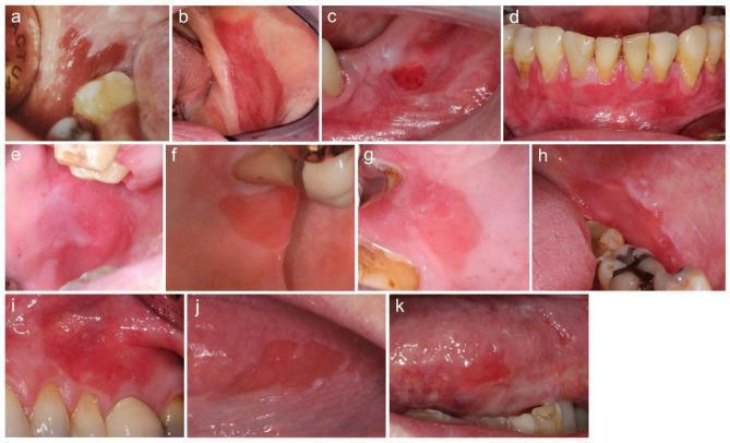
This composite of clinical intraoral photographs (labeled a–k) demonstrates various presentations of oral erythroplakia (OE) across eleven patients. The primary anatomical sites shown include the buccal mucosa, alveolar ridge, and gingiva. The lesions are characterized by persistent, well-demarcated, fiery red macules or patches. Morphologically, the majority of the lesions (e.g., a, b, d, i, j, k) exhibit a uniformly erythematous appearance, while others (e.g., e, g, h) present with minor, interspersed white areas (speckled erythroplakia). All depicted lesions feature a smooth, non-granular surface texture, ranging from matte to shiny. Most OEs show sharp, well-defined borders distinguishing them from the surrounding healthy mucosa, which is a critical diagnostic feature to differentiate them from non-specific inflammatory erythema. Clinically, these images illustrate a high-risk oral potentially malignant disorder (OPMD), highlighting the importance of visual screening for well-defined red patches that lack a clear reactive or inflammatory etiology.
This composite of clinical intraoral photographs (labeled a–k) demonstrates various presentations of oral erythroplakia (OE) across eleven patients. The primary anatomical sites shown include the buccal mucosa, alveolar ridge, and gingiva. The lesions are characterized by persistent, well-demarcated, fiery red macules or patches. Morphologically, the majority of the lesions (e.g., a, b, d, i, j, k) exhibit a uniformly erythematous appearance, while others (e.g., e, g, h) present with minor, interspersed white areas (speckled erythroplakia). All depicted lesions feature a smooth, non-granular surface texture, ranging from matte to shiny. Most OEs show sharp, well-defined borders distinguishing them from the surrounding healthy mucosa, which is a critical diagnostic feature to differentiate them from non-specific inflammatory erythema. Clinically, these images illustrate a high-risk oral potentially malignant disorder (OPMD), highlighting the importance of visual screening for well-defined red patches that lack a clear reactive or inflammatory etiology.

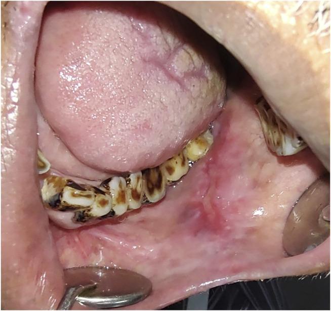
This clinical intraoral photograph demonstrates an erythroplakia patch on the left buccal mucosa. The lesion presents as a well-demarcated, predominantly erythematous (bright red) patch that contrasts sharply with the surrounding paler pink normal oral mucosa. The surface texture appears relatively smooth and velvety, a classic clinical feature of erythroplakia. The primary lesion is located adjacent to the mandibular teeth. Notable secondary findings include significant extrinsic dental staining and calculus buildup on the lower dentition, which may be clinical markers for tobacco or areca nut use—major risk factors for oral potentially malignant disorders (OPMDs). The tongue is visible and appears generally healthy with normal papillation. This image is an educational example used to teach the identification of high-risk premalignant lesions, as erythroplakia carries a significantly higher risk of malignant transformation (MT) compared to leukoplakia, often harboring severe dysplasia or carcinoma in situ upon histological examination.
This clinical intraoral photograph demonstrates an erythroplakia patch on the left buccal mucosa. The lesion presents as a well-demarcated, predominantly erythematous (bright red) patch that contrasts sharply with the surrounding paler pink normal oral mucosa. The surface texture appears relatively smooth and velvety, a classic clinical feature of erythroplakia. The primary lesion is located adjacent to the mandibular teeth. Notable secondary findings include significant extrinsic dental staining and calculus buildup on the lower dentition, which may be clinical markers for tobacco or areca nut use—major risk factors for oral potentially malignant disorders (OPMDs). The tongue is visible and appears generally healthy with normal papillation. This image is an educational example used to teach the identification of high-risk premalignant lesions, as erythroplakia carries a significantly higher risk of malignant transformation (MT) compared to leukoplakia, often harboring severe dysplasia or carcinoma in situ upon histological examination.

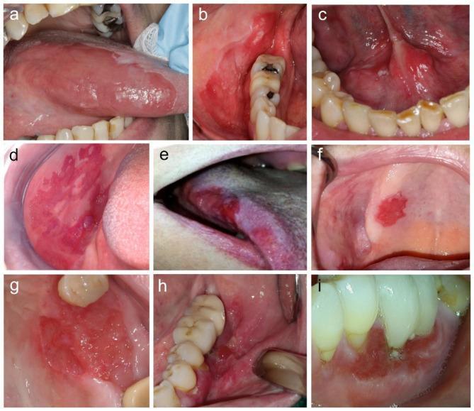
A composite of nine clinical intraoral photographs (labeled a-i) showing various presentations of oral erythroplakia-like squamous cell carcinoma (OELSCC). The images demonstrate a range of erythematous lesions located across various oral sites, including the gingiva, lateral tongue, floor of the mouth, and buccal mucosa. Visually, the lesions are characterized by a predominant bright red color, with some exhibiting minor white leukoplakic patches or focal ulceration. Morphological features include matte to shiny textures and surfaces that range from smooth and flat to granular or uneven. Some lesions are clearly depressed relative to the surrounding healthy mucosa. While most lesions display well-defined, sharp borders—a key clinical feature distinguishing them from diffuse inflammatory conditions—others show more poorly defined margins. This educational compilation illustrates the subtle clinical variations of high-risk red oral lesions that may harbor invasive malignancy, emphasizing the importance of detailed visual assessment in oral pathology and oncology.
A composite of nine clinical intraoral photographs (labeled a-i) showing various presentations of oral erythroplakia-like squamous cell carcinoma (OELSCC). The images demonstrate a range of erythematous lesions located across various oral sites, including the gingiva, lateral tongue, floor of the mouth, and buccal mucosa. Visually, the lesions are characterized by a predominant bright red color, with some exhibiting minor white leukoplakic patches or focal ulceration. Morphological features include matte to shiny textures and surfaces that range from smooth and flat to granular or uneven. Some lesions are clearly depressed relative to the surrounding healthy mucosa. While most lesions display well-defined, sharp borders—a key clinical feature distinguishing them from diffuse inflammatory conditions—others show more poorly defined margins. This educational compilation illustrates the subtle clinical variations of high-risk red oral lesions that may harbor invasive malignancy, emphasizing the importance of detailed visual assessment in oral pathology and oncology.

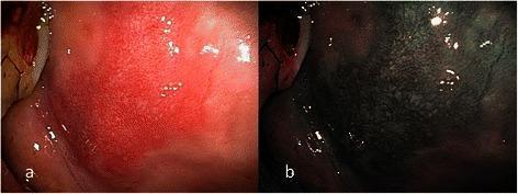
This dual-panel endoscopic image provides a side-by-side comparison of oral erythroplakia on the right buccal mucosa. Image (a) utilizes conventional broadband white light (BWL), revealing a sharply circumscribed, bright red, flat plaque with a velvety texture. Image (b) shows the same lesion under Narrow Band Imaging (NBI), which enhances the visualization of the microvascular architecture. Under NBI, the area exhibits a brownish hue with clearly visible dilated and tortuous intraepithelial papillary capillary loops (IPCL). These vascular changes are classified as IPCL Type II, which are associated with angiogenesis in premalignant lesions. The clinical significance of this finding corresponds to intermediate-grade epithelial dysplasia. The image demonstrates the diagnostic utility of NBI in identifying microvascular morphological patterns—such as vessel winding and crossing—that may be obscured under white light, facilitating the assessment of mucosal lesions in the oral cavity for better triage between benign and dysplastic conditions.
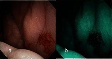
This dual-panel endoscopic image provides a side-by-side comparison of oral erythroplakia on the right buccal mucosa. Image (a) utilizes conventional broadband white light (BWL), revealing a sharply circumscribed, bright red, flat plaque with a velvety texture. Image (b) shows the same lesion under Narrow Band Imaging (NBI), which enhances the visualization of the microvascular architecture. Under NBI, the area exhibits a brownish hue with clearly visible dilated and tortuous intraepithelial papillary capillary loops (IPCL). These vascular changes are classified as IPCL Type II, which are associated with angiogenesis in premalignant lesions. The clinical significance of this finding corresponds to intermediate-grade epithelial dysplasia. The image demonstrates the diagnostic utility of NBI in identifying microvascular morphological patterns—such as vessel winding and crossing—that may be obscured under white light, facilitating the assessment of mucosal lesions in the oral cavity for better triage between benign and dysplastic conditions.

This composite diagnostic image displays an endoscopic examination of oral erythroplakia on the left buccal mucosa. Figure (a) utilizes conventional broadband white light (BWL), revealing a well-demarcated, flat, bright red plaque with a velvety and slightly glistening surface texture. Figure (b) shows the same lesion under Narrow-Band Imaging (NBI), which enhances the visualization of the microvascular architecture. The NBI view demonstrates Type IV intraepithelial papillary capillary loop (IPCL) destruction, characterized by irregular angiogenesis, loss of normal capillary loop structure, and a disorganized vascular arrangement. This microvascular pattern is a significant clinical indicator for advanced pathology. The clinical significance of this finding is demonstrated by the corresponding pathological diagnosis of invasive squamous cell carcinoma, highlighting the role of NBI in identifying high-risk premalignant and malignant lesions during oral cavity screening.
This composite diagnostic image displays an endoscopic examination of oral erythroplakia on the left buccal mucosa. Figure (a) utilizes conventional broadband white light (BWL), revealing a well-demarcated, flat, bright red plaque with a velvety and slightly glistening surface texture. Figure (b) shows the same lesion under Narrow-Band Imaging (NBI), which enhances the visualization of the microvascular architecture. The NBI view demonstrates Type IV intraepithelial papillary capillary loop (IPCL) destruction, characterized by irregular angiogenesis, loss of normal capillary loop structure, and a disorganized vascular arrangement. This microvascular pattern is a significant clinical indicator for advanced pathology. The clinical significance of this finding is demonstrated by the corresponding pathological diagnosis of invasive squamous cell carcinoma, highlighting the role of NBI in identifying high-risk premalignant and malignant lesions during oral cavity screening.

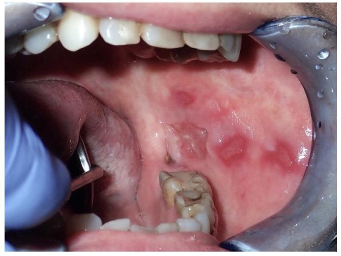
This clinical photograph shows the left buccal mucosa of a 53-year-old female patient, demonstrating multiple multifocal lesions. The visual findings include several well-defined, velvety red patches consistent with erythroplakia, distributed from the posterior to the anterior regions of the cheek. A prominent area of central ulceration is visible, showing a break in the mucosal surface with irregular, slightly raised borders. The surrounding tissue appears mildly edematous. These findings are clinically significant as indicators of potentially malignant disorders, specifically invasive squamous cell carcinoma (OSCC) in this case, which may arise from chronic mucosal trauma. The image illustrates the high-risk nature of erythroplakic and ulcerative oral lesions, highlighting the need for biopsy and histopathological evaluation to distinguish between pre-malignancy and invasive cancer. The target audience includes dental and medical students, as well as oncology specialists focused on early detection of oral cavity malignancies.

This clinical photograph shows the left buccal mucosa of a 53-year-old female patient, demonstrating multiple multifocal lesions. The visual findings include several well-defined, velvety red patches consistent with erythroplakia, distributed from the posterior to the anterior regions of the cheek. A prominent area of central ulceration is visible, showing a break in the mucosal surface with irregular, slightly raised borders. The surrounding tissue appears mildly edematous. These findings are clinically significant as indicators of potentially malignant disorders, specifically invasive squamous cell carcinoma (OSCC) in this case, which may arise from chronic mucosal trauma. The image illustrates the high-risk nature of erythroplakic and ulcerative oral lesions, highlighting the need for biopsy and histopathological evaluation to distinguish between pre-malignancy and invasive cancer. The target audience includes dental and medical students, as well as oncology specialists focused on early detection of oral cavity malignancies.

Finding Sources
Finding Sources
Reading File
Reading File
Finding Sources
Reading File
Reading File
Reading File
Finding Sources
Searching Images

geographic tongue median rhomboid glossitis erythematous candidiasis
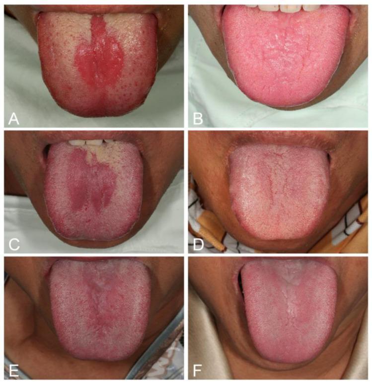
This composite clinical photograph displays six intraoral images of the dorsal tongue, illustrating three cases of Median Rhomboid Glossitis (MRG) as a manifestation of erythematous candidiasis. Panels A, C, and E represent the pre-treatment state, showing well-defined, centrally located erythematous lesions on the posterior dorsal tongue along the midline. These lesions are characterized by atrophy of the filiform papillae, resulting in a smooth, red, rhomboid, or elliptical surface area. Panel C additionally exhibits a focal white coating suggestive of pseudomembranous candidiasis. Panels B, D, and F show the same patients two weeks following combined iron and antifungal (nystatin) therapy, demonstrating complete resolution of the erythema and successful regeneration of the lingual filiform papillae. The images highlight the clinical appearance of atrophic glossitis associated with systemic risk factors like iron deficiency anemia and diabetes mellitus, and the efficacy of targeted antimicrobial and nutritional intervention in restoring normal lingual mucosal texture and coloration.
This composite clinical photograph displays six intraoral images of the dorsal tongue, illustrating three cases of Median Rhomboid Glossitis (MRG) as a manifestation of erythematous candidiasis. Panels A, C, and E represent the pre-treatment state, showing well-defined, centrally located erythematous lesions on the posterior dorsal tongue along the midline. These lesions are characterized by atrophy of the filiform papillae, resulting in a smooth, red, rhomboid, or elliptical surface area. Panel C additionally exhibits a focal white coating suggestive of pseudomembranous candidiasis. Panels B, D, and F show the same patients two weeks following combined iron and antifungal (nystatin) therapy, demonstrating complete resolution of the erythema and successful regeneration of the lingual filiform papillae. The images highlight the clinical appearance of atrophic glossitis associated with systemic risk factors like iron deficiency anemia and diabetes mellitus, and the efficacy of targeted antimicrobial and nutritional intervention in restoring normal lingual mucosal texture and coloration.

This clinical intraoral photograph displays the dorsal surface of a human tongue, showcasing a characteristic presentation of median rhomboid glossitis (MRG). The primary finding is a well-circumscribed, erythematous, rhomboid-shaped area of depapillation located in the midline of the posterior dorsal tongue, immediately anterior to the circumvallate papillae. The lesion appears as a smooth, slightly elevated, and beefy red patch that contrasts with the surrounding light-pink tissue covered in normal filiform papillae. There are no signs of ulceration, fissuring, or exudate within the lesion. The surrounding lingual mucosa shows a mild white coating, possibly representing physiological debris or minor keratosis, while the erythematous patch lacks this coating due to the loss of papillae. This visual is a classic representation of a benign inflammatory or developmental condition often associated with chronic candidal infection, used in dental and medical education to differentiate between geographic tongue, candidiasis, and oral malignancies.
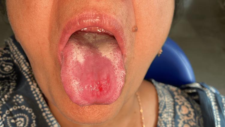
This clinical intraoral photograph displays the dorsal surface of a human tongue, showcasing a characteristic presentation of median rhomboid glossitis (MRG). The primary finding is a well-circumscribed, erythematous, rhomboid-shaped area of depapillation located in the midline of the posterior dorsal tongue, immediately anterior to the circumvallate papillae. The lesion appears as a smooth, slightly elevated, and beefy red patch that contrasts with the surrounding light-pink tissue covered in normal filiform papillae. There are no signs of ulceration, fissuring, or exudate within the lesion. The surrounding lingual mucosa shows a mild white coating, possibly representing physiological debris or minor keratosis, while the erythematous patch lacks this coating due to the loss of papillae. This visual is a classic representation of a benign inflammatory or developmental condition often associated with chronic candidal infection, used in dental and medical education to differentiate between geographic tongue, candidiasis, and oral malignancies.

An intraoral clinical photograph showing the dorsal surface of the tongue of a patient with median rhomboid glossitis. The image demonstrates a well-circumscribed, erythematous, and depapillated area centrally located on the midline of the tongue, anterior to the circumvallate papillae. The lesion exhibits a smooth, atrophic texture compared to the surrounding normal filiform papillae. Within this central erythematous zone, there is a small, focal area of ulceration with minor pinpoint bleeding. Faint, irregular white patches are visible along the lateral and posterior margins of the depapillated area, suggestive of secondary candidal infection (pseudomembranous candidiasis). The surrounding oral mucosa and lips appear clinically normal. This visual presentation is characteristic of a 'kissing lesion' associated with oral thrush, showing evidence of early healing or partial response to antifungal therapy as indicated by the reduction in the size of the central ulcer and thinning of the white coating.
An intraoral clinical photograph showing the dorsal surface of the tongue of a patient with median rhomboid glossitis. The image demonstrates a well-circumscribed, erythematous, and depapillated area centrally located on the midline of the tongue, anterior to the circumvallate papillae. The lesion exhibits a smooth, atrophic texture compared to the surrounding normal filiform papillae. Within this central erythematous zone, there is a small, focal area of ulceration with minor pinpoint bleeding. Faint, irregular white patches are visible along the lateral and posterior margins of the depapillated area, suggestive of secondary candidal infection (pseudomembranous candidiasis). The surrounding oral mucosa and lips appear clinically normal. This visual presentation is characteristic of a 'kissing lesion' associated with oral thrush, showing evidence of early healing or partial response to antifungal therapy as indicated by the reduction in the size of the central ulcer and thinning of the white coating.

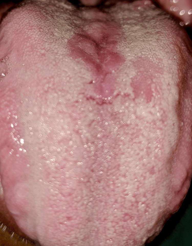
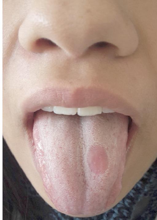
A clinical photograph of a human tongue demonstrating a classic presentation of median rhomboid glossitis, appearing in a slightly paramedian location. The tongue surface shows a pinkish-white background with a rough texture consistent with normal papillation. In the posterior-middle third of the dorsal surface, there is a well-circumscribed, oval-shaped erythematous lesion approximately 1 cm in diameter. This lesion is characterized by a smooth, depapillated surface (atrophy of the filiform papillae) that contrasts sharply with the surrounding healthy tissue. The lesion is positioned slightly to the patient's left of the midline. This visual is typical for oral medicine and dermatology studies focusing on benign inflammatory conditions of the tongue, often associated with chronic candidiasis. The photograph is suitable for medical education on oral pathology and differential diagnosis of lingual lesions.

A clinical photograph of a human tongue demonstrating a classic presentation of median rhomboid glossitis, appearing in a slightly paramedian location. The tongue surface shows a pinkish-white background with a rough texture consistent with normal papillation. In the posterior-middle third of the dorsal surface, there is a well-circumscribed, oval-shaped erythematous lesion approximately 1 cm in diameter. This lesion is characterized by a smooth, depapillated surface (atrophy of the filiform papillae) that contrasts sharply with the surrounding healthy tissue. The lesion is positioned slightly to the patient's left of the midline. This visual is typical for oral medicine and dermatology studies focusing on benign inflammatory conditions of the tongue, often associated with chronic candidiasis. The photograph is suitable for medical education on oral pathology and differential diagnosis of lingual lesions.

Reading File
Finding Sources
Loading Image
Loading Image
Now I have comprehensive information from multiple authoritative textbooks. Let me compile the full answer.

Red Lesions of the Oral Mucosa

Red lesions of the oral mucosa form an important diagnostic category. While some are entirely benign, others carry significant malignant potential and demand biopsy and close follow-up. The table below (from Goldman-Cecil Medicine) summarizes the main causes:
Red or Blue Lesions (Macular / Maculopapular)
  • Squamous cell carcinoma (early)
  • Erythroplakia (epithelial dysplasia)
  • Erythematous (atrophic) candidiasis
  • Median rhomboid glossitis
  • Mucocutaneous diseases (lichen planus, pemphigus, erythema multiforme)
  • Angular cheilitis
  • Telangiectasias and purpuras
  • Kaposi sarcoma (blue to purple)

1. Erythroplakia

Definition: WHO defines erythroplakia as "a fiery red patch that cannot be characterized clinically or pathologically as any other definable lesion."
Clinical features:
  • Red, velvety, possibly eroded area within the oral cavity
  • Usually remains level with or slightly depressed relative to the surrounding mucosa
  • Most commonly found on the soft palate, floor of mouth, and buccal mucosa
  • Usually <1.5 cm; well-demarcated borders
Histology: This is the most alarming red lesion. Approximately 90% of erythroplakias demonstrate severe dysplasia, carcinoma in situ, or minimally invasive carcinoma on biopsy. The reddish color arises from an intense subepithelial inflammatory reaction with vascular dilation and thin, atrophic epithelium that allows the underlying vasculature to show through. (Robbins, Cotran & Kumar Pathologic Basis of Disease)
Speckled erythroplakia: Intermediate forms with combined red and white areas; these also carry high malignant risk.
Risk factors: Tobacco (cigarettes, pipes, cigars, smokeless tobacco), alcohol; usually in adults aged 40-70 years, male preponderance 2:1.
Multiple presentations of oral erythroplakia - well-demarcated fiery red patches on buccal mucosa, alveolar ridge and gingiva
Oral erythroplakia: Multiple well-demarcated, velvety red patches - a high-risk oral potentially malignant disorder (OPMD)

2. Early Squamous Cell Carcinoma (SCC)
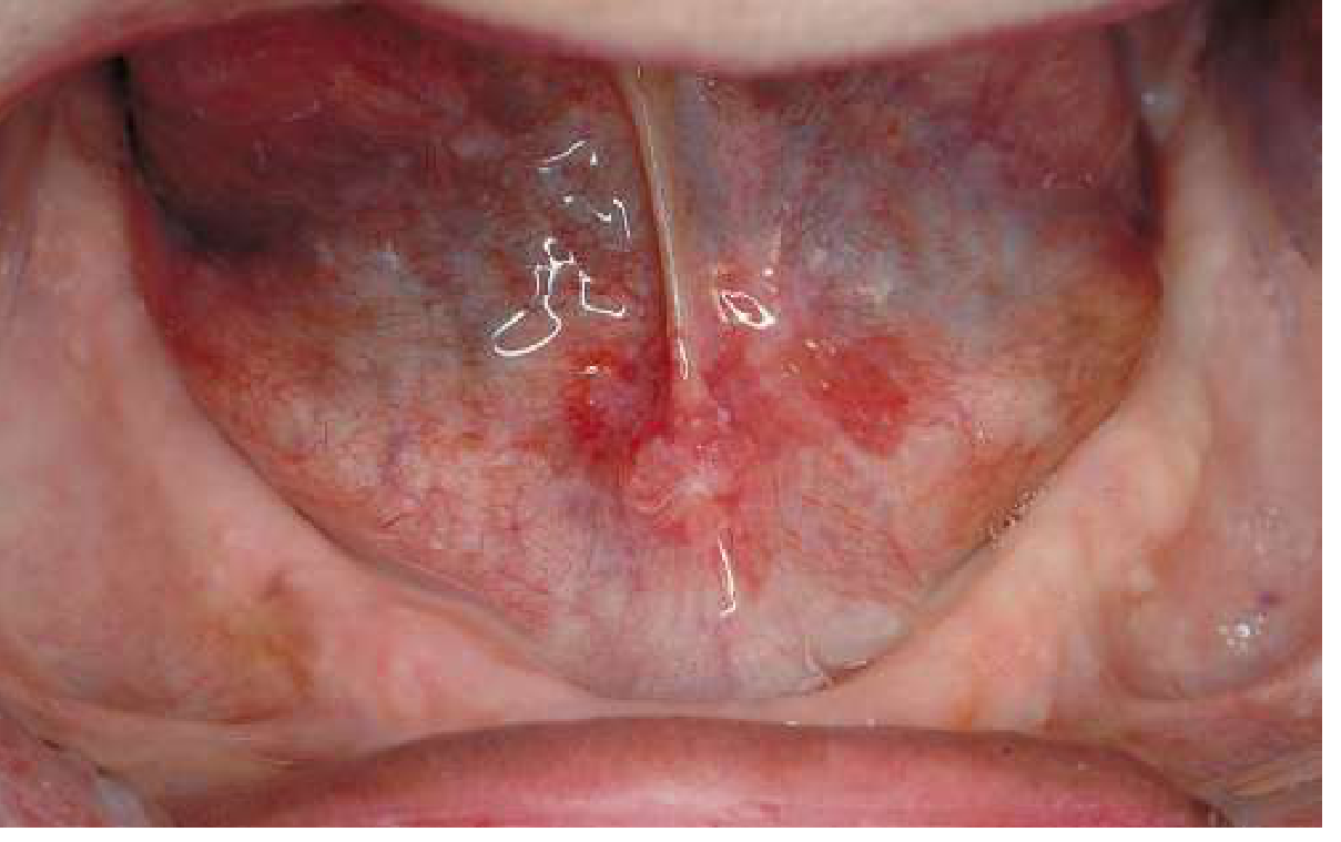
Early SCC may present as an erythroplakic (red) macule or patch, often with induration. The floor of the mouth is a classic site.
Erythroplakia of anterior mouth floor - biopsy proven squamous cell carcinoma
SCC: This area of erythroplakia with slight induration in the anterior mouth floor was confirmed as squamous cell carcinoma on biopsy (Goldman-Cecil Medicine, Fig. 393-3)
Key features pointing toward malignancy: induration, fixation, ulceration, bleeding, cervical lymphadenopathy.

3. Erythematous (Atrophic) Candidiasis

Three forms of oral candidiasis exist: pseudomembranous (thrush), erythematous (atrophic), and hyperplastic.
Erythematous candidiasis is a chronic condition characterized by:
  • Erythema and atrophy of filiform papillae on the dorsal tongue
  • Or patchy, ill-defined erythema on the palate, tongue, or buccal mucosa
  • Symptoms: oral burning, sensitivity to spicy foods
Risk factors: Chronic salivary hypofunction (e.g., Sjögren syndrome, anticholinergic drugs), denture wearers (Candida-infected dentures - erythema confined to denture-bearing area), HIV infection, systemic antibiotics, uncontrolled diabetes, anemia, immunosuppression.
Management: Fluconazole 200 mg day 1, then 100 mg daily for 2 weeks (drug of choice). In severe salivary hypofunction: vaginal nystatin tablets or miconazole tablets dissolved orally (to avoid cariogenic sucrose/glucose in troches). (Goldman-Cecil Medicine)

4. Median Rhomboid Glossitis

  • A red macule in the midline of the posterior dorsal tongue, anterior to the circumvallate papillae
  • Smooth, depapillated, rhomboid/elliptical shape
  • Uniformly benign - one of the few red oral lesions that does not require biopsy for reassurance
  • Associated with localized overgrowth of Candida species
  • May respond to antifungal therapy
Median rhomboid glossitis - erythematous depapillated midline lesion on posterior dorsal tongue
Median rhomboid glossitis: Well-circumscribed smooth erythematous rhomboid patch on midline posterior tongue, characteristically benign

5. Geographic Tongue (Benign Migratory Glossitis)

  • Affects ~2% of the population; idiopathic, benign
  • Well-defined areas of atrophied filiform papillae on dorsal tongue, bordered by arcs of normal/hyperplastic papillae
  • Lesions change location over time ("migratory")
  • Usually asymptomatic; no treatment required
  • Diagnosis by characteristic appearance alone
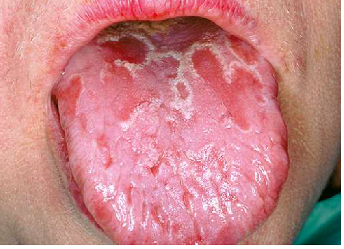
Geographic tongue - patchy depapillation with white-bordered red areas that migrate over time
Geographic tongue (benign migratory glossitis): Distribution of atrophic areas on dorsal tongue changes over time but lesions are asymptomatic (Goldman-Cecil Medicine, Fig. 393-6)

6. Oral Lichen Planus (Erythematous/Erosive Form)

  • Occurs in ~1% of the population
  • Usually bilaterally symmetrical reticular (lattice-like) white lesions with adjacent areas of erythema (atrophy or erosion) or ulcers
  • Atrophic/erosive forms present as red, painful areas with sensitivity to foods
  • Management: topical fluocinolone or clobetasol gel 0.05% three times daily

7. Kaposi Sarcoma

  • Color ranges from blue to purple/red; seen in HIV-infected or immunosuppressed patients
  • Occurs on palate or gingiva as macular, papular, or nodular lesions
  • Biopsy required for diagnosis

8. Vascular Lesions (Telangiectasias, Purpuras)

  • Hereditary hemorrhagic telangiectasia (Rendu-Osler-Weber syndrome): Autosomal dominant; multiple congenital aneurysmal telangiectasias beneath mucosal surfaces of the oral cavity and lips
  • Purpura: Petechiae from thrombocytopenia (e.g., palatal petechiae in infectious mononucleosis)

Summary Table

LesionKey FeaturesMalignant RiskManagement
ErythroplakiaVelvety red patch, well-demarcated~90% dysplasia/CIS/SCCBiopsy mandatory
Early SCCRed/erythroplakic, induratedMalignantSurgery ± RT/chemo
Erythematous candidiasisPatchy erythema, burningNoneAntifungals
Median rhomboid glossitisMidline posterior tongue, smoothNoneAntifungals if symptomatic
Geographic tongueMigratory depapillated patchesNoneReassurance
Lichen planus (erosive)Bilateral, reticular + erythemaLow (1-3%)Topical steroids
Kaposi sarcomaBlue-purple lesions, HIVMalignantSystemic therapy
TelangiectasiasMultiple vascular maculesNoneTreat underlying cause

Key clinical rule: Any red lesion of the oral mucosa that cannot be explained by an obvious reactive cause (candidiasis, trauma, denture irritation) and persists for more than 2-3 weeks after removal of irritants requires biopsy, because erythroplakia carries a far higher risk of harboring carcinoma than leukoplakia.
Sources: Goldman-Cecil Medicine (22e); Robbins, Cotran & Kumar Pathologic Basis of Disease; Robbins & Kumar Basic Pathology; Goldman-Cecil Medicine, Table 393-3.
This is a shared conversation. Sign in to Orris to start your own chat.