
Mastitis detail explanation with respect to veterinary medicine 4th year I want genuine information and practical base treatment for obtaining good marks.
mastitis veterinary medicine bovine classification pathogenesis treatment 4th year
bovine mastitis udder inflammation pathology clinical signs
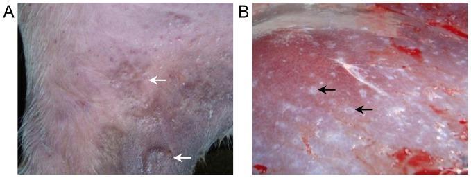
Clinical and gross pathology photographs demonstrating the tissue-level manifestations of Schistosoma japonicum infection. Panel A shows a clinical photograph of the skin on the inner thigh of a bovine model. The image exhibits visible signs of inflammation, characterized by localized erythema and edema. White arrows specifically indicate raised, papular inflammatory reactions at the site of cercarial penetration five days post-infection. Panel B displays a gross pathological view of a liver surface during necropsy. The liver parenchyma shows evidence of chronic schistosome infection. Black arrows highlight numerous, discrete white nodules (granulomas) distributed across the organ surface. These nodules are indicative of egg-induced pathology, where the host immune response forms fibrotic granulomatous lesions around trapped parasite eggs. This comparison illustrates the transition from the acute cutaneous phase of schistosomiasis (swimmer's itch/cercarial dermatitis) to the chronic visceral phase involving hepatic egg pathology.
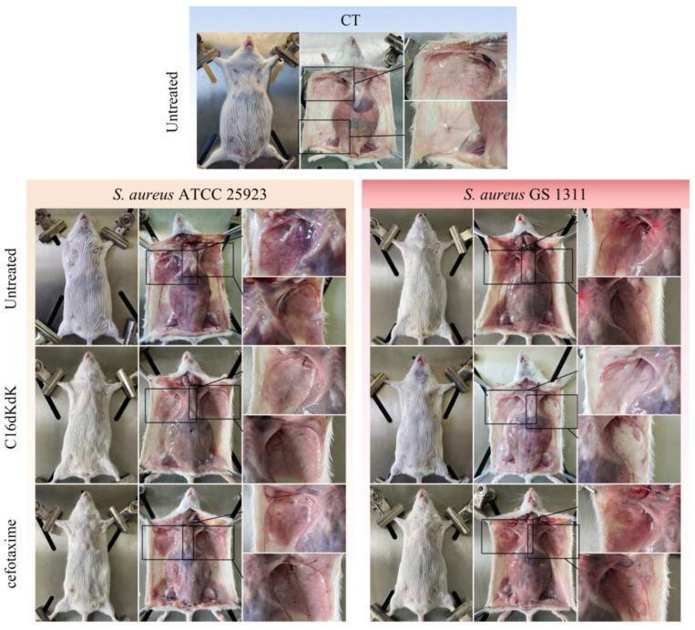
This clinical photograph panel illustrates mammary gland pathology and treatment efficacy in a mouse model of Staphylococcus aureus-induced mastitis. The visual is organized into a healthy control (CT) group and two infection groups (S. aureus ATCC 25923 and S. aureus GS 1311), further subdivided by treatment status: Untreated, C16dKdK (synthetic lipopeptide), and cefotaxime (antibiotic). Each row displays the external appearance in the first column and internal mammary tissue pathology in the second and third (magnified) columns. Untreated infected groups demonstrate clinical signs of acute mastitis, including external redness and swelling, and internal tissue congestion with prominent hemorrhage. Comparative analysis shows that mice treated with C16dKdK exhibit a significant reduction in mammary tissue congestion and absence of visible bleeding, appearing similar to the healthy control. In contrast, the cefotaxime-treated group shows partial recovery but retains areas of persistent congestion and pitting bleeding. The image serves as a visual demonstration of the comparative therapeutic efficacy between novel lipopeptides and traditional antibiotics in resolving infectious inflammation of the mammary glands.
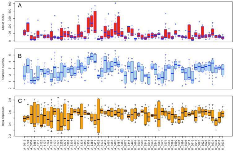
This image consists of three stacked boxplots (A, B, and C) illustrating the ecological diversity of the bovine udder microbiota across individual subjects. The x-axis identifies individual cows, categorized by farm origin (Prefix A or K). Panel A displays the Chao1 index, an estimator of species richness, showing significant variation between individuals with values ranging from near 0 to over 400. Panel B presents the Shannon diversity index, measuring both richness and evenness, with most values concentrated between 1 and 5. Panel C shows the multivariate homogeneity of group dispersions (beta dispersion) based on the Bray–Curtis dissimilarity matrix, reflecting the stability and variation of the microbial community within each cow's quarters. Values here are primarily between 0.4 and 0.8. Collectively, these plots demonstrate the inter-individual variability in microbial composition in the context of veterinary clinical research and mastitis studies, highlighting how richness and diversity are distributed at the individual host level.
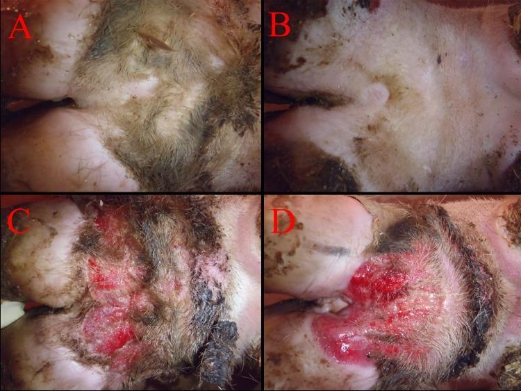
This set of four clinical photographs demonstrates the comparative pathology of digital dermatitis (DD) on the pastern and interdigital region of bovine feet, serving as a model for infectious skin lesions. Panels A and B display control subjects with normal clinical findings: the skin is intact, pale pink to white in color, and possesses a smooth, continuous texture with a normal, undisturbed hair coat. In contrast, panels C and D illustrate severe induced lesions (score 10). These lesions are characterized by marked erythematous inflammation, focal ulceration, and the presence of granular, raw tissue. The affected areas show distinct signs of exudative discharge, causing matted and discolored hair at the periphery. The lesions exhibit irregular borders and apparent tissue depth, consistent with high-grade inflammation and vascular involvement typical of active digital dermatitis. This comparison is used in dermatology and veterinary medicine to teach the visual identification, macroscopic scoring, and clinical manifestation of acute ulcerated skin conditions.
https://www.merckvetmanual.com/reproductive-system/mastitis-…
bovine mastitis treatment intramammary antibiotics
California mastitis test CMT strip cup milk somatic cell count
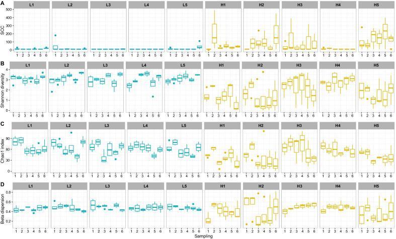
A multi-panel scientific figure displaying four rows of comparative boxplots (A–D) representing longitudinal udder microbiota data from 10 cows across six samplings. The cows are categorized into two groups: Low Somatic Cell Count (L-cows, L1–L5, shown in blue) and High Somatic Cell Count (H-cows, H1–H5, shown in yellow). Row A illustrates Somatic Cell Count (SCC), where L-cows demonstrate consistently low, stable levels while H-cows exhibit significantly elevated and fluctuating SCC values. Row B (Shannon diversity) and Row C (Chao1 index) represent alpha diversity metrics; L-cows show higher species evenness and richness with narrower interquartile ranges compared to the H-cows, which display lower diversity and higher variability. Row D shows 'Multivariate homogeneity of group dispersions' (beta dispersion), indicating that L-cows maintain a more stable microbial community over time, whereas H-cows exhibit greater dispersion and temporal instability. The data illustrates the relationship between inflammatory markers (SCC) and the health/stability of the bovine mammary microbiome, used here as a comparative model for infection and dysbiosis.
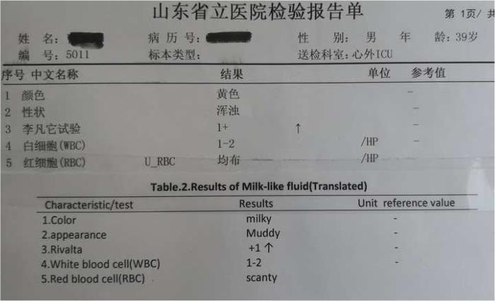
A clinical laboratory report document presented in both original Chinese and translated English, detailing a 'milk-like fluid analysis'. The report header identifies the patient as a 39-year-old male from a Cardiac Surgery Intensive Care Unit (ICU). The data table presents five key parameters: Color (milky/yellow), Appearance (muddy/turbid), Rivalta test (result of +1, indicating the presence of serous exudate protein), White blood cell (WBC) count (1-2 per high-power field), and Red blood cell (RBC) count (described as scanty or homogeneous). This fluid was obtained clinically during a pericardiectomy for localized chronic constrictive pericarditis (CP). The laboratory findings help characterize the fluid gush (chylopericardium vs. inflammatory exudate) encountered during the surgical decompression of the calcified pericardium.
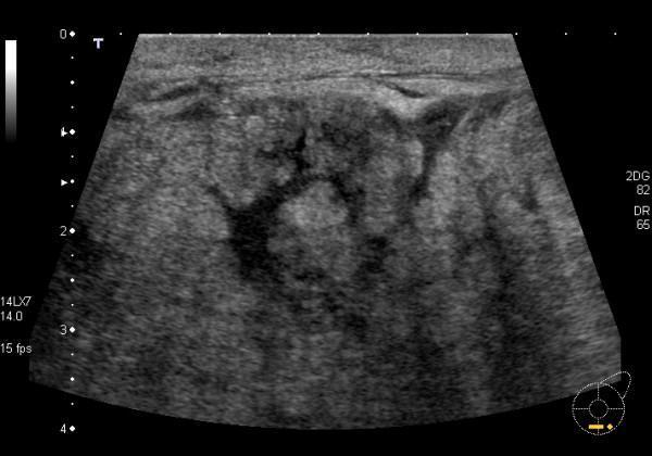
This diagnostic ultrasound image demonstrates the classic sonographic features of acute mastitis in a lactating breast. The image shows prominent skin thickening and increased hyperechogenicity of the subcutaneous tissues. The underlying breast parenchyma exhibits a markedly echogenic and heterogeneous appearance, resulting in a loss of the normal ultrasonic distinction between glandular and adipose tissues. Specifically, dilated, non-compressible milk ducts are visible, appearing as hypoechoic tubular structures amidst the dense stroma. Furthermore, visible skin lymphatics and interstitial fluid signify active inflammation and edema. These features—parenchymal echogenicity, ductal distension, and cutaneous changes—are key educational indicators used to differentiate inflammatory processes from localized abscesses or normal lactational changes in a clinical setting.
Mastitis in Veterinary Medicine - Comprehensive 4th Year Guide
1. DEFINITION
2. ETIOLOGY (Causative Agents)
A. Bacterial Causes (Most Common - >137 species documented)
| Category | Organisms | Type |
|---|---|---|
| Gram-positive cocci | Staphylococcus aureus (major), CoNS, Streptococcus agalactiae, S. uberis, S. dysgalactiae | Contagious & Environmental |
| Gram-negative bacilli | E. coli, Klebsiella spp., Enterobacter spp. (Coliforms) | Environmental |
| Others | Mycoplasma bovis, Corynebacterium bovis, Arcanobacterium pyogenes, Bacillus spp. | Contagious/Environmental |
- S. agalactiae - obligate intramammary pathogen; lives only in the udder; highly contagious
- S. aureus - most difficult to treat due to intracellular survival, biofilm formation, and AMR
- Coliforms - environmental, peak at periparturient period; cause acute/peracute mastitis
B. Fungal / Mycotic Causes
- Candida spp., Aspergillus fumigatus, Prototheca zopfii (algal mastitis)
- Typically follow prolonged or improper antibiotic use; treatment not generally successful
C. Viral Causes
- BHV-1, BHV-4, FMD virus, Parainfluenza-3, Cowpox, Pseudocowpox
- Bovine viral diarrhea virus (BVDV), BIV, BLV act indirectly via immunosuppression
3. PREDISPOSING FACTORS
- Cow-level: Poor teat end condition, parity (older cows), stage of lactation, dry period, periparturient immunosuppression
- Machine-related: Faulty milking machine (irregular vacuum, liner slippage), improper milking technique
- Environmental: Poor hygiene, wet/dirty bedding, overcrowding
- Managerial: No teat dipping, poor dry cow therapy, failure to cull chronic cases
4. PATHOGENESIS
- Entry - Pathogens enter via the teat canal (streak canal). The teat sphincter and keratin lining are the first defense barriers.
- Colonization - Bacteria adhere to mammary epithelium, multiply in milk cistern and alveolar spaces.
- Toxin release - Bacterial toxins (hemolysins, leukotoxins, endotoxins in coliforms) damage epithelium.
- Inflammatory cascade - Lipopolysaccharide (LPS from gram-negatives) triggers IL-1, IL-6, TNF-α release. Prostaglandins and arachidonic acid metabolites cause vasodilation, increased vascular permeability.
- Leukocyte infiltration - Massive influx of neutrophils into milk → somatic cell count (SCC) rises sharply (normal <200,000 cells/mL; mastitis >500,000 cells/mL).
- Tissue damage - Neutrophil-derived reactive oxygen species and proteases destroy alveolar epithelium, reduce milk synthesis.
- Fibrosis - Chronic infections lead to irreversible fibrosis, reduced secretory tissue, and permanent agalactia.
5. CLASSIFICATION
A. Based on Clinical Presentation
| Type | Features |
|---|---|
| Peracute | Sudden onset, severe udder edema/pain/heat, high fever (40-41°C), systemic toxemia, recumbency, can be fatal - classic with coliforms |
| Acute | Sudden onset, marked udder changes, altered milk, fever, reduced appetite/milk yield |
| Subacute | Mild udder changes, slightly altered milk (flakes, clots), no fever - common with S. uberis |
| Chronic | Recurrent flare-ups, fibrosis, hardening of quarters, persistent elevated SCC - classic with S. aureus |
| Subclinical | No visible changes; only elevated SCC and bacteriological changes in milk - most economically significant as it is invisible |
B. Based on Etiology
- Bacterial, Fungal/Mycotic, Viral, Algal (Prototheca)
C. Histopathological Classification
D. Based on Pathogen Ecology
- Contagious (cow-to-cow spread, primarily at milking): S. aureus, S. agalactiae, Mycoplasma bovis
- Environmental (from cow's environment): Coliforms, S. uberis, S. dysgalactiae
6. CLINICAL SIGNS
Local Signs (Udder/Milk)
- Udder: Swelling, heat, pain, redness, induration, atrophy (chronic)
- Milk changes: Watery/serous milk, clots, flakes, blood-tinged milk, altered color/consistency
- Quarter asymmetry: Atrophy of chronic quarters visible at inspection
Systemic Signs (Peracute/Acute)
- Pyrexia (40-41.5°C)
- Anorexia, depression
- Reduced rumen motility
- Tachycardia, tachypnea
- Dehydration, recumbency in severe cases
- Endotoxic shock (coliform mastitis)
7. DIAGNOSIS
A. Physical Examination
- Strip cup test: First 3-4 streams of milk into a dark-bottomed cup; check for clots, flakes, changed consistency - quick, bedside test
- Forestripping: Done before every milking; reveals subclinical changes
B. California Mastitis Test (CMT) - Most Practical Field Test
- Principle: Detects elevated SCC. The reagent (anionic detergent) lyses leukocyte cell membranes, releasing DNA which forms a gel.
- Scoring:
| Score | Reaction | SCC (cells/mL) | Interpretation |
|---|---|---|---|
| 0 (Negative) | No precipitation | <200,000 | Normal |
| Trace | Slight precipitation, disappears | 150,000-500,000 | Suspicious |
| 1+ | Distinct precipitation, no gel | 400,000-1,500,000 | Subclinical |
| 2+ | Gel forms, moves to center | 800,000-5,000,000 | Clinical |
| 3+ | Thick gel, does not flow | >5,000,000 | Severely infected |
C. Milk Culture & Sensitivity (Gold Standard)
- Aseptically collected milk sample → bacterial culture
- Identifies causative organism → guides antibiotic selection
- Gram stain for rapid preliminary identification
D. Somatic Cell Count (SCC)
- Electronic cell counting (Fossomatic)
- Bulk tank SCC: <200,000 cells/mL = acceptable; >400,000 = problem herd
- Individual cow SCC >200,000 suggests subclinical mastitis
E. Other Tests
- Whiteside test: NaOH-based field test (less popular)
- Surf field mastitis test: Modified CMT
- Bromothymol blue test: pH-based test
- Electrical conductivity: Automated inline mastitis detection
- Ultrasound: Identifies abscesses, fibrotic areas in chronic mastitis
- Histopathology: Biopsy for chronic/atypical cases
8. TREATMENT - Practical Protocol
Principles of Treatment
- Identify causative organism whenever possible
- Begin treatment early for best outcomes
- Choose appropriate drug, dose, route, and duration
- Follow milk/meat withdrawal times
- Support therapy for systemic cases
A. Intramammary (IMM) Therapy - First Line
| Drug | Product Example | Use |
|---|---|---|
| Penicillin G + Novobiocin | MastiCillin, Albadry | Gram-positive (Strep, Staph) |
| Amoxicillin | Amoxicillin IMM | Gram-positive, broad spectrum |
| Cloxacillin | Orbenin, Cloxapen | S. aureus (beta-lactamase stable) |
| Cefquinome | Cobactan IMM | Broad spectrum, 3rd gen cephalosporin |
| Pirlimycin | Pirsue | S. aureus, Streptococci |
| Ceftiofur | Spectramast LC/DC | Broad spectrum, short withdrawal |
- Strip quarter completely (empty milk)
- Clean and disinfect teat end with alcohol swab
- Partially insert cannula (partial insertion reduces teat canal trauma)
- Infuse product
- Dip teat with post-dip solution
- Note: Teat canal massage toward cistern helps drug distribution
B. Systemic (Parenteral) Therapy
| Drug | Dose & Route | Indication |
|---|---|---|
| Penicillin G procaine | 22,000 IU/kg IM BID | Gram-positive mastitis |
| Ampicillin | 5-10 mg/kg IM/IV BID | Broad spectrum |
| Ceftiofur sodium | 2.2 mg/kg IM SID | Broad spectrum, coliform mastitis |
| Enrofloxacin | 5 mg/kg SC SID (cattle) | Gram-negative, mycoplasma |
| Tetracycline | 10-20 mg/kg IV SID | Mycoplasma mastitis |
| Gentamicin | 4-6 mg/kg IV/IM SID | Gram-negative coliform (extra-label use) |
C. Anti-Inflammatory / Supportive Therapy (Critical for Peracute Cases)
| Drug | Dose | Purpose |
|---|---|---|
| Flunixin meglumine | 2.2 mg/kg IV/IM SID | NSAID of choice for endotoxemia; inhibits PGE2, reduces fever, pain |
| Meloxicam | 0.5 mg/kg IV/SC | Selective COX-2 inhibitor, good tissue penetration |
| Dexamethasone | 0.02-0.04 mg/kg IM | Anti-inflammatory for severe cases (caution - immunosuppressive, can trigger abortion) |
| IV fluids | Hartmann's/LRS 20-40 L | Dehydration, endotoxic shock (coliform mastitis) |
| Oxytocin | 20 IU IV/IM | Promotes milk let-down and drainage, flush out bacteria |
| Vitamin C | 10-30 g IV | Antioxidant support, especially useful in coliform mastitis |
| Calcium borogluconate | 400 mL IV | If concurrent milk fever/hypocalcemia (common periparturient) |
D. Dry Cow Therapy (DCT) - Most Economically Important Prevention Strategy
- Treat ALL quarters with long-acting IMM antibiotic at dry-off
- Teat sealants (Orbeseal, Teatseal) provide physical barrier
| Drug | Product | Duration |
|---|---|---|
| Cloxacillin benzathine | Orbenin DC | 8-12 weeks |
| Ampicillin + Cloxacillin | Ampiclox DC | 6-8 weeks |
| Cefquinome | Cobactan DC | 6-8 weeks |
E. Treatment Outcomes by Pathogen
| Pathogen | Prognosis | Comments |
|---|---|---|
| S. agalactiae | Excellent | Almost 100% cure with penicillin |
| Streptococcus spp. | Good | 70-80% cure |
| S. aureus | Poor-Fair | <40% bacteriological cure; biofilm, intracellular survival |
| E. coli | Variable | Self-limiting mild cases; severe/peracute has 30-50% death/cull rate |
| Klebsiella | Poor | Worst prognosis among coliforms; 2x more likely to die/cull |
| Mycoplasma bovis | Very poor | Essentially untreatable; cull affected cows |
| Fungal/Prototheca | Very poor | Treat with iodine infusion; cull is usually recommended |
F. When NOT to Treat (Selective Dry Therapy / Culling)
- Chronic S. aureus mastitis (>3 clinical episodes)
- Mycoplasma bovis positive (risk to whole herd)
- Prototheca/fungal mastitis
-
3 quarters affected chronically
- Non-productive or low-value animals
9. PREVENTION AND CONTROL - The 5-Point Plan (National Mastitis Council)
- Proper milking hygiene: Pre-dip, dry with individual towels, post-dip (iodophor 0.5% or chlorhexidine 0.5%)
- Milking machine maintenance: Regular servicing, correct vacuum levels (40-50 kPa), liner replacement
- Dry cow therapy: Treat ALL cows at drying off
- Cull chronic cases: Eliminate reservoir animals
- Good record keeping: Monitor SCC trends, identify problem animals early
- Pre-dip: 30-second contact time, then dry
- Post-dip: Apply immediately after unit removal, 2/3 teat coverage
10. ECONOMIC SIGNIFICANCE
- A single clinical mastitis case costs approximately $400-$500 USD (treatment + milk loss + labor)
- Subclinical mastitis reduces milk yield by 10-25% per affected quarter
- SCC >400,000 cells/mL results in milk rejection/price penalties
- Mastitis is responsible for ~17% of dairy cow cullings
11. MASTITIS IN OTHER SPECIES
| Species | Common Cause | Notes |
|---|---|---|
| Buffalo | Similar to cattle; S. aureus, Streptococcus | Higher prevalence of subclinical form |
| Goat | Staphylococcus aureus, Caprine arthritis encephalitis virus (CAEV) | CAEV causes chronic indurative mastitis |
| Sheep | Mannheimia haemolytica, Staphylococcus | Can be peracute gangrenous |
| Mare (Horse) | Streptococcus zooepidemicus, S. aureus | Uncommon; usually at weaning |
| Sow (Pig) | MMA syndrome (E. coli, Klebsiella) | Mastitis-Metritis-Agalactia complex postpartum |
12. KEY EXAM POINTS (High-Yield for 4th Year)
- CMT is the most practical field-side screening test for subclinical mastitis
- Bulk tank SCC is the most important herd-level monitoring tool
- S. agalactiae is the most sensitive to penicillin and nearly 100% curable
- S. aureus is the hardest to treat due to intracellular survival and biofilm
- Coliform mastitis is the most dangerous (endotoxemia, shock, death)
- Oxytocin + frequent milking is a key adjunct treatment
- Flunixin meglumine is the NSAID of choice for endotoxemia in mastitis
- Dry cow therapy is the single most effective mastitis control strategy
- Mycoplasma mastitis - no cell wall, beta-lactams useless, cull recommended
- The teat canal (streak canal) is the primary defense against intramammary infection
- SCC normal = <200,000 cells/mL in individual cows
- Prognosis: S. agalactiae > Streptococcus > S. aureus > Coliforms > Klebsiella > Mycoplasma/Fungal (in order of decreasing treatment success)
13. RECENT EVIDENCE (PubMed 2023-2025)
- A meta-analysis on S. aureus AMR in Iran (PMID: 37302281) confirmed high-level antibiotic resistance, reinforcing the need for culture-guided therapy before treatment.
- A systematic review on S. uberis AMR (PMID: 37844492) highlights increasing resistance in environmental streptococci, justifying sensitivity testing.
- A 2025 network meta-analysis on dry-off interventions (PMID: 40073588) found that combining internal teat sealants with antibiotics at dry-off gives the best protection against new intramammary infections.
Quick Reference Diagram: Pathogenesis Flow
Teat canal penetration
        ↓
Bacterial colonization in milk cistern
        ↓
Toxin/LPS release
        ↓
Cytokine cascade (IL-1, IL-6, TNF-α)
        ↓
Neutrophil infiltration → ↑SCC
        ↓
Epithelial damage + ↓milk production
        ↓
Chronic: Fibrosis → Agalactia
Acute: Systemic SIRS → Endotoxemia → Shock
Glander details explanation I want hindenglish language for batter understand core of topic
Glanders disease Burkholderia mallei veterinary pathogenesis clinical signs treatment diagnosis horses
Glanders horse nasal discharge nodules skin farcy Burkholderia mallei
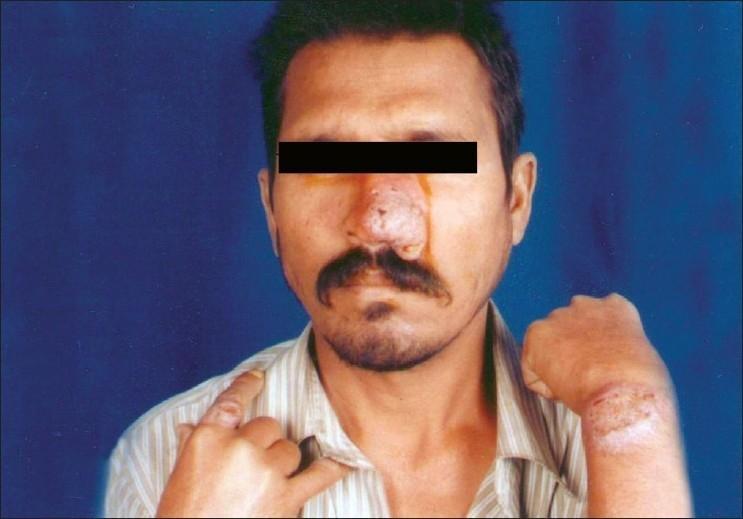
Content Type: Clinical Photograph. This image displays multiple skin lesions on a 45-year-old male. The primary visual finding is a large, infiltrated, nodular plaque covering the majority of the nasal bridge and tip, appearing raised with areas of surface irregularity and mild serosanguinous discharge. Secondary lesions include a well-demarcated, hypopigmented, and scaly plaque on the dorsum of the left wrist and a similar infiltrated nodule on the right index finger. Medical Context: These clinical manifestations are consistent with Post-Kala-azar Dermal Leishmaniasis (PKDL) in an HIV-positive patient, characterized by disseminated nodules and plaques containing Leishman-Donovan bodies. Educational Significance: The photograph illustrates the atypical and more aggressive presentation of cutaneous leishmaniasis in immunocompromised individuals. Target Audience: Dermatologists, infectious disease specialists, and medical students. Keywords: Infiltrated plaque, nodular lesion, hypopigmentation, nasal lesion, cutaneous leishmaniasis, HIV-coinfection, tropical medicine.
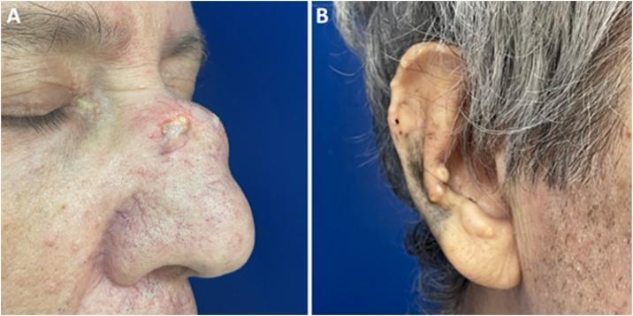
This clinical photograph consists of two panels illustrating cutaneous manifestations of tophaceous gout in a 62-year-old patient. Panel A displays the nasal region in a profile view, featuring a large, firm, erythematous nodule on the nasal dorsum. The lesion is notable for its central ulceration and yellowish-white amorphous discharge, characteristic of urate crystal deposits (tophus). Surrounding skin shows telangiectasia and smaller satellite papules. Panel B provides a lateral view of the right external ear, demonstrating multiple small, firm, yellowish-white papules and nodules along the helix and antihelix. These lesions are discrete, non-ulcerated, and represent classical gouty tophi in a common anatomical site. The image serves as an educational reference for identifying extra-articular manifestations of chronic hyperuricemia and the varied morphology of tophi, ranging from small auricular papules to large, ulcerating facial nodules.
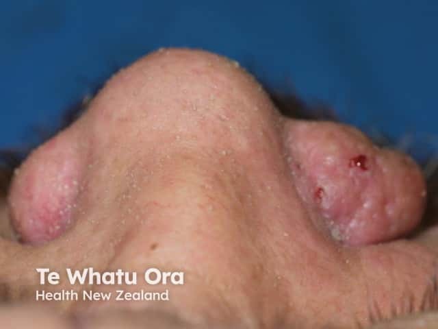
Clinical photography of the nasal and perinasal facial skin in visible light, captured as a close‑up frontal view. The image depicts bilateral perinasal/nasal skin with raised pink to erythematous nodules on the nasal ala and adjacent cheek. Lesions are firm, well circumscribed but with irregular, possibly ulcerated surfaces and focal crusting. The surrounding skin appears mildly scarred and sun‑exposed, consistent with chronic actinic damage. The right nasal lesion shows a slightly larger, rounded dome shaped nodule with an attenuated pearly/pink appearance and subtle surface erosion; the left lesion is smaller and less well defined. No deep tissue involvement is ascertainable from this external view. There is no dermatoscopic detail. The clinical significance is high: new or enlarging cutaneous nodules on the nose region raise suspicion for malignant skin neoplasms such as basal cell carcinoma or squamous cell carcinoma, though benign mimics (sebaceous hyperplasia, keratoacanthoma) are possible. The image is valuable for educational purposes, differential diagnosis discussion, and biopsy planning. Potential clinical use cases include triage in primary care, dermatology assessment, radiology correlation for facial skin cancer staging, and teaching of lesion morphology. This representation aids pattern recognition and correlates with sun‑related skin cancer risk and perinasal oncologic workup and management.
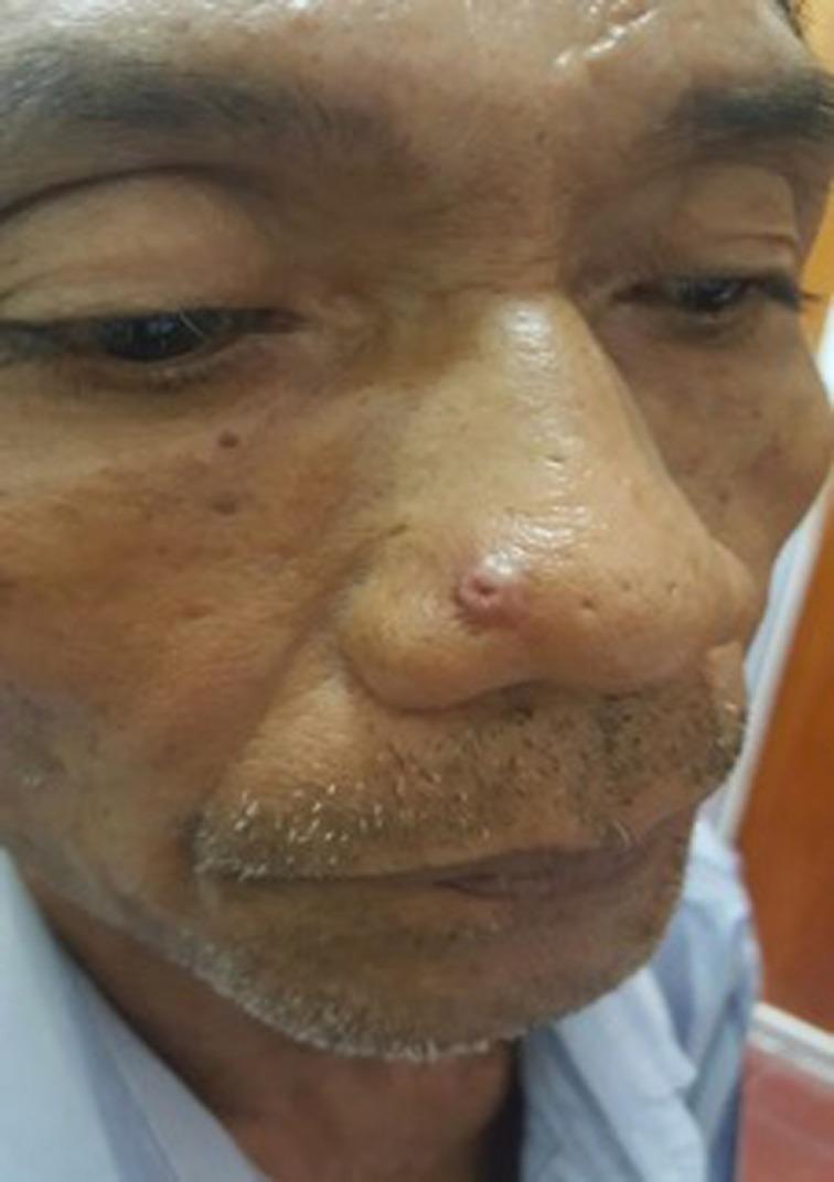
This clinical photograph shows a close-up frontal view of a patient's face, focusing on a solitary, erythematous nodule located on the right nasal sidewall. The primary lesion is approximately 0.5 cm in diameter, well-demarcated, and exhibits a characteristic central ulceration or crateriform appearance. The surrounding nasal skin shows mild erythema and several smaller, non-specific pigmented macules. The background facial skin displays signs of solar elastosis and actinic damage, including uneven texture and dyschromia. This visual presentation is clinically significant for the differential diagnosis of cutaneous neoplasms, particularly keratoacanthoma or basal cell carcinoma, though the pathological diagnosis in this specific case is angiolymphoid hyperplasia with eosinophilia (ALHE). The image serves as an educational resource for identifying umbilicated or ulcerated skin nodules on sun-exposed facial areas in dermatology and oncology curricula.
https://www.merckvetmanual.com/generalized-conditions/glande…
"Burkholderia mallei" glanders diagnosis treatment
🐴 GLANDERS (Ander) - Complete Veterinary Guide
Hinglish mein - 4th Year ke liye
1. DEFINITION (Parichay)
- Caused by: Burkholderia mallei (pehle Pseudomonas mallei kehte the)
- Iska skin form "FARCY" kehlaata hai
- India mein yeh Notifiable Disease hai - matlab case hone par government ko report karna compulsory hai
- OIE (World Organisation for Animal Health) listed disease hai
Yaad rakho: Glanders = Respiratory + Nasal + Skin nodules wali disease in horses. Farcy = Sirf skin/lymphatic form ka naam.
2. CAUSATIVE AGENT (Kaaran)
| Property | Detail |
|---|---|
| Organism | Burkholderia mallei |
| Gram stain | Gram-NEGATIVE bacillus (rod shape) |
| Motility | NON-MOTILE (non-motile hona iska distinct feature hai) |
| Oxygen | Strictly aerobic (oxygen chahiye) |
| Intracellular | Facultative intracellular parasite (cell ke andar bhi survive karta hai) |
| Origin | Pathoadaptive clone hai B. pseudomallei se |
| Survival | Direct sunlight, desiccation, aur common disinfectants se kill ho jaata hai |
| Bioterrorism | B. mallei ko potential bioterrorism agent classify kiya gaya hai kyunki untreated septicemia mein 40-95% mortality humans mein |
- Pyocyanin - electron transfer interfere karta hai
- Lecithinase - cell lysis karta hai
- Collagenase - connective tissue destroy karta hai
- Lipase & Hemolysin - tissue damage
3. HOST RANGE (Kaun kaun bimaar hota hai)
| Species | Susceptibility |
|---|---|
| Mule (Khacchar) | Sabse zyada susceptible - Peracute, rapid death |
| Donkey (Gadha) | Highly susceptible - usually fatal, acute form |
| Horse (Ghoda) | Relatively resistant - Chronic form (farcy) develop karta hai, carrier ban sakta hai |
| Camel, Tiger, Lion | Uncommon but usually fatal if infected |
| Human | Zoonotic infection possible - occupational risk |
| Cattle, Pig, Dog | Natural resistance hoti hai |
Trick: "MD Fatally Die" - Mule = Die fastest. Horse = chronic survivor.
4. TRANSMISSION (Failna kaise hai)
Infected animal
       ↓
Nasal discharge / Skin secretions / Pus
       ↓
Direct contact  ←→  Contaminated feed/water/trough
       ↓
Entry via:
  → Nasal mucosa (sabse common)
  → Skin abrasion/wounds
  → Mouth (ingestion)
  → Aerosol (laboratory mein)
- Contagious spread milking time pe ya shared water/feed troughs se hota hai
- Carrier horses (occult glanders) - jo dikhne mein normal lagte hain but infected hain - sabse dangerous source of spread
- Infected stallion → breeding ke time mein spread
5. PATHOGENESIS (Andar kya hota hai)
- Pehle firm hota hai
- Phir soften hota hai (liquefaction necrosis)
- Phir rupture ho jaata hai
- Rupture ke baad ulcer banta hai - ulcer se honey-like discharge aata hai
- Lymphatic vessels mein beads jaisi nodules banti hain = "Farcy buds"
- Lymphatic vessels thickened, cord-like ho jaate hain = "Farcy pipes"
- Multiple organ nodules
- Septicemia → Multi-organ failure → Death
6. FORMS / CLASSIFICATION (Kitni tarah ka hota hai)
A. NASAL FORM (Naak waala form)
- Sabse common
- Unilateral ya bilateral nasal discharge - pehle watery, phir mucopurulent (thick yellowish-green)
- Nasal mucosa par nodules bante hain → rupture → ulcers → heal ho ke stellate scars (taare jaisi daag) banti hain
- Nasal septum perforation ho sakti hai
- Submaxillary lymph nodes enlarge ho jaate hain - ek taraf (rope jaisa, hard)
B. PULMONARY FORM (Phepde waala form)
- Bacteria inhale hone se
- Chronic dry cough, labored breathing, weight loss
- Lungs mein nodules bante hain jo necrotic (dead tissue) ho jaate hain
- X-ray: Multiple nodular lesions in lungs
- Usually chronic course - weeks to months
C. CUTANEOUS FORM = FARCY (Chara ki bimari)
- Skin aur lymphatics involve hoti hain
- Farcy buds: Skin aur subcutaneous tissue mein nodules along lymphatic vessels
- Farcy pipes: Lymphatic vessels thickened, cord jaisi feel hoti hain (palpate kar sakte ho)
- Nodules rupture ho ke ulcers banaate hain with honey/oily discharge
- Mostly medial aspect of hind limbs aur neck par
D. ACUTE SEPTICEMIC FORM
- Donkeys aur mules mein sabse common
- High fever (40-41°C), complete anorexia, severe depression
- Rapid deterioration → Death within 3-4 days
- Nodules dikhne ka time bhi nahi milta
E. CHRONIC FORM
- Horses mein
- Waxing and waning course - kabhi theek lagta hai kabhi bura
- Months to years tak chal sakta hai
- Carrier state possible - Occult Glanders
7. CLINICAL SIGNS - Quick Summary Table
| Sign | Form |
|---|---|
| Thick yellowish-green nasal discharge | Nasal |
| Ulcers with stellate scars in nasal mucosa | Nasal |
| Hard, rope-like submaxillary lymph nodes | Nasal |
| Cough, difficulty breathing, weight loss | Pulmonary |
| Skin nodules along lymphatics (farcy buds) | Cutaneous/Farcy |
| Cord-like thickened lymphatics (farcy pipes) | Cutaneous/Farcy |
| High fever, rapid death | Acute/Septicemic |
| Emaciation, debilitation, chronic course | Chronic |
8. DIAGNOSIS (Pehchaanna kaise hai)
A. CLINICAL DIAGNOSIS (Dekhke)
- Nasal discharge + nasal ulcers + stellate scars = strongly suspect glanders
- Farcy buds along medial hind limb lymphatics
- Submaxillary lymph node enlargement
Caution: Occult glanders (carrier animals) mein koi sign nahi hote - tabhi specific tests important hain
B. MALLEIN TEST - Most Important Practical Diagnostic Test ⭐
| Type | Method | Reading |
|---|---|---|
| Ophthalmic (Intrapalpebral) - Gold Standard | 0.1 mL mallein intradermal injection in lower eyelid | After 24-48 hours: Positive = Purulent conjunctivitis + eyelid swelling |
| Subcutaneous | 2-3 mL mallein SC in neck | Positive = Fever + local swelling + general reaction within 8-24 hrs |
| Intradermal | Small dose intradermal in neck skin | Swelling >35mm = positive |
C. SEROLOGICAL TESTS
| Test | Details |
|---|---|
| CFT (Complement Fixation Test) | Standard OIE prescribed test; most widely used; Official test for international trade |
| ELISA | More sensitive than CFT; good for screening |
| Rose Bengal Plate Test | Quick screening |
| Indirect Hemagglutination Test | Also used |
| Luminex bead-based test | Modern, high throughput |
D. BACTERIOLOGICAL CULTURE
- Sample: Nasal discharge, pus from nodules/ulcers
- Culture on Nutrient agar, MacConkey agar
- BSL-3 laboratory required (bahut dangerous organism hai - biosafety Level 3 chahiye)
- Confirmatory diagnosis
E. PCR
- Most sensitive and specific
- B. mallei-specific gene targets detect karta hai
- Quick result
F. DIFFERENTIAL DIAGNOSIS (Kya kya lagta hai par hai nahi)
| Disease | How to Differentiate |
|---|---|
| Strangles (Streptococcus equi) | Lymph node abscess, no nasal ulcers, mallein test negative |
| Melioidosis (B. pseudomallei) | Soil-borne organism, PCR differentiation |
| Epizootic Lymphangitis (Histoplasma farciminosum) | Fungal, slower progress, different culture result |
| Ulcerative Lymphangitis (Corynebacterium pseudotuberculosis) | Fetlock region mainly, different organism |
| Sporotrichosis | Fungal, no nasal involvement |
9. TREATMENT (Ilaaj)
Veterinary Treatment - IMPORTANT EXAM POINT ⭐
Horses mein treatment GENERALLY PROHIBITED AND CONTRAINDICATED hai!
- Zoonotic risk - infected horse ke paas rehna aur treat karna = human infection ka risk
- Treatment bacteriological cure reliably nahi deta
- Carrier state develop ho sakta hai - jo aur dangerous hai
- OIE and most countries ka protocol = CULL (euthanize) infected animals
Agar treatment karna hi pade (Research/Special cases)
| Drug | Route | Duration |
|---|---|---|
| Doxycycline | Oral | Long term (60-150 days) |
| TMP-SMX (Trimethoprim-Sulfadiazine) | Oral | Long term |
| Amoxicillin-Clavulanate | Oral | 60-150 days |
| Ciprofloxacin | - | Alternative |
| Streptomycin | - | Alternative |
| Meropenem | IV | Severe cases |
Human Treatment (Zoonosis mein)
- Same drugs as above
- Localized disease: TMP-SMX ya Doxycycline 60-150 days
- Severe/Septicemic: IV Imipenem/Meropenem + Doxycycline combination
10. CONTROL AND PREVENTION (Rokna kaise hai)
- Newly purchased animals
- Animals returning from travel
- Before equine events/shows
- Formalin 2% ya Phenol 5% ya Bleach effective hain
- B. mallei direct sunlight aur desiccation se bhi mar jaata hai
Vaccination
- Koi effective vaccine available NAHI hai presently
- Yeh ek major research gap hai (2025 review bhi yahi confirm karta hai - Torres AG, PLoS NTD 2025)
Movement Control
- Infected area se equid movement ban
- International import/export: CFT testing compulsory
11. ZOONOTIC IMPORTANCE (Insaanon ko kab hota hai)
- Risk group: Veterinarians, horse handlers, stable workers, slaughterhouse workers, laboratory personnel
- Entry route: Skin wounds/abrasions, nasal mucosa, eye, aerosol (lab mein)
- Human clinical forms: Localized cutaneous, pulmonary, chronic, septicemic
- Mortality: Untreated septicemic form mein 40-95% mortality
- Bioterrorism: B. mallei ko biological weapon ke roop mein World War I aur II mein use kiya gaya tha - officially bioterrorism agent listed hai
- PPE (gloves, mask, goggles) pehno infected animals handle karte waqt
- All samples BSL-3 lab mein process hon
- Any suspected human case immediately report karo
12. EPIDEMIOLOGY (Kahaan-Kahaan hai)
- India: Sporadic cases - notifiable disease under Prevention and Control of Infectious and Contagious Diseases in Animals Act 2009
- Endemic areas: Middle East (UAE, Bahrain), Pakistan, India, Brazil, Southeast Asia
- Eradicated areas: USA (last case 1934), Western Europe, Australia
- Reemerging: Bahrain, UAE mein recent outbreaks reported
13. EXAM KE LIYE KEY POINTS (High-Yield) ⭐⭐⭐
| Point | Answer |
|---|---|
| Causative agent | Burkholderia mallei |
| Gram stain | Gram-negative, non-motile bacillus |
| Most susceptible | Mule > Donkey > Horse |
| Horse mein form | Chronic / Farcy |
| Cutaneous form naam | FARCY |
| Skin nodules naam | Farcy buds |
| Thickened lymphatics naam | Farcy pipes |
| Most important diagnostic test | Mallein test (Intrapalpebral) |
| OIE prescribed serological test | CFT (Complement Fixation Test) |
| Treatment in horses | CONTRAINDICATED / PROHIBITED |
| Control method | Test and Cull |
| Vaccine available? | NO |
| Zoonosis? | YES - very dangerous |
| Bioterrorism potential? | YES |
| India mein status | Notifiable Disease |
| Differential for farcy | Epizootic lymphangitis, Ulcerative lymphangitis, Sporotrichosis |
| Stellate scars kahan banti hain | Nasal mucosa par (healed ulcers se) |
QUICK MEMORY TRICKS (Yaad karne ke tricks)
- "MALLEI = MULE KILLER" - Mules mein most fatal
- "FARCY = FAR from cure" - Chronic skin form, incurable in horses
- "No Vax, No Treat, Only Cull" - Glanders management mantra
- "B. mallei - NON-MOTILE" - Whereas B. pseudomallei MOTILE hai - difference yaad rakho
- "Mallein = Tuberculin of horses" - Same DTH principle, different organism
Recent Evidence: 2025 ka ek review (PMID: 40498787) confirm karta hai ki aaj bhi glanders ke liye koi approved vaccine ya definitive treatment nahi hai - yeh ek "ancient and emergent disease" hai. Aur 2025 mein ek paper (PMID: 41200364) glanders ka bioterrorism history cover karta hai jo exams mein zaroor poochha jaata hai.
Canine distemper detail explanation
canine distemper virus pathogenesis clinical signs diagnosis treatment vaccination veterinary
canine distemper hard pad hyperkeratosis nasal footpad dog inclusion bodies
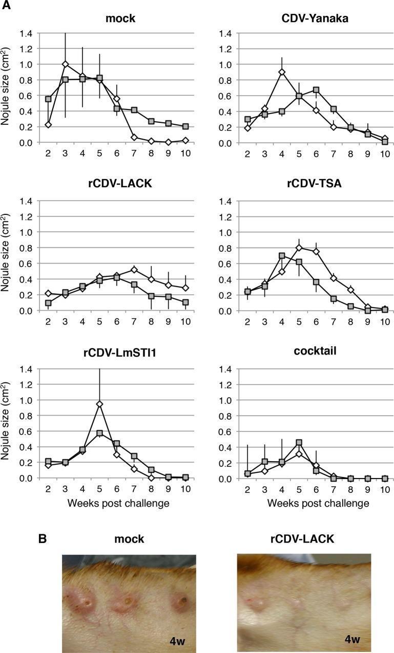
This figure demonstrates the protective efficacy of recombinant canine distemper virus (rCDV) vaccines against Leishmania major challenge in a canine model. Panel A consists of six line graphs plotting nodule size (cm²) over a 10-week post-challenge period across different vaccine groups: mock, CDV-Yanaka, rCDV-LACK, rCDV-TSA, rCDV-LmSTI1, and a cocktail vaccine. The mock group shows rapid nodule growth peaking between weeks 3–5, while the rCDV-LACK and cocktail groups demonstrate significant suppression of nodule development, with the cocktail group showing the fastest resolution. Panel B presents clinical photographs of canine ears at 4 weeks post-challenge. The mock-immunized dog exhibits multiple large, raised, inflamed, and centrally ulcerated nodules (crater-like lesions) typical of cutaneous leishmaniasis. In contrast, the rCDV-LACK-immunized dog shows significantly smaller, non-ulcerated, and less inflamed nodules. This comparison illustrates the immunogenic effect of LACK-expressing viral vectors in reducing clinical disease severity in cutaneous leishmaniasis models.
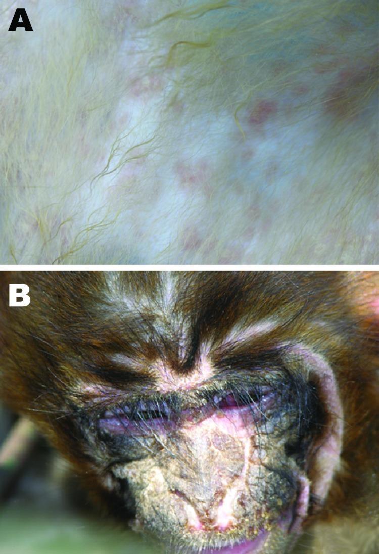
Two-panel clinical photograph illustrating the manifestations of Canine Distemper Virus (CDV) in a rhesus monkey specimen, serving as a non-human primate model for viral pathogenesis. Panel A shows a close-up of the integumentary system, demonstrating a maculopapular rash. The rash consists of discrete, rosy to purple macules approximately 2–4 mm in diameter, distributed across the pale skin beneath the fur. Panel B illustrates severe ocular and facial involvement. Notable findings include suppurative conjunctivitis with thick purulent exudate matted in the fur and eyelids, significant periorbital inflammation, and ulcerated, crusting lesions on the nasal bridge and perinasal skin. These clinical signs reflect the systemic and epithelial tropism of the virus, mimicking human measles-like symptoms. The image is used in medical pathology to highlight cross-species viral transmission and clinical diagnostic signs of paramyxovirus infections.
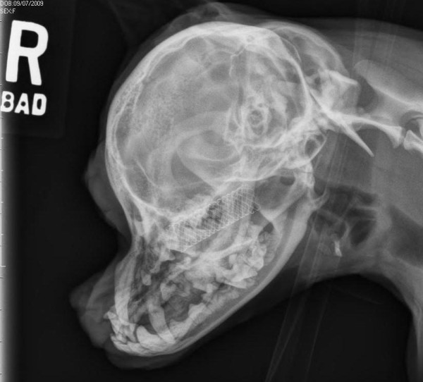
**Imaging Modality:** Veterinary diagnostic radiography (X-ray). **Anatomical Region:** Lateral view of a canine skull (brachycephalic conformation). **Observed Pathology/Intervention:** A radiopaque, metallic, self-expanding mesh stent is visible within the nasopharyngeal or nasal cavity region. The stent is cylindrical with a crisscross lattice pattern, positioned dorsal to the hard palate and extending caudally toward the nasopharynx. **Characteristic Visual Features:** * **Skeletal Structures:** The skull demonstrates characteristic brachycephalic features, including a shortened maxillary and nasal apparatus and a rounded calvarium. Dentition is visible with some crowding typical of the breed type. * **Device Positioning:** The stent appears well-expanded and maintains its structural integrity without evidence of migration, fracture, or collapse. It occupies the airway space, likely placed to address nasopharyngeal stenosis or upper airway obstruction. * **Soft Tissue/Airway:** The pharyngeal airway remains patent caudal to the stent. No significant periosteal reaction or gross bony lysis is noted in the immediate surrounding facial bones. **Key Diagnostic Features:** Presence of an intraluminal metallic prosthesis in the upper respiratory tract of a dog, confirming post-procedural monitoring of a nasopharyngeal stent.
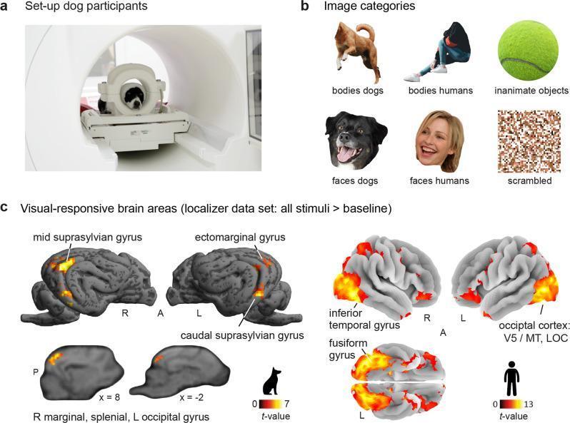
An educational infographic summarizing a comparative functional magnetic resonance imaging (fMRI) study on visual processing in dogs and humans. Panel A shows the experimental setup with a dog positioned inside a 3T MRI scanner using a custom-made head coil. Panel B illustrates the six stimulus categories: canine bodies, human bodies, inanimate objects (tennis ball), canine faces, human faces, and scrambled control images. Panel C displays statistical parametric maps of visual-responsive brain areas (all stimuli > baseline) projected onto species-specific cortical surfaces. In dogs, activation is localized to the mid suprasylvian gyrus, ectomarginal gyrus, caudal suprasylvian gyrus, and marginal/splenial regions, with t-values ranging from 0 to 7. In humans, activation is prominent in the inferior temporal gyrus, fusiform gyrus, and occipital cortex (including V5/MT and the lateral occipital cortex), with t-values ranging from 0 to 13. The image demonstrates the comparative neuroanatomy of the ventral visual stream and the specialization of higher-order sensory cortices across species for animate and inanimate stimulus perception.
https://www.merckvetmanual.com/infectious-diseases/canine-di…
"canine distemper virus" diagnosis treatment
"canine distemper" pathogenesis neurological vaccination
🐶 CANINE DISTEMPER - Complete Veterinary Guide
Hinglish mein - 4th Year ke liye
1. DEFINITION (Parichay)
- Worldwide distribution - sab jagah paaya jaata hai
- Unvaccinated puppies sabse zyada susceptible
- Measles (human) aur Rinderpest (cattle) ka close relative hai
- Multi-systemic disease - respiratory, GI, nervous system, skin sab affect hote hain
2. ETIOLOGY (Karan - Kaun karta hai bimaar?)
| Property | Detail |
|---|---|
| Virus | Canine Distemper Virus (CDV) |
| Family | Paramyxoviridae |
| Genus | Morbillivirus (measles, rinderpest isi genus mein) |
| Type | Enveloped, Single-stranded RNA virus (ss-RNA) |
| Shape | Pleomorphic (spherical to filamentous) |
| Stability | Fragile - environment mein jaldi mar jaata hai |
| Serotypes | Only 1 serotype (but multiple strains/lineages) |
| Inclusions | Both intranuclear AND intracytoplasmic inclusions banta hai (Morbillivirus ka characteristic) |
Key Protein Features:
- H Protein (Hemagglutinin) - Host cell se attachment karta hai (SLAM/nectin-4 receptors pe)
- F Protein (Fusion) - Cell membrane fusion karta hai; highly conserved
- No Neuraminidase - Morbillivirus mein neuraminidase ABSENT hoti hai (Respirovirus/Rubulavirus mein hoti hai) - Exam MCQ!
3. HOST RANGE (Kaun kaun bimaar hota hai)
| Family | Species |
|---|---|
| Canidae | Dog, Wolf, Fox, Jackal, Coyote |
| Mustelidae | Ferret (most susceptible!), Mink, Weasel, Badger, Otter |
| Procyonidae | Raccoon, Red Panda |
| Ursidae | Bears |
| Felidae | Lion, Tiger, Leopard (large cats) |
| Primates | Rhesus monkey (experimentally) |
| Hyaenidae | Hyena |
Important: Domestic cat ko CDV naturally affect nahi karta - woh resistant hain. Ferret sabse zyada susceptible hai.
4. TRANSMISSION (Failna kaise hai)
Infected dog
      ↓
Aerosol droplets (nasal/ocular discharge, saliva, urine, feces)
      ↓
Inhalation (MAIN ROUTE)
      ↓
Susceptible dog ke respiratory tract mein enter
- Primary route: Respiratory aerosols - khaansne, naak saaf karne se
- Virus environment mein sensitive hai - thodi si garmi/light se mar jaata hai
- Infected dog months tak virus shed kar sakta hai
- Fomites (kapde, utensils) se bhi spread possible
- Transplacental (pregnant female se puppy ko) - congenital distemper
5. PATHOGENESIS - Step by Step (Andar kya hota hai)
Phase 1: RESPIRATORY ENTRY (Days 1-4)
Virus inhale hota hai
       ↓
Upper respiratory tract epithelium se contact
       ↓
SLAM receptor (CD150) wale cells target hote hain
- Macrophages
- Lymphocytes (tonsils, bronchial lymph nodes)
       ↓
Local multiplication shuru
       ↓
VIREMIA (virus blood mein aa jaata hai)
Phase 2: LYMPHOID DEPLETION (Days 3-7)
Virus systemically spread hota hai
       ↓
Lymphoid organs attack hote hain:
- Spleen, Thymus, Lymph nodes, Bone marrow, Peyer's patches
       ↓
T & B lymphocytes destroy hote hain
       ↓
SEVERE IMMUNOSUPPRESSION
       ↓
FIRST FEVER peak (38.5-40°C) - Diphasic fever ka 1st phase
Phase 3: SECOND VIREMIA (Days 8-14)
2 outcomes possible based on immune response:

(A) STRONG immune response → Virus clear ho jaata hai → Recovery
           ↓
(B) WEAK immune response → Second viremia
           ↓
Virus epithelial cells, CNS, skin attack karta hai
via NECTIN-4 receptor (epithelial cells pe)
           ↓
SECOND FEVER peak - higher than first
Phase 4: ORGAN INVOLVEMENT (Days 10-30+)
Respiratory: Bronchitis, Pneumonia
        ↓
GI: Diarrhea, vomiting
        ↓
Skin: Pustular dermatitis, Footpad hardening
        ↓
CNS: Demyelination → Neurological signs
     (Eosinophilic plaques in white matter)
Diphasic Fever - Samjhao Examiners ko ⭐
Day 3-4:  1st fever (Virus lymphoid organs mein)
Day 5-6:  Fever thoda kam
Day 8-14: 2nd fever (Virus epithelial/CNS mein spread)
6. CLINICAL SIGNS - System-wise (Lakshan)
A. SYSTEMIC SIGNS
- Diphasic fever (38.5-40.5°C) - hallmark sign
- Depression, lethargy, anorexia
- Weight loss, cachexia
B. RESPIRATORY SIGNS
- Nasal discharge - pehle serous (paani jaisa), phir mucopurulent (thick, yellowish-green)
- Ocular discharge - bilateral, mucopurulent, conjunctivitis
- Coughing, sneezing
- Bronchopneumonia - labored breathing
- Secondary bacterial pneumonia (immunosuppression ki wajah se)
C. GASTROINTESTINAL SIGNS
- Vomiting
- Diarrhea (yellow/greenish, can be hemorrhagic)
- Dehydration
- Anorexia
D. NEUROLOGICAL SIGNS (Worst prognosis wala part) ⭐
| Sign | Description |
|---|---|
| Myoclonus | Repetitive muscle twitching/spasm - PATHOGNOMONIC sign |
| Chewing gum fits | Jaw chamchana jaisa movement - characteristic |
| Enamel hypoplasia | Puppy mein dant ki enamel kharab - CDV infection in utero/early life |
| Seizures | Focal ya generalized |
| Ataxia | Chalne mein lardkharna |
| Circling | Ek direction mein ghoomna |
| Nystagmus | Aankhon ki abnormal movement |
| Paraparesis/Paralysis | Hind limb weakness or complete paralysis |
| Old Dog Encephalitis | Years baad chronic progressive CNS disease |
Myoclonus (chamchane jaisa twitching) = CDV ka pathognomonic sign - Yeh kisi aur disease mein itna characteristic nahi hota
E. CUTANEOUS SIGNS - HARDPAD DISEASE
- Hyperkeratosis of footpads - Panje sख्त (hard) ho jaate hain - isliye "Hardpad Disease" kehte hain
- Hyperkeratosis of nasal planum - Naak ki tip bhi hard/crusty ho jaati hai
- Pustular dermatitis - Abdomen, inner thighs pe pustules (pus wale daane)
- Skin dry, scaly, flaky
F. OPHTHALMIC SIGNS
- Conjunctivitis, keratitis
- Optic neuritis - blindness
- Chorioretinitis
7. LESIONS (Pathological - Exam mein diagram/describe karo)
Gross Lesions:
- Lungs: Consolidation, grey-red discoloration (bronchopneumonia)
- Lymph nodes: Enlarged, hemorrhagic
- Brain: Congested, edematous
- Footpads: Hard, hyperkeratotic
- GI: Hemorrhagic enteritis
Microscopic/Histopathological Lesions: ⭐
| Finding | Location |
|---|---|
| Intranuclear + Intracytoplasmic inclusion bodies | Epithelial cells, neurons, astrocytes |
| Perivascular cuffing | CNS (lymphocytes surrounding blood vessels) |
| Demyelination | White matter of brain + spinal cord |
| Eosinophilic plaques | White matter (CDV antigen deposits) |
| Interstitial pneumonia | Lungs |
| Lymphoid depletion | Spleen, lymph nodes, thymus |
Key Fact: CDV Morbillivirus hai, isliye BOTH intranuclear AND intracytoplasmic inclusions bante hain. Measles bhi same karta hai. (Doosre Paramyxoviruses sirf cytoplasmic inclusions banate hain) - Important MCQ
8. DIAGNOSIS (Pehchaan kaise karein)
A. CLINICAL DIAGNOSIS
- Unvaccinated puppy/young dog
- Diphasic fever
- Multisystemic signs (respiratory + GI + neuro)
- Myoclonus (pathognomonic)
- Hardpad (hyperkeratotic footpads)
- Mucopurulent ocular + nasal discharge
B. LABORATORY DIAGNOSIS
| Test | Sample | Notes |
|---|---|---|
| RT-PCR | Nasal/conjunctival swab, blood, urine, CSF | Most sensitive and specific - Gold standard; detects viral RNA |
| ELISA | Blood (serum) | Antibody detection; may be negative early in disease |
| IFA (Immunofluorescence) | Blood smear, conjunctival scraping | CDV antigen detect karta hai in WBCs |
| Inclusion body detection | Conjunctival scraping, blood smear | Eosinophilic inclusion bodies in cells |
| Virus isolation | Difficult, specialized lab required | Not practical for routine |
| Histopathology | Post-mortem brain/lung tissue | Inclusion bodies, demyelination confirm |
C. HEMATOLOGY FINDINGS
- Lymphopenia (lymphocytes kam - immunosuppression)
- Leukopenia (early in disease)
- Thrombocytopenia
- Eosinophilic inclusion bodies in RBCs, WBCs ke andar
D. CSF Analysis (Neurological cases mein)
- Increased protein
- Pleocytosis (cells ka badhna)
- CDV-specific antibodies CSF mein
E. DIFFERENTIAL DIAGNOSIS (Yeh nahi hai, yeh hai)
| Condition | Kaise alag karein |
|---|---|
| Kennel Cough (Bordetella) | Sirf respiratory - no neurological signs, no fever |
| Parvovirus | Sirf GI (bloody diarrhea, severe vomiting), no neuro/resp |
| Rabies | Neuro signs + aggression + history of bite; no resp/GI |
| Hepatitis (CAV-1) | Liver involvement, "blue eye", jaundice |
| Leptospirosis | Renal failure, jaundice, no neuro |
| Toxoplasmosis | Serology se distinguish |
| Epilepsy | No systemic signs, no fever |
9. TREATMENT (Ilaaj)
Sabse Important Point: CDV ke against koi specific antiviral drug nahi hai - Treatment purely symptomatic and supportive hai!
A. SUPPORTIVE THERAPY (Foundation)
| Treatment | Purpose |
|---|---|
| IV fluids (Ringer's Lactate/Normal Saline) | Dehydration correct karo |
| Electrolyte supplementation | Na, K, Cl balance |
| Vitamin supplements (B-complex, Vit C) | Immune support |
| High protein, easily digestible diet | Nutritional support |
| NSAIDs (Meloxicam) | Fever control, anti-inflammatory |
B. RESPIRATORY SIGNS ke liye
| Drug | Purpose |
|---|---|
| Doxycycline / Ampicillin | Secondary bacterial pneumonia rokne ke liye (antibiotics virus ko nahi maarte, bacteria ko maarte hain) |
| Amoxicillin-Clavulanate | Broad spectrum antibiotic |
| Nebulization | Mucolytic agents - balgam loose karo |
| Cough suppressants | Symptomatic relief |
| Nasal decongestants | Breathing comfortable banao |
C. GI SIGNS ke liye
| Drug | Purpose |
|---|---|
| Metronidazole | Secondary bacterial infections |
| Antiemetics (Metoclopramide, Ondansetron) | Vomiting rokna |
| Probiotics | Gut flora restore |
| NPO (nil per oral) briefly | Severe vomiting mein gut rest |
D. NEUROLOGICAL SIGNS ke liye ⭐
| Drug | Purpose |
|---|---|
| Phenobarbital | Seizure control (1st choice anticonvulsant) |
| Diazepam (Valium) | Acute seizures ke liye; status epilepticus |
| Potassium Bromide | Add-on anticonvulsant |
| Prednisolone | Demyelination kam karne ke liye, CNS inflammation control |
| B-complex vitamins | Nerve regeneration support |
| Omega-3 fatty acids | Neuroprotective |
2025 ka ek study (PMID: 40833346) confirm karta hai ki Phenobarbital + Prednisolone combination neurological signs mein beneficial hai.
E. SKIN/HARDPAD ke liye
- Emollient creams (petroleum jelly) footpads pe
- Antibiotics for secondary skin infections
- Medicated shampoos
F. NURSING CARE
- Isolation from other dogs (highly contagious)
- Warm, dry environment
- Regular cleaning of nasal/ocular discharge
- Prevent pressure sores in recumbent animals
10. PROGNOSIS (Theek hoga ya nahi?)
| Condition | Prognosis |
|---|---|
| Sirf Respiratory form | Good (70-80% recovery) |
| Sirf GI form | Good |
| Neurological signs ++ | Guarded to Poor |
| Myoclonus severe | Poor - usually permanent |
| Peracute CDV | Very poor - death likely |
| Old Dog Encephalitis | Grave - progressive, fatal |
Overall: 50% subclinical infections, mild cases recover. Severe neuro cases = poor prognosis. Survivors ke respiratory/GI signs theek ho jaate hain but neurological damage permanent ho sakti hai.
11. PREVENTION - VACCINATION ⭐⭐ (Sabse Important)
Vaccine Types Available:
- Modified Live Vaccine (MLV) - Most commonly used, best protection
- Killed/Inactivated vaccine - Less immunogenic, not preferred
- Recombinant vaccines - Available in some countries (Canarypox vector)
Standard Vaccination Schedule:
| Age | Vaccine | Notes |
|---|---|---|
| 6-8 weeks | CDV + CPV + CAV (Combination) | First dose |
| 10-12 weeks | Booster | Important! |
| 14-16 weeks | Booster | Last puppy dose |
| 12-16 months | Adult booster | 1 year baad |
| Every 3 years | Revaccination | Adult dogs |
Combination Vaccines (Common Products):
- DHPP = Distemper + Hepatitis + Parainfluenza + Parvovirus
- DHPPiL = + Leptospira bhi
- DA2PP = Distemper + Adenovirus-2 + Parvovirus + Parainfluenza
Maternal Antibody Interference: ⭐
- Puppy ke andar maaki antibodies hoti hain - yeh vaccine ka kaam rok deti hain
- Isliye series of vaccinations zaroori hain 6-16 weeks tak
- Vaccination Gap = Window period jab maternal antibody itni nahi ki protect kare, vaccine bhi kaam nahi kare - yeh period dangerous hai
- Isliye environment mein infected dogs se bachao is period mein
12. EPIDEMIOLOGY
- Worldwide distribution
- Unvaccinated dogs + puppies (6 weeks - 6 months) sabse zyada affected
- Shelters, kennels, pet shops mein outbreaks common
- Seasonal peak: Winter months mein zyada
- Urban areas mein vaccination ki wajah se control hua hai, rural areas mein abhi bhi problem
- Wildlife mein (wolves, lions) significant conservation threat
13. RECENT RESEARCH (2023-2026)
- 2023 study (PMID: 36645301) ne discover kiya ki LRP6 ek naaya functional receptor hai attenuated CDV ke liye - vaccine strain ke entry mechanism samajhne mein helpful
- 2026 study (PMID: 41501035) - ek dual epitope-targeting synthetic antibody ne lethal CDV infection se protection diya - future passive immunotherapy ka direction
- Phenobarbital + Prednisolone combination neurological CDV mein beneficial confirmed (PMID: 40833346)
14. EXAM KE LIYE KEY POINTS ⭐⭐⭐ (High-Yield Summary)
| Question | Answer |
|---|---|
| Causative agent | Canine Distemper Virus |
| Family | Paramyxoviridae |
| Genus | Morbillivirus |
| Human equivalent | Measles virus (same genus, similar disease) |
| Nucleic acid | ss-RNA |
| Enveloped? | YES |
| Neuraminidase? | NO (Morbillivirus mein absent) |
| Inclusions | Both intranuclear + intracytoplasmic |
| Pathognomonic sign | Myoclonus (muscle twitching) |
| Fever type | Diphasic fever |
| Skin form name | Hardpad Disease (footpad hyperkeratosis) |
| CNS lesion | Demyelination |
| Most susceptible | Unvaccinated puppies 6wks-6months |
| Most susceptible non-dog | Ferret |
| Does it affect cats? | NO |
| Specific antiviral? | NONE |
| Treatment | Supportive only |
| Best diagnostic test | RT-PCR |
| Anticonvulsant of choice | Phenobarbital |
| Vaccine type | MLV (Modified Live) - preferred |
| First vaccine age | 6-8 weeks |
| Why multiple puppy vaccines? | Maternal antibody interference |
QUICK MEMORY TRICKS
- "HARD PAD = HARD TO CURE" - Hardpad distemper ka naam + treatment mushkil
- "Morbillivirus = Measles ka bhai" - CDV aur Measles same genus
- "No Neuraminidase in Morbilli" - Morbillivirus unique hai - HN nahi, sirf H protein
- "Myoclonus = Must think CDV" - Repetitive twitching = CDV first suspect
- "Diphasic Fever = Double Trouble" - 2 fever peaks = 2 phases of spread
- "Both Inclusions - Both Nuclear + Cytoplasmic" - Morbillivirus unique inclusion pattern
- "DHPP = Dog's Hero Protection Plan" = D(istemper) + H(epatitis) + P(arvo) + P(arainfluenza)
FMD detail as per veterinary medicine
foot and mouth disease FMD veterinary pathogenesis clinical signs serotypes diagnosis treatment prevention cattle
foot and mouth disease cattle vesicles oral lesions foot blister FMD cow
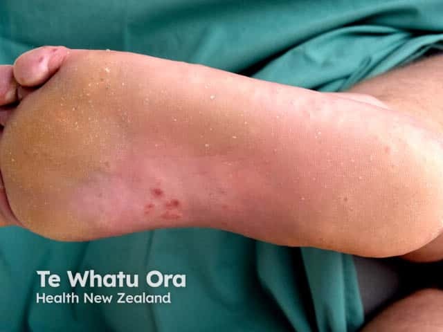
Clinical photography of a pediatric foot plantar surface demonstrates vesicular lesions consistent with hand-foot-mouth disease (HFMD) involving the instep and toes. The image captures small, 1–3 mm vesicles and papules with clear vesicular roofs, some with adjacent erythema and mild edema. Several lesions appear isolated while others are clustered along pressure-bearing areas of the instep and toe pads. In early stages, vesicles are translucent; as they evolve, superficial erosions and crusts may form. The surrounding skin remains largely intact, and there is no evidence of purulence. No mucosal involvement is evident in this view, though HFMD commonly involves oral mucosa and presents with fever and malaise in children. Etiology is enteroviral, predominantly Coxsackievirus A16 or enterovirus 71. The image is valuable for dermatologic morphology education, aiding recognition of acral vesiculopapular eruptions and differentiation from varicella, pompholyx, eczema, or contact dermatitis. It supports clinical correlation with systemic symptoms and oral findings, guides triage, infection control, and patient counseling. For researchers, it illustrates lesion topography on the plantar surface and helps quantify lesion density and progression over time. This photograph emphasizes the characteristic distribution on the feet with toe involvement and instep involvement, reinforcing a HFMD diagnosis in pediatric patients.
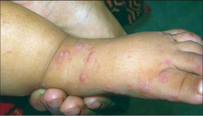
Clinical photograph of a pediatric patient's lower extremity, focusing on the dorsum of the foot and distal lower leg. The image shows a scattered papulovesicular eruption characteristic of Hand, Foot, and Mouth Disease (HFMD). The lesions consist of multiple small (approximately 3-10mm) erythematous papules and clear to gray-tinged fluid-filled vesicles. Many lesions exhibit a distinct peripheral erythematous halo. The vesicles are varied in morphology, appearing round, oval, or elongated. There is a higher concentration of lesions across the dorsal surface of the foot and extending toward the anterior ankle and distal leg. No secondary changes such as crusting, ulceration, or honey-colored scaling are visible. The clinical presentation is consistent with a viral exanthem, specifically Coxsackievirus or Enterovirus infection, which typically manifests as vesiculobullous lesions in specific acral and oral distributions.
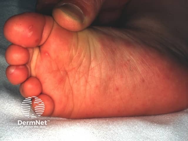
Clinical photograph of the plantar foot in a pediatric patient showing vesicular and maculopapular lesions on the plantar surface with surrounding erythema. The image captures a typical hand-foot-and-mouth disease (HFMD) presentation, caused by enteroviruses such as Coxsackie A16 or enterovirus 71. Lesions are vesicular or vesiculopapular, commonly affecting acral sites including the palms and soles and may extend to the toes; oral mucosal involvement with painful ulcers is common but not always visible in a single image. Morphology includes small, shallow vesicles on an erythematous base that may rupture and crust over time. Clinically this pattern supports HFMD diagnosis, distinguishes it from varicella and other vesicular eruptions, and emphasizes the pediatric infectious-disease context. Imaging modality is non-invasive clinical photography with a close-up plantar view. Significance includes pediatric contagiousness, self-limited course, and the need for supportive care and hydration. This image is valuable for dermatology and infectious disease education, aiding differential diagnosis, clinical correlation with fever and oral lesions, and examination of vesicular acral eruptions for pediatric case studies and medical training.
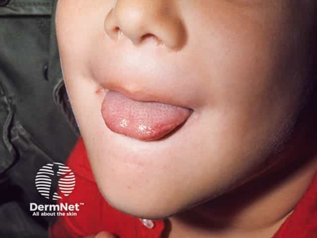
This is a high‑resolution, in vivo clinical photograph of the oral cavity in a pediatric patient with hand‑foot‑mouth disease (HFMD). The focus is the tongue tip, revealing multiple small, translucent vesicles on the anterior tongue surface with shallow erosions and a mildly erythematous mucosal base. The surrounding mucosa is moist, with preserved color and no obvious edema or ulcerations beyond the vesicular sites. The lesions are aphthoid in appearance and compatible with enterovirus–related HFMD, most commonly Coxsackie A16 or enterovirus 71. This oral pattern often accompanies vesicles on buccal mucosa, gingiva, palate, and, in some cases, perioral skin, reflecting the characteristic mucocutaneous involvement of HFMD. Clinically, HFMD typically presents after a febrile prodrome with malaise, irritability, and oral pain that can impair feeding in young children. Differential diagnoses include herpangina (posterior pharyngeal involvement), herpetic gingivostomatitis, aphthous stomatitis, and varicella with oral lesions. The image is valuable for medical education, illustrating vesicular-to-ulcerative mucosal lesions, aiding diagnosis, differential reasoning, and clinical correlation with systemic symptoms. It supports case-based learning, documentation in pediatric infectious diseases, and patient counseling regarding transmission and disease course. High-quality oral mucosa imaging like this enhances recognition of early HFMD lesions, supporting timely isolation and supportive therapy decisions.
https://www.merckvetmanual.com/infectious-diseases/foot-and-…
"foot-and-mouth disease" pathogenesis vaccine diagnosis
"foot and mouth disease" FMDV Asia serotype control
🐄 FOOT AND MOUTH DISEASE (FMD) - Complete Veterinary Guide
Hinglish mein - 4th Year ke liye
1. DEFINITION (Parichay)
- Synonyms: Aftosa, Aphthous Fever, Khur-Paka Munh-Paka (Hindi), Khurpa
- OIE Category A disease - International trade ban hoti hai infected countries mein
- India mein: Notifiable disease - endemic hai
- World's most economically important livestock disease
- Morbidity almost 100% in non-immune herds
- Mortality usually low (adults mein <5%) BUT young animals mein higher
2. ETIOLOGY (Karan)
| Property | Detail |
|---|---|
| Virus | Foot and Mouth Disease Virus (FMDV) |
| Family | Picornaviridae |
| Genus | Aphthovirus |
| Species | Aphthovirus vesiculae |
| Type | Non-enveloped (envelope NAHI hai), single-stranded RNA (+ssRNA) |
| Size | Very small - ~28 nm diameter |
| Shape | Icosahedral capsid |
| Genome | ~8.4 kb (+ssRNA) - sabse large among picornaviruses |
SEROTYPES - MOST IMPORTANT ⭐⭐⭐
| Serotype | Distribution |
|---|---|
| O | Sabse common worldwide; India mein prevalent |
| A | Worldwide |
| C | Rare; possibly EXTINCT in wild |
| Asia-1 | Asia, Middle East - India mein important |
| SAT-1 | Southern African Territories |
| SAT-2 | Southern African Territories |
| SAT-3 | Southern African Territories |
India mein mainly O, A, aur Asia-1 circulate hote hain
KEY Properties of Virus:
- Acid labile - pH 6.0 se neeche ya 9.0 se upar quickly inactivate ho jaata hai (pH 6.8 se neeche unstable)
- NO cross-immunity between serotypes - Ek serotype ki vaccine doosre se protect nahi karti - Exam MCQ!
- Highly stable in environment - 3 months tak survive kar sakta hai
- Inactivated by: Heat (>79°C), UV light, common disinfectants (NaOH, citric acid, Na carbonate)
- Buoyant density: 1.43 g/mL in cesium chloride
3. HOST RANGE (Kaun kaun affect hota hai)
Domestic Animals:
| Animal | Severity |
|---|---|
| Cattle (Gaye/Bail) | Most severely affected - dramatic signs |
| Pig (Suar) | Very dramatic foot lesions; HUGE virus shedder |
| Buffalo (Bhains) | Similar to cattle |
| Sheep (Bhed) | Often mild/subclinical - silent spreader! |
| Goat (Bakri) | Usually subclinical |
Wild Animals (Susceptible):
- African buffalo, Bison, Giraffe, Deer, Antelope
- Bactrian camel, Asian elephant (order Proboscidea - also susceptible!)
- Hedgehogs, Capybara, Warthog
- Virus isolated from 70+ mammalian species
Resistant Animals:
- Horse (Ghoda) - RESISTANT (Solipeds ko nahi hota - important MCQ!)
- Dog, Cat - resistant
- Humans - very rarely affected (zoonotic potential but minimal)
"Cloven-hoof = FMD susceptible; Single hoof (horse) = RESISTANT" - Golden Rule
4. TRANSMISSION (Kaise failta hai)
Primary Routes:
- Infected pigs particularly ENORMOUS amount of aerosol produce karte hain
- Wind se kilometers tak travel kar sakta hai
- UK 1981 epidemic: France se English Channel cross karke aerosol se spread hua!
- Infected animals ke saath physical contact
- Saliva, nasal discharge, milk, semen, urine, feces, skin lesion fluid
- Vehicles, equipment, clothing, footwear
- Feed, water troughs
- Veterinarians, farm workers ke kapde
- Raw/undercooked meat, milk (heat treated milk safe hai)
- Bones, hides, wool
- Cattle: Up to 2-3 years carrier ban sakte hain (nasopharyngeal tonsils mein persist)
- Sheep: Bhi carrier (tonsillar crypts mein)
- Pigs: NO carrier state - recover ke baad completely clear ho jaate hain
Carrier cattle/sheep
       ↓
Upper respiratory tract mein persistent infection
       ↓
Virus nasal secretions mein
       ↓
Environment contamination
       ↓
New animals infect
5. PATHOGENESIS - Step by Step ⭐
Phase 1: PRIMARY INFECTION (Incubation 2-14 days)
FMDV nasopharynx/upper respiratory tract mein enter karta hai
(Cattle: Respiratory route primary)
(Pig: Oral exposure - mouth se primary)
       ↓
Pharyngeal epithelium mein PRIMARY REPLICATION
       ↓
1-2 days mein: PRIMARY VIREMIA shuru
       ↓
Virus blood mein circulate karta hai
       ↓
FEVER shuru (38.5-40°C)
Phase 2: VESICLE FORMATION
Virus non-haired epithelium target karta hai:
- Tongue, buccal mucosa, dental pad, gums
- Coronary band, interdigital cleft
- Teats (lactating animals)
       ↓
Integrin receptors (αvβ3, αvβ6) se attach karta hai
       ↓
INTRACELLULAR REPLICATION - Cytoplasm mein
       ↓
Cell death → serous fluid accumulate
       ↓
VESICLE (Blister) formation
       ↓
1-2 din mein vesicle RUPTURE
       ↓
ULCER/EROSION banta hai
       ↓
Secondary bacterial infection
       ↓
Healing: 7-14 days mein (uncomplicated cases)
Phase 3: MYOCARDITIS (Young animals mein)
Viremia ke dauran
       ↓
Cardiac muscle cells infect ho jaate hain
       ↓
Myocardial necrosis
       ↓
"Tiger Heart" lesion (pathognomonic)
       ↓
Sudden death in young calves/piglets/lambs
Species-wise Pathogenesis Difference:
| Species | Entry | Carrier State | Virus Shed |
|---|---|---|---|
| Cattle | Respiratory tract | YES (2-3 years) | Moderate |
| Pig | Oral mainly | NO | Enormous (amplifier host!) |
| Sheep | Respiratory | YES (tonsils) | Low - silent spreader |
6. CLINICAL SIGNS - Species wise
A. CATTLE (Sabse important - maximum marks)
- Fever 40-41°C
- Depression, anorexia
- Milk production mein sudden drop
- Shivering, dull appearance
- Tongue - Most common site; large vesicles (2-3 cm), rupture ke baad ulcers
- Hard palate, dental pad, lips, gums, muzzle
- Characteristic sign: Profuse salivation (Ptyalism) - daant pikne ki awaaz (smacking noise) - highly characteristic!
- Animal khana nahi khaata - anorexia
- Vesicles rupture ke baad: shallow, painful erosions
- Coronary band (khur ke upar ki band) pe vesicles
- Interdigital cleft (do khur ke beech) pe vesicles
- Rupture → painful ulcers
- Lameness - severe, reluctant to move, prefer lying down
- Stamping of feet (pair patak te hain)
- Severe cases: Hoof sloughing (pura khur gir jaata hai)
- Lactating cattle ki teats pe vesicles
- Milk production crash - sudden agalactia
- Secondary mastitis
- Oral lesions: 2 weeks mein heal
- Foot lesions: Thoda zyada time lagte hain
- Scars bante hain
B. PIGS (Suar)
- Most dramatic foot lesions - cattle se zyada severe
- Coronary band rupture → hoof detachment
- Lameness severe
- Oral lesions: tongue, snout pe
- Young piglets: Sudden death without vesicles (myocarditis)
- Pigs = "Amplifier host" - enormous virus shedding via aerosol
C. SHEEP/GOATS
- Usually MILD or SUBCLINICAL - bahut important epidemiologically!
- Lameness may be first sign
- Oral lesions sometimes present
- Teats affected in lactating animals
- Young lambs: sudden death (myocarditis)
- Silent spreader - symptoms nahi toh detect nahi hote, but virus failaate rehte hain
D. YOUNG ANIMALS (Calves, Piglets, Lambs) ⭐
- MYOCARDITIS - viremia ke dauran heart infect ho jaata hai
- Sudden death bina vesicles ke
- "Tiger Heart" lesion at post mortem - characteristic white/yellow stripes on myocardium = coagulation necrosis
- Mortality can reach 70% in young animals during severe outbreaks
7. LESIONS (Pathological Changes)
Gross (Naked eye) Lesions:
| Location | Lesion |
|---|---|
| Tongue | Vesicles → Ulcers → Erosions |
| Oral mucosa | Vesicles, erosions |
| Coronary band | Vesicles, ulcers |
| Interdigital space | Vesicles, ulcers |
| Teats | Vesicles |
| Heart (young animals) | "Tiger Heart" - white/yellow streaks = myocardial necrosis |
| Rumen/reticulum (cattle) | Occasionally vesicles |
Microscopic Lesions:
- Intraepithelial vesicles - serous fluid collect hota hai epithelium ke andar
- Acantholysis - epithelial cell separation
- Neutrophilic infiltration in vesicle fluid
- Myocardial necrosis (young animals)
- No intranuclear/intracytoplasmic inclusion bodies - (Picornavirus mein hote nahi)
8. DIAGNOSIS
A. CLINICAL DIAGNOSIS
- Cloven-hoofed animal
- Sudden onset fever
- Profuse salivation
- Vesicles in mouth + feet + teats
- Severe lameness
- High morbidity in herd
Any vesicular disease in cloven-hoofed animal = FMD suspect karo aur report karo immediately
B. LABORATORY CONFIRMATION (MANDATORY - Clinical alone insufficient)
- Vesicle fluid (best sample - highest viral load)
- Epithelium from unruptured/freshly ruptured vesicle (transport media mein)
- Serum (blood)
- Nasal/esophageal swab
- Transport: Glycerol buffer (50%) at 4°C ya frozen
Caution: BSL-3 laboratory required for FMDV work in FMD-free countries
Diagnostic Tests:
| Test | Purpose | Notes |
|---|---|---|
| RT-PCR (Real-time) | Virus detection | Fastest, most sensitive - Gold standard; identifies serotype |
| ELISA (Antigen) | Virus antigen detection + serotyping | OIE prescribed test |
| ELISA (Antibody) | Antibody detection | Differentiates infected vs vaccinated animals |
| Virus Isolation | Cell culture (BHK-21, bovine thyroid cells) | Gold standard but slow (2-3 days) |
| Complement Fixation Test (CFT) | Antigen detection + serotyping | Older test, still used |
| Virus Neutralization Test | Serotyping, antibody titers | Reference test |
| NSP (Non-Structural Protein) ELISA | Differentiate infected vs vaccinated (DIVA) | Vaccinated animals NSP antibody nahi banate |
DIVA Strategy (Differentiating Infected from Vaccinated Animals):
- NSP (3ABC) ELISA use karo
- Vaccinated but not infected: Sirf structural protein antibodies
- Infected (even if vaccinated): NSP antibodies bhi bante hain
C. DIFFERENTIAL DIAGNOSIS ⭐ (Important - Vesicular diseases mein confusion)
| Disease | How to Differentiate |
|---|---|
| Vesicular Stomatitis (VS) | Horses bhi affected (FMD mein nahi); Lab test se confirm |
| Swine Vesicular Disease (SVD) | Sirf pigs; horses bhi susceptible; Lab confirm |
| Vesicular Exanthema of Swine | Sirf pigs; serology |
| Malignant Catarrhal Fever (MCF) | No foot lesions; lymph node enlargement; corneal opacity |
| Mucosal Disease (BVD) | No foot lesions typically; different lab tests |
| Bluetongue | Insect-borne; tongue cyanosis; no vesicles |
| Rinderpest | Erosive stomatitis; no foot lesions; now ERADICATED |
| Contagious Ecthyma (Orf) | Sheep/goats; proliferative lesions; no foot involvement |
"Any vesicular disease in cattle-pigs = MUST lab-confirm; Cannot diagnose clinically alone"
9. TREATMENT (Ilaaj)
FMD ke against koi specific antiviral nahi hai - Treatment SYMPTOMATIC hai
A. GENERAL PRINCIPLE:
- FMD-free countries: Treatment PROHIBITED - Stamping out policy (sare animals cull karo)
- Endemic countries (like India): Symptomatic treatment allowed + vaccination
B. SYMPTOMATIC TREATMENT:
| Treatment | Purpose |
|---|---|
| Rest, isolation | Stress reduce karo |
| Soft, easily chewable feed | Khana kha sake kyunki mouth mein dard hai |
| Fresh, clean water | Dehydration rokna |
| NSAIDs (Flunixin meglumine, Meloxicam) | Fever, pain, anti-inflammatory |
| Mouth wash | Dilute antiseptic (1% alum solution, Borax glycerine) - oral lesions clean karo |
| Foot bath | 4% formalin ya copper sulphate solution - foot lesions treat karo |
| Antibiotics (Oxytetracycline, Penicillin) | Secondary bacterial infections rokne ke liye - Viral disease hai toh direct kaam nahi karta |
| Vitamin supplements | Recovery support |
| IV fluids | Severe dehydration mein |
C. FOOT CARE:
- Regular cleaning of foot lesions
- Bandaging if hoof detachment ho
- Copper sulphate foot bath daily
- Trimming of detached hoof if needed
D. YOUNG ANIMALS (Calves/Lambs/Piglets):
- Immediate separation from mothers
- Colostrum/milk supplementation (bottle feeding)
- Supportive care for myocarditis - no specific treatment
- Poor prognosis for myocarditis cases
10. PREVENTION AND CONTROL ⭐⭐
STRATEGY 1: VACCINATION (Endemic countries like India)
| Property | Detail |
|---|---|
| Type | Formalin-inactivated BHK-21 cell culture virus |
| Adjuvant | Aluminum hydroxide + Saponin OR Oil adjuvant (ISA 206) |
| Serotypes | Must match circulating serotypes |
| Vaccine | Serotypes covered |
|---|---|
| FMD-CP (Control Programme) | O + A |
| Trivalent vaccine | O + A + Asia-1 |
| Raksha Ovac | O + A + Asia-1 |
| Aftovax | O + A + Asia-1 |
| Age | Schedule |
|---|---|
| Calves: 4 months old | First dose |
| 6 months | Booster |
| Then every 6 months | Biannual revaccination |
- Killed vaccine - killed vaccine hai, immunity 6 months se zyada nahi chalta → isliye biannual (6 monthly) vaccination zaroori
- No cross-protection between serotypes - Serotype O ki vaccine Asia-1 se protect nahi karti
- No protection against all strains even within a serotype
- Recombinant/VLP vaccines research mein hain (2025 study - PMID 40559776 shows promising E. coli expressed VLP vaccine)
STRATEGY 2: STAMPING OUT (FMD-free countries)
- Detect - Any vesicular disease suspect → immediate report
- Confirm - Lab diagnosis (RT-PCR/ELISA)
- CULL - All infected + in-contact animals euthanize
- Disinfect - Premises, equipment, vehicles
- Quarantine - Movement ban 3 km radius
- Surveillance - 30 day observation period
- Declare free - No new cases in 30 days
STRATEGY 3: MOVEMENT CONTROL
- Import ban on animals + animal products from infected countries
- Surveillance zones, protection zones
- Animal movement ban during outbreak
STRATEGY 4: BIOSECURITY
- Visitor control at farm
- Vehicle disinfection at farm entry
- Dedicated equipment per farm
- Proper disposal of slaughterhouse waste
- Cooking of animal products before feeding to pigs
Disinfectants Effective Against FMDV:
| Disinfectant | Concentration |
|---|---|
| Sodium Hydroxide (NaOH) | 2% - very effective |
| Citric acid | 0.2% |
| Sodium carbonate (Washing soda) | 4% |
| Formalin | 1-2% |
| Sodium hypochlorite (Bleach) | 1-2% |
| Quaternary ammonium compounds | NOT effective against FMDV |
11. EPIDEMIOLOGY
Global Status:
- FMD-free countries: USA, Canada, Australia, New Zealand, Western Europe
- Endemic: India, Pakistan, Bangladesh, most of Africa, Middle East, Southeast Asia
- 2025 review (PMID: 39689813) confirms Asia mein O, A, Asia-1 dominant hain
India mein FMD:
- Endemic - year round cases hote rehte hain
- Serotypes O, A, Asia-1 circulate karte hain
- FMD-Control Programme (FMD-CP) government chal raha hai biannual vaccination ke saath
- Huge economic impact: Milk production loss, draft power loss, export ban
Economic Impact:
- Single clinical case: Direct + indirect losses = lakhs of rupees
- Trade restrictions: FMD-affected countries livestock/products export nahi kar sakte
- UK 2001 outbreak: £8 billion loss - 10 million animals culled!
- India mein annual loss: Thousands of crores
12. ZOONOTIC IMPORTANCE
- Rarely transmitted to humans
- Human infection: Contact with infected animals or raw milk ingestion
- Human symptoms: Fever, vesicles in mouth (tongue, lips), skin of hands/feet
- Self-limiting in humans - mild disease
- Not a significant public health concern compared to animal impact
13. COMPARISON: FMD vs VESICULAR STOMATITIS (Important Table)
| Feature | FMD | Vesicular Stomatitis (VS) |
|---|---|---|
| Virus | Aphthovirus (Picornaviridae) | Vesiculovirus (Rhabdoviridae) |
| Cattle | Affected | Affected |
| Pigs | Affected | Affected |
| Horse | RESISTANT | SUSCEPTIBLE |
| Vector | None (direct contact) | Insect-borne (sandfly) |
| Serotypes | 7 | 2 (Indiana, New Jersey) |
| Mortality | Low (<5% adults) | Very low |
| OIE Status | Category A | Less restricted |
"Horse affected = NOT FMD; Horse safe = Could be FMD" - Key differentiator
14. EXAM KEY POINTS ⭐⭐⭐
| Question | Answer |
|---|---|
| Causative virus | FMD Virus (FMDV) |
| Family | Picornaviridae |
| Genus | Aphthovirus |
| Enveloped? | NO - Non-enveloped |
| Genome | +ssRNA |
| Serotypes | 7 (O, A, C, Asia-1, SAT1, SAT2, SAT3) |
| India mein prevalent serotypes | O, A, Asia-1 |
| Most common worldwide | Serotype O |
| Cross-immunity between serotypes? | NO |
| Possibly extinct serotype | C |
| Host range | Cloven-hoofed animals (Artiodactyla) |
| Horse susceptible? | NO - Resistant |
| Most severe foot lesions | Pigs |
| Most virus shed | Pigs (amplifier host) |
| Silent spreader | Sheep (often subclinical) |
| Carrier state kisme? | Cattle, Sheep (NOT pigs) |
| Carrier site | Nasopharyngeal/oropharyngeal tonsils |
| Characteristic oral sign | Profuse salivation (Ptyalism) |
| Young animal lesion | Tiger Heart (myocarditis) |
| Best diagnostic sample | Vesicle fluid |
| Gold standard test | RT-PCR |
| OIE prescribed test | ELISA |
| Infected vs Vaccinated test | NSP ELISA (3ABC) |
| Vaccine type | Inactivated (Killed) |
| Vaccination frequency India | Every 6 months (biannual) |
| FMD-free policy | Stamping out + slaughter |
| pH stability | Inactivated below 6.0 or above 9.0 |
| Best disinfectant | NaOH 2% |
| OIE category | Category A (List A disease) |
| Economic significance | World's most economically important livestock disease |
QUICK MEMORY TRICKS
- "O A C Asia SAT 1-2-3" = 7 serotypes yaad karo (Oac Asia SATeen)
- "PIGS AMPLIFY, SHEEP HIDE" = Pigs most virus shed, sheep subclinical spreader
- "NO HORSE = FMD" = Horse resistant, horse affected = Vesicular Stomatitis
- "Tiger Heart = Young Dead" = Myocarditis in young = Tiger heart at PM
- "No cross immunity" = Har serotype alag - vaccine match hona chahiye
- "pH 6-9 = Safe Zone" = FMDV pH 6 se neeche, 9 se upar mar jaata hai
- "PICORNA = PIg CORNer ka disease" (Picornaviridae - suar mein dramatic)
- "Biannual India" = India mein har 6 month vaccine - killed vaccine short duration
Recent Evidence: 2025 Asia review (PMID: 39689813) confirms Asia mein FMD O, A, Asia-1 dominant serotypes hain aur regional control programs ke bawajood disease persist kar rahi hai. 2025 VLP vaccine study (PMID: 40559776) E. coli mein produced Virus-Like Particles novel vaccine ke roop mein promising results show kar raha hai - future mein better vaccines aa sakte hain.