
Acute kidney injury
"acute kidney injury"[MeSH Terms] AND management
acute kidney injury pathophysiology classification diagram

This pathophysiology diagram consists of a mathematical graph and its corresponding equations, illustrating the relationship between kinetic Glomerular Filtration Rate (GFRK) and the rate of change in serum creatinine concentration (∂Crt/∂GFRK) during Acute Kidney Injury (AKI). The graph displays two curves, red and blue, positioned entirely in the fourth quadrant (negative y-values). This visualizes the physiological principle that creatinine concentration and GFR move in opposite directions. The x-axis represents GFRK (independent variable), and the y-axis represents the partial derivative ∂Crt/∂GFRK. The red curve reflects standard clinical parameters, while the blue curve represents more extreme clinical scenarios (e.g., higher initial creatinine of 9.0 mg/dL and altered fluid distribution volumes). Both curves demonstrate a steep negative slope at low GFRK values, which gradually flattens and approaches the x-axis as GFRK increases. This illustrates that the sensitivity of creatinine changes is significantly higher at low renal function levels, recapitulating patterns seen in chronic kidney disease within an acute clinical context.
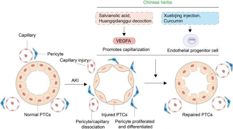
A pathophysiology diagram illustrating the progression of proximal tubular cell (PTC) morphology and vascular changes during Acute Kidney Injury (AKI) and subsequent repair mediated by Traditional Chinese Medicine (TCM). The diagram displays three states: 'Normal PTCs', 'Injured PTCs', and 'Repaired PTCs'. In the normal state, healthy cuboidal epithelial cells form the tubule, surrounded by intact capillaries and associated pericytes. Upon AKI, the PTCs flatten and thin, accompanied by capillary injury, pericyte-capillary dissociation, and pericyte proliferation/differentiation. The repair phase highlights two therapeutic pathways: 1) Salvianolic acid and Huangqidanggui decoction inducing VEGFA to promote capillarization, and 2) Xuebijing injection and Curcumin acting on endothelial progenitor cells. The final stage shows 'Repaired PTCs' where the tubular architecture is restored and capillary-pericyte density is increased. This diagram serves as an educational summary of renal vascular regeneration mechanisms and nephroprotective interventions.
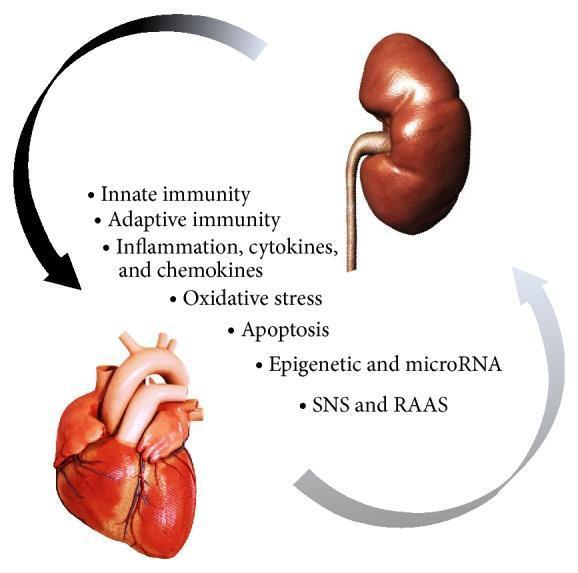
This pathophysiology diagram illustrates the bidirectional 'cardiorenal connectors' involved in cardiorenal syndrome type 3 (acute renocardiac syndrome). The visual features a human kidney positioned superiorly and a human heart positioned inferiorly. Two curved arrows create a circular flow between the organs: a primary dark arrow directed from the kidney to the heart, and a lighter return arrow from the heart to the kidney. Central to the diagram is a list of direct physiologic mechanisms that mediate organ crosstalk during acute kidney injury (AKI). These include innate and adaptive immunity; inflammation involving cytokines and chemokines; oxidative stress; cellular apoptosis; epigenetic factors and microRNA; and neurohormonal activation via the sympathetic nervous system (SNS) and the renin-angiotensin-aldosterone system (RAAS). The diagram is designed for intermediate to advanced medical education, emphasizing the multisystemic nature of renal-induced cardiac dysfunction.
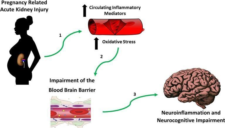
A pathophysiology diagram illustrating the hypothesized mechanistic link between Pregnancy-Related Acute Kidney Injury (PR-AKI) and long-term neurocognitive impairment. The flowchart follows a three-step sequence: (1) Initial PR-AKI, represented by a silhouette of a pregnant person with an highlighted kidney, leads to an increase in systemic circulating inflammatory mediators and oxidative stress, shown as a blood vessel cross-section with upward-trending arrows. (2) These systemic changes result in the impairment of the blood-brain barrier (BBB). A microscopic illustration of the BBB shows disrupted tight junctions and increased permeability, indicated by yellow triangular symbols passing through the endothelial lining into the neural tissue. (3) This compromised barrier facilitates neuroinflammation, which is depicted leading to the final stage: a human brain labeled with neurocognitive impairment. This diagram serves as a conceptual model for understanding the extra-renal complications of obstetric kidney disease and its potential impact on maternal brain health postpartum.
Acute Kidney Injury (AKI)
Definition
- Serum creatinine (SCr) rise ≥0.3 mg/dL within 48 hours, OR
- SCr rise ≥1.5× baseline within the prior 7 days, OR
- Urine output <0.5 mL/kg/h for >6 hours
KDIGO Staging
| Stage | Serum Creatinine | Urine Output |
|---|---|---|
| 1 | 1.5-1.9× baseline OR ≥0.3 mg/dL increase | <0.5 mL/kg/h for 6-12 h |
| 2 | 2.0-2.9× baseline | <0.5 mL/kg/h for ≥12 h |
| 3 | ≥3.0× baseline OR SCr ≥4.0 mg/dL OR initiation of RRT OR (in patients <18 yrs) eGFR <35 mL/min/1.73 m² | <0.3 mL/kg/h for ≥24 h OR Anuria ≥12 h |
Epidemiology
- Complicates 5-7% of acute-care hospital admissions and up to 30% of ICU admissions
- Incidence in the US has grown more than fourfold since 1988; estimated at ~500/100,000 population per year - higher than the yearly incidence of stroke
- ICU mortality in patients with AKI exceeds 50% in many series
- AKI increases long-term risk of CKD progression and cardiovascular disease - Harrison's, 22nd ed.
Etiology and Classification
1. Prerenal AKI (~40-70% of cases)
- Hypovolemia: hemorrhage, GI losses (vomiting, diarrhea), burns, diuretic overuse
- Reduced cardiac output: heart failure, cardiogenic shock, massive PE
- Decreased effective arterial volume: cirrhosis, nephrotic syndrome, sepsis
- Drugs that impair autoregulation: NSAIDs (impair afferent arteriolar dilation), ACE inhibitors / ARBs (impair efferent vasoconstriction)
- Urine sodium <20 mmol/L
- FeNa <1%
- Urine osmolality >500 mOsm/kg
- BUN:creatinine ratio >20
2. Intrinsic Renal AKI
- Ischemic ATN: from prolonged prerenal states, sepsis, major surgery, post-cardiac arrest
- Nephrotoxic ATN: aminoglycosides, contrast agents, myoglobin (rhabdomyolysis), hemoglobin, cisplatin, amphotericin B
- Tubular cell death - ischemia or toxin-mediated apoptosis/necrosis, especially in the S3 segment of the proximal tubule and medullary thick ascending limb (vulnerable due to high O₂ demand and marginal supply)
- Cast obstruction - disruption of actin cytoskeleton leads to integrin relocation, cell detachment, and sloughed cells binding Tamm-Horsfall protein forming obstructing casts
- Back-leak - loss of E-cadherin and tight junction proteins (ZO-1, occludin) allows filtrate to leak back into interstitium, reducing measured GFR
- Endothelial injury - cell swelling, upregulation of adhesion molecules, impaired NO production, vascular congestion in the outer medulla
- Rapidly progressive glomerulonephritis (RPGN), anti-GBM disease, ANCA vasculitis, IgA nephropathy
- Drug-induced (beta-lactams, NSAIDs, proton pump inhibitors, sulfonamides), infectious, autoimmune
- Classic triad: fever, rash, eosinophilia (present in minority of cases)
- Renal artery/vein thrombosis, thrombotic microangiopathy (TTP/HUS), malignant hypertension, scleroderma renal crisis, DIC, vasculitis - Comprehensive Clinical Nephrology, 7th ed.; Harrison's, 22nd ed.
3. Postrenal AKI
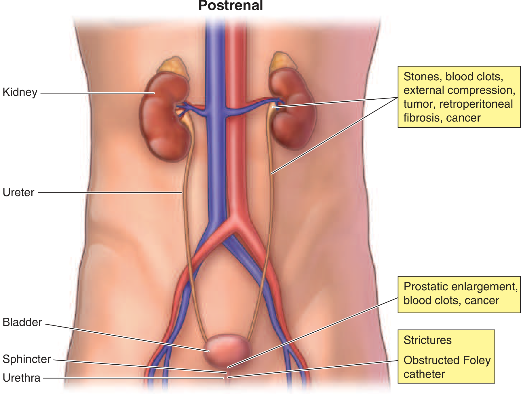
- Ureteric: calculi, blood clots, sloughed renal papillae (intraluminal); neoplasia (intramural); retroperitoneal fibrosis, cancer, abscess (external compression)
- Bladder neck/urethra: benign prostatic hypertrophy (most common cause in men), neurogenic bladder, anticholinergic drugs, cancer, strictures, obstructed Foley catheter
Diagnostic Evaluation
History and Physical Examination
| AKI Type | Clues |
|---|---|
| Prerenal | Vomiting, diarrhea, bleeding, poor intake, diuretic use, heart failure, cirrhosis; tachycardia, hypotension, dry mucous membranes, low JVP |
| Intrinsic - ATN | Recent hypotension, sepsis, nephrotoxin exposure, rhabdomyolysis, contrast |
| Intrinsic - AIN | Drug exposure (PPIs, NSAIDs, antibiotics), fever, rash |
| Intrinsic - GN | Hematuria, proteinuria, systemic disease signs (lupus, vasculitis), hemoptysis (Goodpasture) |
| Postrenal | Obstructive symptoms (hesitancy, weak stream, overflow incontinence), suprapubic pain, pelvic/retroperitoneal malignancy |
Laboratory Studies
| Test | Prerenal | ATN |
|---|---|---|
| Urine Na | <20 mEq/L | >40 mEq/L |
| FeNa | <1% | >2% |
| Urine osmolality | >500 mOsm/kg | ~300 mOsm/kg (isosthenuria) |
| BUN:Cr ratio | >20 | ~10-15 |
| Urine sediment | Normal/hyaline casts | Muddy brown granular casts, RTECs |
- FeNa can be <1% in contrast nephropathy, myoglobinuric AKI, and early obstruction - misleadingly "prerenal" pattern
- FEurea <35% is more reliable in patients on diuretics
Urinalysis and Urine Microscopy
- Hematuria + RBC casts → glomerulonephritis
- WBC casts, eosinophiluria → AIN
- Granular "muddy brown" casts → ATN
- Waxy/broad casts → advanced CKD
Imaging
- Renal ultrasound is the first-line imaging: assesses kidney size, echogenicity, hydronephrosis (postrenal AKI)
- Doppler to assess renal artery/vein flow if vascular etiology suspected
Kidney Biopsy
Novel Biomarkers
- NGAL (Neutrophil Gelatinase-Associated Lipocalin): rises within 2-3 h of AKI onset
- KIM-1 (Kidney Injury Molecule-1): tubular injury marker
- IL-18: associated with duration and progression of AKI (TRIBE-AKI data: highest quintile of IL-18 had 6.8× higher risk of AKI after cardiac surgery)
- TIMP-2 × IGFBP7 (cell cycle arrest markers): FDA-cleared for AKI risk stratification
- Urinary cystatin C and α1-microglobulin: predict need for RRT - Brenner and Rector's The Kidney, 2-Volume Set
Complications
| System | Complication |
|---|---|
| Fluid/electrolyte | Hypervolemia, hyponatremia, hyperkalemia, hyperphosphatemia, hypocalcemia |
| Acid-base | Metabolic acidosis (anion gap) |
| Hematologic | Anemia (multifactorial), uremic platelet dysfunction, bleeding |
| Cardiovascular | Arrhythmias (from hyperkalemia), pericarditis, pericardial effusion, volume overload/pulmonary edema |
| Neurologic | Uremic encephalopathy, asterixis, seizures |
| Infectious | Impaired immunity; infections both precipitate and complicate AKI |
| Nutritional | Hypercatabolism, protein-energy wasting |
Management
General Principles
- Treat the underlying cause
- Optimize hemodynamics - correct intravascular volume (but avoid fluid overload)
- Discontinue nephrotoxins - NSAIDs, aminoglycosides, contrast, ACE inhibitors/ARBs in low-flow states
- Dose-adjust renally cleared drugs
- Avoid further injury - maintain perfusion, monitor for sepsis
Prerenal AKI
- Volume resuscitation with isotonic crystalloids (balanced crystalloids preferred over normal saline to reduce hyperchloremic acidosis)
- Treat underlying cause: blood transfusion for hemorrhage, inotropes/vasopressors for cardiogenic/vasodilatory shock, diuretics in decompensated heart failure
- Note: Oliguria alone is NOT an indication for fluid bolus - only true intravascular hypovolemia is - Harrison's, 22nd ed.
Intrinsic AKI - ATN
- No specific reversal therapy once established; focus is supportive
- Treat sepsis aggressively
- Dopamine at low doses has NOT been shown to be beneficial and carries risk of arrhythmias and bowel ischemia - should NOT be used specifically for AKI prevention/treatment
- Rhabdomyolysis: aggressive IV fluid (may need 10 L/day initially); alkalinizing urine (sodium bicarbonate added to 0.45% saline) may reduce cast formation; target urine output 200-300 mL/h
Intrinsic AKI - Specific Causes
- GN/vasculitis: immunosuppression (steroids, cyclophosphamide, rituximab), plasmapheresis for anti-GBM and ANCA disease
- AIN: discontinue offending drug; glucocorticoids considered if AKI persists (no RCT evidence)
- Scleroderma renal crisis: ACE inhibitors
- TTP: therapeutic plasma exchange (medical emergency)
- Atypical HUS: eculizumab (complement blockade)
Postrenal AKI
- Prompt relief of obstruction:
- Lower tract: urethral or suprapubic catheterization
- Upper tract: percutaneous nephrostomy or ureteral stent
- Expect post-obstructive diuresis after relief; may need IV fluid/electrolyte replacement
Supportive Measures
Volume Management
- Restrict fluid and sodium in oliguric AKI with volume overload
- Furosemide: bolus up to 200 mg, then IV drip (10-40 mg/h) ± thiazide for refractory volume overload
- Diuretics do NOT improve the natural history of AKI but help avoid dialysis in some
Electrolytes and Acid-Base
- Hyperkalemia: dietary restriction, eliminate K+-retaining drugs, sodium bicarbonate, insulin/glucose, albuterol, calcium gluconate (for membrane stabilization), kayexalate, or dialysis
- Metabolic acidosis: treat if pH <7.20 or HCO₃⁻ <15 mmol/L with IV/oral sodium bicarbonate
- Hyperphosphatemia: phosphate binders (calcium carbonate, sevelamer, lanthanum)
- Hypocalcemia: treat symptomatically
Nutrition (KDIGO Guidelines)
- Total energy intake: 20-30 kcal/kg/day
- Protein: 0.8-1.0 g/kg/day (non-catabolic, no dialysis) → 1.0-1.5 g/kg/day (on dialysis) → up to 1.7 g/kg/day (hypercatabolic on CRRT)
- Supplement trace elements and water-soluble vitamins in dialysis patients
Renal Replacement Therapy (RRT)
Indications (Emergent/"AEIOU")
- Acidosis: refractory metabolic acidosis (pH <7.1)
- Electrolytes: refractory hyperkalemia
- Intoxications: certain dialyzable toxins (methanol, ethylene glycol, salicylates, lithium)
- Overload: refractory pulmonary edema/volume overload
- Uremia: encephalopathy, asterixis, uremic pericarditis, uremic bleeding
Modalities
| Mode | Features |
|---|---|
| Intermittent HD (IHD) | 3-4 h sessions, 3-4×/week; most common; risk of hypotension |
| CRRT (CVVH, CVVHD, CVVHDF) | Continuous; preferred in hemodynamically unstable patients; avoids rapid fluid/osmotic shifts |
| Sustained Low-Efficiency Dialysis (SLED) | Hybrid; 8-12 h; used in ICU |
| Peritoneal Dialysis | Requires peritoneal access; less efficient; used if vascular access impossible |
AKI and Long-Term Outcomes
- AKI significantly increases the risk of subsequent CKD progression and ESKD
- Patients with AKI superimposed on CKD are at highest risk of maladaptive repair and non-recovery
- AKI is a risk factor for future cardiovascular disease
- AKI-CKD continuum: each episode of AKI accelerates CKD progression and each stage of CKD increases AKI risk
- Harrison's Principles of Internal Medicine, 22nd Edition (2025) - Chapter 321, Waikar & Bonventre
- Comprehensive Clinical Nephrology, 7th Edition - Chapter 70, Paine, Jefferson & Velez
- Brenner and Rector's The Kidney, 2-Volume Set - Chapters 15-16 (AKI biomarkers)
- Sabiston Textbook of Surgery, 21st Edition - Chapter 26 (postoperative AKI, KDIGO table)
- Campbell-Walsh-Wein Urology, 3-Volume Set - Chapter 48