
Hhow to interpret chest x ray
I'll start with a quick overview while I pull up the medical library for a thorough answer.
Quick Answer: Chest X-ray (CXR) interpretation follows a systematic approach - check technical quality first (rotation, inspiration, exposure), then work through a mnemonic like ABCDE (Airway, Breathing/lungs, Cardiac, Diaphragm/Digestive, Everything else/bones & soft tissue). Look for symmetry, compare sides, and always correlate with clinical findings.
Searching sources...
Reading File
Now let me search the medical library and web in parallel for comprehensive CXR interpretation content.
Finding Sources
Searching the Web
systematic approach chest x-ray interpretation radiology
Good - found a dedicated section in the textbook. Let me read it along with additional searches.
Reading File
Finding Sources
Reading File
Finding Sources
Reading File
I have an image URL from the textbook. Let me fetch it and continue reading.
Loading Image
Reading File
Reading File
Loading Image
Excellent - I have a great lateral chest anatomy diagram and comprehensive textbook content. Now I have everything needed for a thorough, well-structured answer.
How to Interpret a Chest X-Ray (CXR)
A systematic approach ensures nothing is missed. The steps below follow the framework from Pfenninger and Fowler's Procedures for Primary Care with clinical pearls throughout.
Step 1: Before You Interpret - Check Technical Quality (RIP)
Always assess these three factors first. A technically flawed film can mimic or hide pathology.
| Factor | How to Check | What it Means |
|---|---|---|
| R - Rotation | Measure distance from spinous processes to medial clavicular heads on each side. Should be equal (< 2-3 mm difference is acceptable). | Rotation shifts the mediastinum and distorts cardiac silhouette |
| I - Inspiration | Count posterior ribs where they join the spine. A minimally adequate inspiration shows 9 ribs. | Poor inspiration causes apparent opacities that mimic pulmonary edema (CHF) |
| P - Penetration/Exposure | Intervertebral spaces should disappear within the cardiac shadow and NOT be visible below the diaphragm. | Overexposed = film too black (misses lung lesions); Underexposed = film too white (false opacities) |
Always note technique limitations before interpreting - these introduce disclaimers that protect both the patient and clinician.
Step 2: PA vs AP vs Portable
- PA (Posteroanterior) - standard view; beam travels back-to-front; patient stands upright, takes a deep breath. Most accurate for cardiac size.
- AP (Anteroposterior) - portable/bedside; beam travels front-to-back. Cardiomegaly definitions differ - AP views magnify the heart, so don't apply PA cardiac size criteria.
- Lateral view - essential in ambulatory adults. Reveals retrocardiac and retrosternal (anterior clear space) lesions that the PA view misses.
Step 3: Systematic Review - ABCDE Approach
A - Airway
- Is the trachea midline? Deviation suggests tension pneumothorax, large effusion (pushes trachea away), or fibrosis/collapse (pulls trachea toward).
- Check the carina - normally the angle of bifurcation is <70°. Widening (>70°) suggests left atrial enlargement.
- Are the main bronchi visible and symmetric?
B - Breathing / Lungs
This is the most content-rich step. Use a "ping-pong" left-right comparison to detect asymmetries.
Hila:
- In 70% of normals, left hilum is higher than the right. The right hilum is never normally higher than the left.
- Bilateral hilar enlargement = sarcoid, lymphoma, TB, primary pulmonary hypertension.
- Unilateral enlargement = malignancy, TB, infection.
Lung parenchyma:
- Vascular markings normally stop 3-5 mm short of the chest wall.
- Cephalization of flow (enlarged upper lobe vessels) = early CHF.
- Silhouette sign: when a lesion shares the same density as an adjacent border (heart, aorta, diaphragm), that border is "erased." This localizes the lesion:
- Right heart border erased → right middle lobe pathology
- Left heart border erased → lingula pathology
- Diaphragm border erased → lower lobe pathology
Nodules vs Masses:
- Nodule = 5-30 mm diameter
- Mass = >30 mm diameter
- Small lesions 2-10 mm are usually benign calcified granulomas; follow with serial imaging
Pleura:
- Pneumothorax: absent vascular markings extending to the inner thoracic wall (no lung markings reaching the rib edges)
- Pleural effusion: blunting of costophrenic angles; the meniscus sign. Causes include infection, malignancy, CHF, renal failure, hypoalbuminemia, pancreatitis, subphrenic abscess.
- Untreated chronic effusions can loculate and form adhesions.
C - Cardiac Silhouette
Size:
- Cardiothoracic (CT) ratio on PA view: cardiac transverse diameter / thoracic diameter at the same level. Normal = <0.5 (50%).
- If >0.5 on a true PA view = cardiomegaly. (Note: AP views make the heart appear larger - do not use the same cutoff.)
- A "thin heart" in COPD (hyperinflation flattens and elongates it) is not pathological.
Specific findings:
- Enlarged pulmonary artery segment = extra hump on left cardiac border → suggests pulmonary hypertension
- Pneumopericardium = black line around the heart border
- On the lateral view: left ventricular enlargement casts a shadow >2 cm posterior to the inferior vena cava shadow
Borders to identify:
- Right heart border = right atrium
- Left heart border = left ventricle (superior portion = left atrial appendage)
D - Diaphragm
- Right diaphragm is normally higher than left by 2-20 mm (liver pushes it up; gastric bubble is under the left).
- Abnormal elevation of a hemidiaphragm: atelectasis, phrenic nerve palsy, effusion, post-lobectomy.
- Air under the diaphragm (free intraperitoneal air) = surgical emergency until proven otherwise (perforated viscus).
- Check costophrenic angles - should be sharp. Blunting = ~200-300 mL of pleural fluid.
E - Everything Else (Bones, Soft Tissues, Mediastinum, Devices)
Mediastinum:
- Normal width = <8 cm in adults (measured at aortic knob level). If >25% of thoracic diameter at the carina = widened.
- Widened mediastinum: aortic dissection, pericardial tamponade, lymphoma, thymoma, germ cell tumor, thyroid mass.
- "Thin" mediastinum has no clinical significance.
- Note: children <5 years normally have a wide-looking mediastinum and large thymic shadow.
Bones:
- Ribs - fractures, metastatic lesions, notching (coarctation of aorta causes rib notching on the inferior rib margins, 3rd-8th ribs bilaterally)
- Vertebrae - compression fractures, lytic/sclerotic lesions
- Clavicles and shoulder joints
Soft tissues:
- Subcutaneous emphysema
- Breast shadows (can mimic lung opacification)
- Foreign bodies, implanted devices
Lines and tubes (on ICU/portable films):
- ETT tip: should be 3-5 cm above the carina
- Central venous catheter: tip at the SVC-RA junction
- NGT: below the diaphragm, midline
- Chest drains: in the pleural space
Step 4: The Lateral View
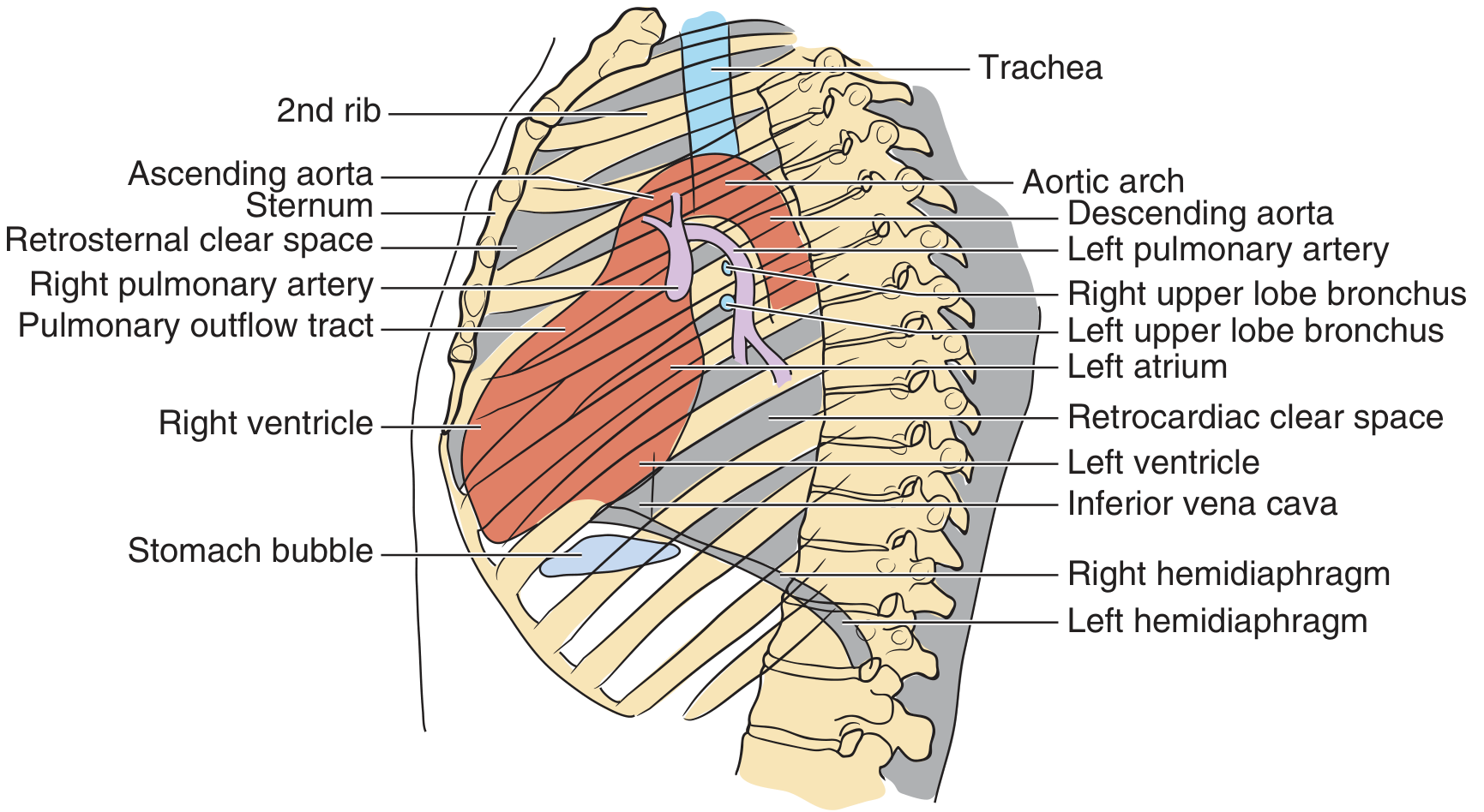
Check four key areas on the lateral:
- Spine - disc spaces, vertebral body density. Normally the vertebrae should appear progressively darker (more lucent) as you go inferiorly. If they get whiter toward the base = posterior lung pathology (e.g., lower lobe consolidation).
- Diaphragms - both should be visible separately. Loss of posterior diaphragm contour = lower lobe consolidation.
- Anterior clear space (retrosternal) - should be clear. Fullness = anterior mediastinal mass (thymoma, teratoma, thyroid, terrible lymphoma - "4 T's").
- Retrocardiac space - should be clear. Opacification = left lower lobe consolidation or effusion.
Step 5: Top 10 "Normal" Benchmarks to Memorize
From Pfenninger & Fowler's Procedures for Primary Care:
- Integrate history and physical - always correlate clinically
- Clavicular heads equidistant from spinous processes (no rotation)
- At least 9 posterior ribs visible on good inspiration
- Intervertebral spaces NOT visible below the diaphragm
- Before age 5, a large thymus can widen the mediastinum normally
- Adults: mediastinum ≤ 8 cm wide
- Left hilum higher than right (normal in 70%)
- Right diaphragm higher than left
- Lesions >10 mm may benefit from a second opinion or CT
- "Chance favors the prepared mind" - experience + pattern recognition builds skill
Common CXR Patterns and Their Diagnoses
| Pattern | Key Features | Think of... |
|---|---|---|
| Consolidation (lobar) | Homogeneous opacity, air bronchograms, silhouette sign | Pneumonia, pulmonary infarction |
| Bilateral perihilar ("bat-wing") haziness | Central opacities, cephalization | Pulmonary edema (CHF) |
| Reticulonodular pattern | Fine "net-like" or nodular shadows | Interstitial lung disease, sarcoid, TB miliary |
| Unilateral whiteout | Entire hemithorax opaque | Massive effusion, collapse, pneumonia |
| Hyperinflation + flat diaphragms | Low flat diaphragms, increased AP diameter | COPD, acute asthma |
| Pneumothorax | Absent lung markings at periphery, visible visceral pleural edge | Trauma, spontaneous, iatrogenic |
| Hilar enlargement | Bilateral symmetric | Sarcoid; unilateral = malignancy/TB |
Summary Workflow
1. Clinical context (symptoms, old films)
2. RIP - check Rotation, Inspiration, Penetration
3. PA + Lateral views both assessed
4. ABCDE: Airway → Breathing → Cardiac → Diaphragm → Everything else
5. Ping-pong comparison: left vs right lung fields
6. Apply silhouette sign to localize lesions
7. Final interpretation: normal / probably insignificant / questionable / abnormal
8. Plan: CT, repeat film, consultation, or immediate action
- Pfenninger and Fowler's Procedures for Primary Care, 3rd Edition, Chapter 231
This is a shared conversation. Sign in to Orris to start your own chat.