
Oral manifestation of HIV AIDS 7 marker question general medicine
Reading File
Now searching the medical library for oral manifestations of HIV/AIDS:
Finding Sources
Finding Sources
Good - found Robbins and Harrison's. Let me read the relevant sections:
Reading File
Finding Sources
Finding Sources
Excellent - found a table and image in Robbins. Let me read the full table and also get the comprehensive HIV oral section from Cummings and Tintinalli:
Reading File
Reading File
Reading File
I now have comprehensive data. Let me also get the Harrison's section for the HIV oral manifestations and fetch key images:
Finding Sources
Finding Sources
Now let me get the KJ Lee classification and the image for oral Kaposi sarcoma:
Reading File
Loading Image
I now have all the information needed for a comprehensive 7-marker answer. Let me also fetch the hairy leukoplakia image from Sleisenger:
Finding Sources
Reading File
I now have all the information needed for a thorough 7-marker answer. Here is the comprehensive response:
Oral Manifestations of HIV/AIDS
(7-marker answer - General Medicine)
Classification
Oral lesions in HIV/AIDS are grouped into three categories based on the EC-Clearinghouse/WHO classification:
| Group | Definition |
|---|---|
| Group 1 | Strongly associated with HIV (appear frequently, help staging) |
| Group 2 | Less commonly associated with HIV |
| Group 3 | Seen in HIV, but may occur in other conditions too |
Group 1 - Strongly Associated (High-Yield)
1. Oral Candidiasis (Thrush)
- Most common oral manifestation of HIV/AIDS; affects >80% of AIDS patients.
- Caused by Candida albicans (part of normal flora in ~50% of the population; overgrows with immunosuppression).
- Forms:
- Pseudomembranous (Thrush): White, gray, curd-like plaques on tongue and buccal mucosa - can be scraped off to reveal an erythematous base. This is the hallmark finding.
- Erythematous: Red patches on hard/soft palate or dorsum of tongue.
- Hyperplastic: Adherent white plaques that cannot be scraped off.
- CD4 correlation: Thrush increases sharply when CD4 <500 cells/mm³.
- Diagnosis: KOH smear showing pseudohyphae.
- Treatment: Fluconazole 100 mg OD for 7-14 days (first-line); clotrimazole/nystatin (second-line).
- Robbins Pathologic Basis of Disease
2. Oral Hairy Leukoplakia (OHL)
- Caused by Epstein-Barr virus (EBV) reactivation in HIV-infected individuals.
- Occurs in 3-12% of HIV-positive patients.
- Site: Lateral border of the tongue (most characteristic location).
- Appearance: White, fluffy ("hairy"), corrugated/shaggy thickenings that cannot be scraped off (unlike thrush).
- Histology: Hyperparakeratosis, acanthosis, characteristic "balloon cells" in upper spinous layer, EBV detectable in basal epithelium.
- Significance: Its presence in an otherwise asymptomatic patient is a strong indicator of HIV with moderate-to-severe immunosuppression. May portend development of full AIDS.
- Key distinction from candidiasis: OHL is adherent; lesion persists after antifungal therapy.
- Does not undergo malignant transformation.
- Cummings Otolaryngology; Robbins Pathologic Basis of Disease
3. HIV-Associated Periodontal Disease
Staged into three levels (KJ Lee / Cummings classification):
(a) Linear Gingival Erythema (LGE)
- Fiery red band of marginal gingiva - out of proportion to the degree of dental plaque.
- Prone to hemorrhage; persists despite dental plaque removal.
- Often involves Candida and gram-negative organisms.
(b) Necrotizing Ulcerative Gingivitis/Periodontitis (NUG/NUP)
- Spectrum of the same pathology, with progressive gingival recession, bleeding, tissue sloughing, pain, malodor, and loss of interdental papillae.
- Rapid and painful destruction of bone and soft tissue.
(c) Enhanced chronic periodontitis
- Accelerated progression of otherwise-common periodontal disease.
Gingival disease that is necrotizing or ulcerative in a previously healthy young adult is highly suggestive of HIV and warrants serologic testing.
- KJ Lee's Essential Otolaryngology; Goldman-Cecil Medicine
4. Kaposi Sarcoma (KS) of the Oral Cavity
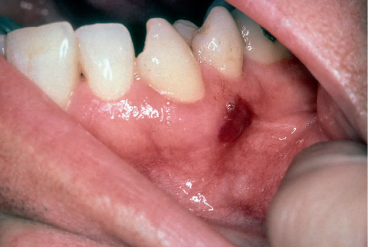
Oral Kaposi Sarcoma in an HIV-infected patient - Andrews' Diseases of the Skin Clinical Atlas
- AIDS-defining illness. Caused by HHV-8 (Human Herpesvirus-8).
- Oral cavity is a common early site in AIDS-associated KS (in contrast to classic KS).
- A solitary genital or oral lesion may be the first manifestation of AIDS-KS.
- Appearance: Nontender, well-circumscribed, slightly raised violaceous (purple/red) lesion on the palate or gingiva.
- Diagnosis: Requires biopsy (spindle cell proliferation with slit-like vascular spaces).
- Treatment: Topical therapy is palliative; systemic chemotherapy for advanced disease.
- Tintinalli's Emergency Medicine; Campbell-Walsh Urology; Harrison's Principles
Group 2 - Less Commonly Associated
5. Recurrent Aphthous Stomatitis (RAS) / Aphthous Ulcers
- Painful oral ulcerations that recur; can be major, minor, or herpetiform type.
- Major aphthous ulcers in HIV are larger, deeper, and more persistent than in immunocompetent patients.
- Common in HIV/AIDS patients; listed as Group 1 lesion in some classifications.
- Cummings Otolaryngology
6. Herpes Simplex Virus (HSV) Stomatitis
- HSV-1 seropositivity approaches 95% in the HIV-positive community.
- Herpetic lesions are particularly persistent when CD4 <100 cells/μL.
- Appearance: Painful vesicular oral and perioral lesions that rupture and crust.
- Diagnosis: Multinucleated giant cells on Tzanck smear; viral culture.
- Treatment: Oral acyclovir/valacyclovir; IV acyclovir for immunocompromised patients with severe disease.
- Cummings Otolaryngology; Tintinalli's Emergency Medicine
7. Salivary Gland Disease & Other Notable Lesions
Salivary gland disease:
- Bilateral parotid enlargement (cystic lymphoid hyperplasia / lymphoepithelial cysts) - common ENT/oral finding in HIV.
- Can cause xerostomia (dry mouth), which secondarily promotes oral candidiasis.
Other important oral lesions in HIV:
- Non-Hodgkin Lymphoma: Rapidly enlarging oral lesion, commonly involving Waldeyer's ring; can mimic lymphadenopathy or benign ulceration.
- CMV oral ulcerations: CMV co-infects 90% of HIV patients; can cause oral ulcers, especially with advanced immunosuppression.
- Oral HPV/warts: Common, plantar, flat, oral, and genital warts all increase with HIV; warty keratoses at the angle of the mouth are characteristic of HPV in AIDS patients.
- Deep fungal infections (histoplasmosis, cryptococcosis): Oral lesions may be the first manifestation of disseminated histoplasmosis in HIV/AIDS patients.
Summary Table for Exam
| Lesion | Pathogen | Key Feature | CD4 Level |
|---|---|---|---|
| Oral candidiasis (thrush) | Candida albicans | Scrapable white plaques | <500/mm³ |
| Hairy leukoplakia | EBV | Non-scrapable, lateral tongue | <200/mm³ |
| Linear gingival erythema | Mixed (Candida + bacteria) | Fiery red gingival band | Variable |
| NUG/NUP | Mixed | Rapid bone/tissue destruction | Variable |
| Kaposi sarcoma | HHV-8 | Violaceous palatal lesion | AIDS-defining |
| Aphthous ulcers | None (immune dysregulation) | Painful, recurrent ulcers | Variable |
| HSV stomatitis | HSV-1 | Vesicular, perioral | <100/mm³ (persistent) |
Key exam pearl: The single most common oral manifestation of HIV/AIDS is oral candidiasis (thrush). Oral hairy leukoplakia (lateral tongue, EBV, non-scrapable, balloon cells on histology) is the most specific and diagnostically important. Both together in a young immunocompetent-appearing patient should prompt HIV testing.
- Sources: Robbins & Cotran Pathologic Basis of Disease; Cummings Otolaryngology Head and Neck Surgery; KJ Lee's Essential Otolaryngology; Tintinalli's Emergency Medicine; Goldman-Cecil Medicine
This is a shared conversation. Sign in to Orris to start your own chat.