
Radial nerve femoral nerve sciatic nerve ulnar nerve etc VS cranial nerve all things cover to create a good concept ?
peripheral nerve anatomy radial ulnar sciatic femoral diagram
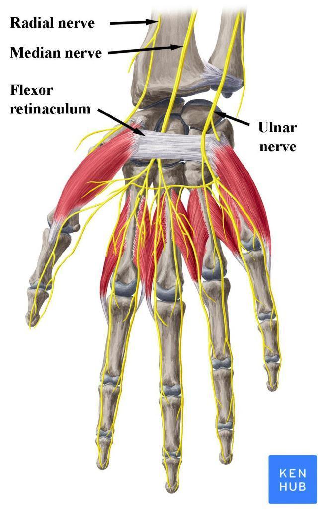
An anatomical illustration of the human hand and wrist (anterior palmar view), highlighting the peripheral nerves and the carpal tunnel. The image depicts the skeletal structure of the carpus, metacarpals, and phalanges, overlaid with musculature and major nerves in yellow. Key structures are labeled: the median nerve is shown passing deep to the flexor retinaculum (transverse carpal ligament), demonstrating its entry into the carpal tunnel. The ulnar nerve is positioned medially (ulnar side), passing superficial to the flexor retinaculum, while the superficial branch of the radial nerve is shown laterally near the radius. Distal to the wrist, the nerves are shown branching into digital nerves to provide sensory and motor innervation to the fingers. This diagram is clinically relevant for explaining the pathophysiology of Carpal Tunnel Syndrome, where the median nerve undergoes compression beneath the flexor retinaculum. Target audience: medical students and clinicians studying upper limb anatomy and entrapment neuropathies.
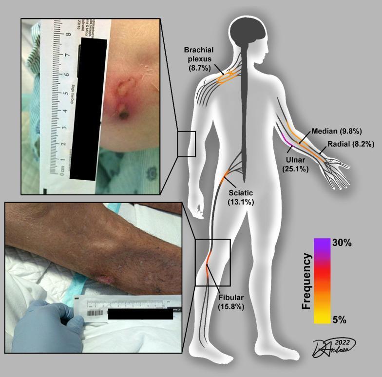
This medical infographic illustrates common peripheral nerve injury (PNI) sites in survivors of severe COVID-19. The central element is an anatomical diagram of a human figure highlighting several nerves with color-coded injury frequencies. The ulnar nerve (25.1%) is the most frequent site, followed by the common fibular (15.8%), sciatic (13.1%), median (9.8%), brachial plexus (8.7%), and radial (8.2%) nerves. A color-gradient scale (5% yellow to 30% purple) correlates with these frequencies. Two inset clinical photographs demonstrate stage-related skin pressure sores (decubitus ulcers) at critical compression points. The top inset shows an elbow ulcer with central necrosis and surrounding erythema, while the bottom inset depicts a pressure sore over the fibular head on a limb with darker skin pigmentation. Both clinical images include a surgical ruler for scale. The visual summarizes how mechanical loading during prolonged ICU hospitalization and prone positioning can lead to both skin breakdown and underlying axonal nerve injury, emphasizing the clinical relevance of monitoring bony prominences.
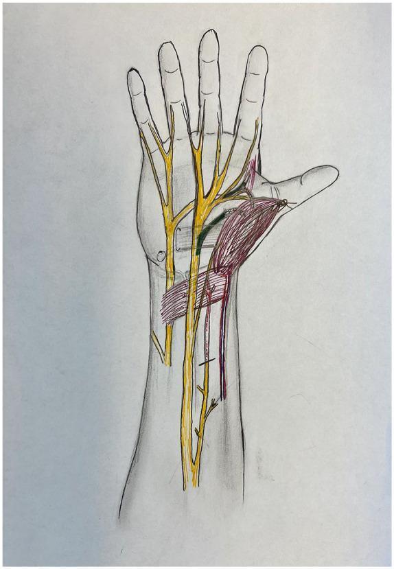
Anatomical diagram of the human hand and distal forearm in a palmar view, illustrating peripheral nerve anatomy and muscle transposition for pronator quadratus opponensplasty. The diagram features color-coded structures overlaid on a skeletal and soft tissue outline. Major peripheral nerves, including the median and ulnar nerves, are highlighted in yellow, showing their proximal courses and distal branching into digital nerves. The recipient anterior interosseous nerve (AIN) motor branch is highlighted in red, indicating its path toward the transposed muscle. The donor recurrent median nerve and donor deep ulnar nerve branches are highlighted in green, demonstrating potential donor sites for nerve coaptation to restore thenar function. The pronator quadratus muscle is shaded in red, shown in a transposed position toward the thumb (thenar eminence) to simulate its role as an opponens substitute. This illustration is designed for surgical planning in cases of chronic carpal tunnel syndrome or atrophic thenar muscle dysfunction, focusing on the spatial relationships required for effective nerve transfer and muscle mobilization.
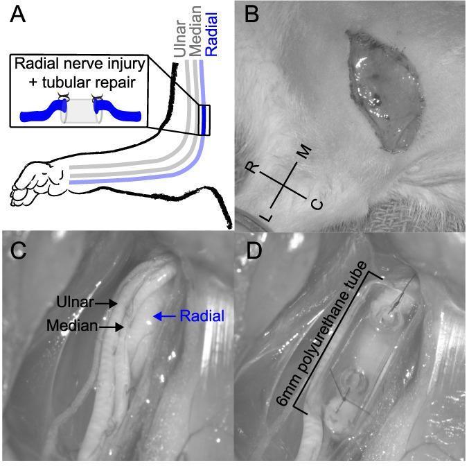
A multi-panel series illustrating a peripheral nerve injury and repair procedure in an animal model (rat forelimb), used for neuroanatomical and microsurgical education. Panel A is a schematic diagram showing the anatomical pathway of the ulnar, median, and radial nerves in the right forelimb, with an inset detailing a tubular nerve repair technique. Panel B shows a clinical photograph of a skin incision proximal to the elbow with orientation markers (R-L, M-C). Panel C provides a high-magnification intraoperative view of the isolated ulnar, median, and radial nerves, with the radial nerve specifically targeted for intervention. Panel D demonstrates the microsurgical tubular repair of a completely transected radial nerve. The proximal and distal nerve stumps are shown sutured 1 mm into the opposite ends of a 6-mm saline-filled polyurethane tube, leaving a 4-mm internal gap. This visual serves to demonstrate experimental peripheral nerve regeneration models, microsurgical suturing techniques, and the use of conduit-based bridging for segmental nerve defects.
cranial nerves overview diagram all 12
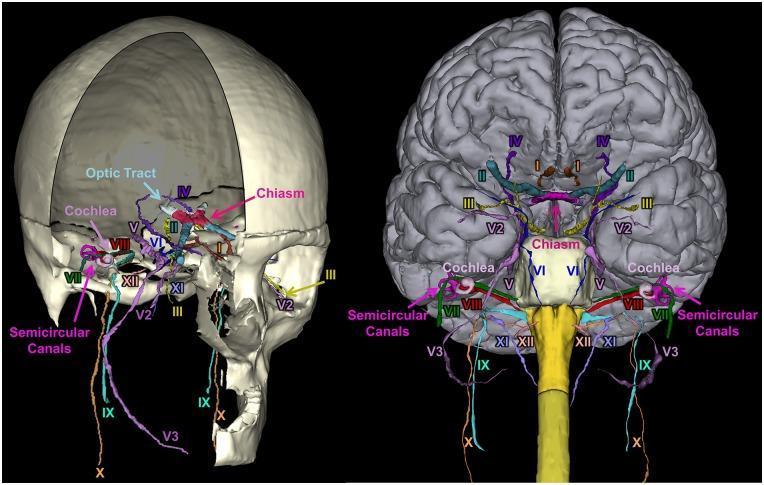
A 3D anatomical reconstruction showcasing the segmentation and spatial orientation of the 12 pairs of human cranial nerves (CN I–XII). The image presents two views: a lateral perspective within a partially transparent skull (left) and an anterior-inferior view relative to the brain and brainstem (right). Each nerve is color-coded for identification: CN I (Olfactory), CN II (Optic) with the optic chiasm and tract, CN III (Oculomotor), CN IV (Trochlear), CN V (Trigeminal) including V2 (maxillary) and V3 (mandibular) divisions, CN VI (Abducens), CN VII (Facial), CN VIII (Vestibulocochlear), CN IX (Glossopharyngeal), CN X (Vagus), CN XI (Accessory), and CN XII (Hypoglossal). Key inner ear structures, including the snail-shaped cochlea and the loop-like semicircular canals, are visualized in relation to the facial and vestibulocochlear nerves. This diagnostic-quality illustration demonstrates the complex neuroanatomical pathways through the skull base and their origins at the brainstem, serving as a pedagogical tool for neurology, radiology, and neurosurgery training.
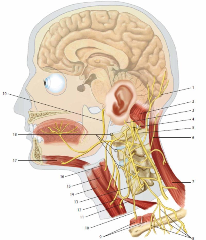
This medical anatomical diagram provides a lateral view of the human head and neck, focusing on the cervical plexus and its integration with cranial nerves. Key neural structures include the lesser occipital nerve (1), greater auricular nerve (3), and transverse cutaneous nerve of the neck (6), demonstrating the sensory distribution of the cervical plexus. The phrenic nerve (10) is shown descending inferiorly anterior to the prevertebral muscles (18). Cranial nerves X (vagus, 4), XI (accessory, 5), and XII (hypoglossal, 19) are illustrated to show their spatial relationships with cervical spinal branches. Muscular landmarks include the sternocleidomastoid (2, 9), trapezius (7), and infrahyoid muscles such as the omohyoid (11, 15) and thyrohyoid (16). The ansa cervicalis (13) is depicted innervating the sub-hyoid musculature. The diagram also illustrates the sagittal section of the brain and oral cavity, highlighting the innervation of the tongue and the relationship between the cervical spine vertebrae and emerging nerve roots. This resource is intended for medical education regarding the peripheral nervous system and regional anatomy of the neck.
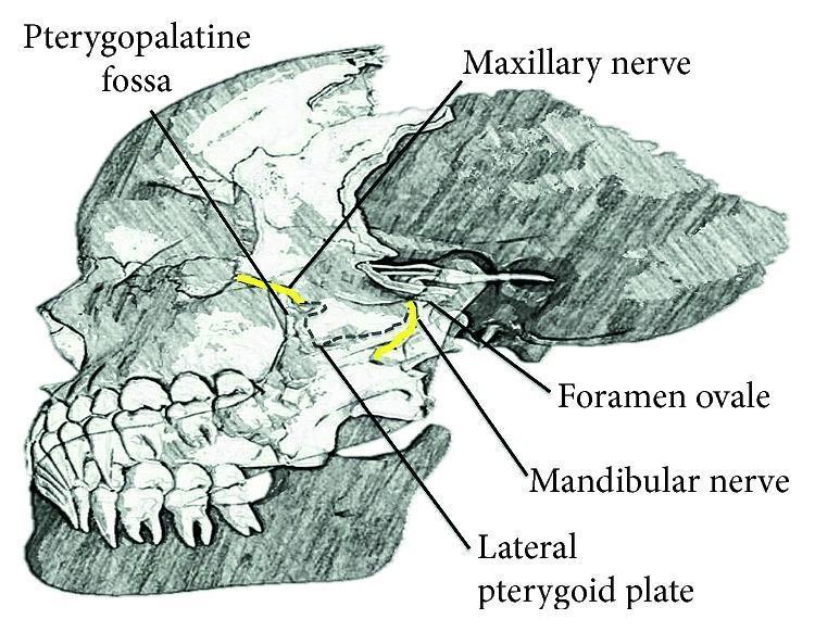
Anatomical diagram of a lateral view of the human skull, specifically focusing on the deep facial nerves and related osteological landmarks. The illustration highlights the trigeminal nerve (CN V) divisions using yellow color-coding. The maxillary nerve (V2) is shown exiting the cranial cavity through the foramen rotundum to enter the pterygopalatine fossa, located anterior to the lateral pterygoid plate. The mandibular nerve (V3) is depicted exiting via the foramen ovale, descending posterior to the lateral pterygoid plate. Key labels include the Pterygopalatine fossa, Maxillary nerve, Foramen ovale, Mandibular nerve, and Lateral pterygoid plate. The diagram serves as an educational resource for understanding the regional anatomy required for performing nerve blocks and identifying neural pathways within the infratemporal and pterygopalatine regions.
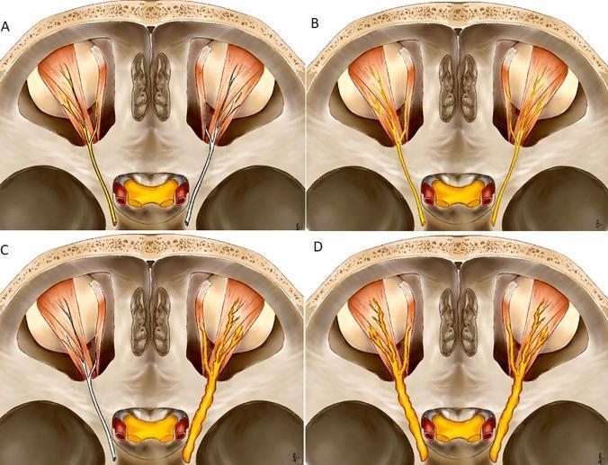
This medical illustration depicts four distinct patterns of cranial nerve enhancement (CNE) in an axial anatomical diagram of the skull base and orbits, focusing on the trigeminal and oculomotor regions. (A) Unilateral linear: Post-contrast enhancement (represented in yellow) along the course of a single nerve without an increase in its diameter. (B) Bilateral linear: Symmetrical enhancement of both nerves, maintaining a thin, smooth morphology. (C) Unilateral thickened: Pathological enhancement of one nerve accompanied by increased diameter and an irregular or nodular contour, contrasting with the normal contralateral nerve (white). (D) Bilateral thickened: Symmetrical enhancement and significant enlargement of both nerves with nodular irregularities. These patterns are used in neuroradiology to differentiate between various pathologies, such as viral neuritis (linear patterns) and neoplastic processes like perineural tumor spread or neurolymphomatosis (thickened patterns). The diagram serves as a diagnostic educational tool for classifying imaging findings in magnetic resonance imaging (MRI) and computed tomography (CT) of the cranial nerves.
sciatic nerve anatomy course distribution leg
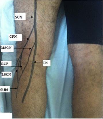
This clinical photograph shows a surface anatomy projection of the sciatic nerve (ScN) and its branches on the posterior aspect of a patient's left knee and leg. The illustration demonstrates the bifurcation of the sciatic nerve into the medial tibial nerve (TN) and the lateral common peroneal nerve (CPN). From the CPN, the lateral sural cutaneous nerve (LSCN) and the fibular communicating branch (RCF) are shown branching off. On the medial side, the medial sural cutaneous nerve (MSCN) descends from the tibial nerve. These branches converge to form the sural nerve (SuN) in the lower leg. The diagram serves as an educational tool for understanding the peripheral nerve distribution in the popliteal fossa and posterior compartment of the leg, specifically highlighting the pathways relevant to conditions like external popliteal sciatic nerve (EPSN) injuries or peripheral nerve tumors.
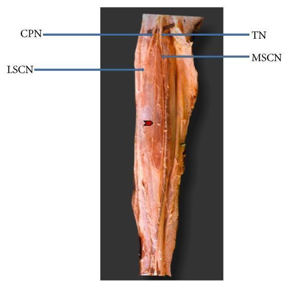
This clinical anatomical photograph displays a cadaveric dissection of the posterior lower leg, specifically focusing on the branching patterns of the nerves in the popliteal fossa and posterior calf. The image illustrates the division of the sciatic nerve into the tibial nerve (TN) and common peroneal nerve (CPN). From the tibial nerve, the medial sural cutaneous nerve (MSCN) is seen descending vertically. The lateral sural cutaneous nerve (LSCN) is labeled as a branch of the CPN. A significant anatomical variation is highlighted by a red arrowhead: an additional accessory branch originating from the common peroneal nerve superior to the origin of the LSCN. This branch follows a descending longitudinal course but remains independent, showing no communication or anastomosis with the medial sural cutaneous nerve. This image serves as an educational resource for studying peripheral nerve anatomy and the morphological variations of the sural nerve complex, which is clinically relevant for nerve grafts, biopsies, and understanding compression neuropathies.
nerve injury clinical signs wrist drop claw hand foot drop
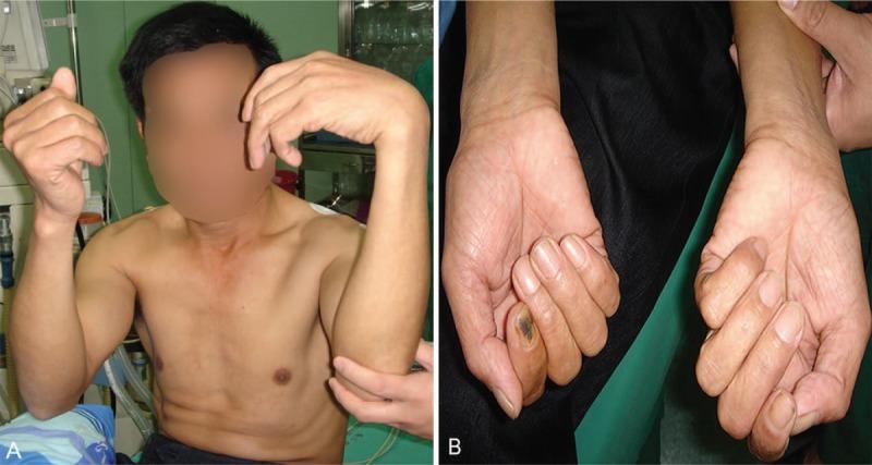
Clinical photograph of a 50-year-old male presenting with multi-nerve palsy of the left upper limb involving the radial, median, and ulnar nerves. Panel A shows a side-by-side comparison of the upper extremities; the left hand exhibits a characteristic 'wrist drop' and 'claw-shaped hand' deformity due to extensor and intrinsic muscle weakness. Panel B provides a close-up comparative view of the palmar surfaces. The left hand (right of image) demonstrates significant muscle atrophy, particularly in the thenar and hypothenar eminences, alongside flattening of the forearm flexor group. This visual evidence highlights the motor consequences of prolonged compression (Saturday night palsy/autogenous compression), demonstrating the classic postural signs of combined peripheral nerve injury in the hand and wrist.
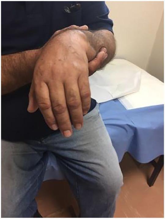
A clinical photograph of a male patient demonstrating 'wrist drop' or 'drop hand,' a classic sign of radial nerve palsy. The patient's right hand is shown held in a characteristic posture where the wrist is flexed downward and cannot be actively extended against gravity. The fingers are in a state of passive flexion at the metacarpophalangeal and interphalangeal joints, giving the hand a limp, flaccid appearance. The skin of the forearm and hand appears normal in color and texture, with no obvious swelling or ecchymosis visible in this view. This clinical manifestation is frequently associated with trauma, such as humeral fractures or shoulder dislocations that result in compression or injury to the radial nerve. The image serves as a high-yield educational resource for identifying peripheral nerve injuries during a neurological or orthopedic examination.
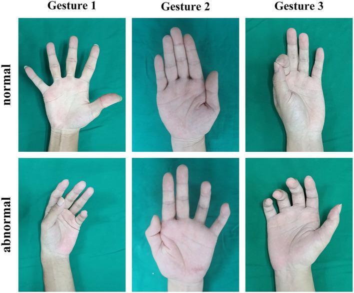
Clinical photographs depicting a comparison between normal and abnormal hand gestures used in the physical examination for peripheral nerve injuries. The image is organized in a 2x3 grid. Column 1 (Gesture 1) compares a normal fully extended and abducted hand against a 'wrist drop' or 'finger drop' appearance, typically associated with radial nerve injury. Column 2 (Gesture 2) shows a normal flat palm with fingers adducted against an abnormal posture with inability to fully extend or adduct the medial digits, often seen in ulnar nerve pathology. Column 3 (Gesture 3) demonstrates a normal 'O' sign or precision grip versus an abnormal attempt where the distal phalanges of the thumb and index finger fail to form a circle, indicative of median nerve injury (specifically anterior interosseous nerve). These images illustrate key clinical diagnostic signs including muscle weakness, atrophy, and characteristic postural deviations resulting from traumatic or compressive neuropathies of the upper limb.
Peripheral Nerves vs Cranial Nerves - Complete Concept Guide
1. The Big Picture: Nervous System Classification
- 31 pairs of spinal nerves - arise from the spinal cord
- 12 pairs of cranial nerves - arise from the brain
"Thirty-one pairs of spinal nerves arise from the spinal cord in the peripheral nervous system, compared to 12 pairs of cranial nerves that arise from the brain." - General Anatomy and Musculoskeletal System (THIEME Atlas of Anatomy), p. 98
2. Key Distinction: Peripheral (Spinal) Nerves
- Somatic motor (to voluntary muscles)
- Somatic sensory (from skin, joints, muscles)
- Autonomic fibers
3. Major Peripheral Nerves - Upper Limb
Radial Nerve (C5-C8, T1)
- Origin: Posterior cord of brachial plexus
- Course: Winds around the posterior humerus in the radial groove (spiral groove)
- Motor supply: All extensors of the wrist and fingers, supinator, brachioradialis, triceps
- Sensory: Dorsum of hand (first dorsal web space)
- Key clinical sign: Wrist drop - inability to extend the wrist at the MCP joints
- Common injury site: Midshaft humerus fracture (spiral groove), "Saturday night palsy" (prolonged arm compression)
- Important nuance: Interphalangeal extension is preserved (done by interossei via ulnar nerve through extensor expansions) - do not confuse this with signs of regeneration - Das Manual on Clinical Surgery, p. 143
Median Nerve (C6-T1)
- Origin: Two roots - one from lateral cord (C5,6,7) and one from medial cord (C8,T1)
- Course: Descends lateral to brachial artery, crosses in midarm, passes between heads of pronator teres, enters carpal tunnel deep to flexor retinaculum
- Motor supply: Pronators, wrist flexors (FCR, palmaris longus), FDS, FDP (lateral half), FPL, thenar muscles (LOAF: Lumbricals 1&2, Opponens pollicis, Abductor pollicis brevis, Flexor pollicis brevis)
- Sensory: Volar thumb/index/middle/lateral ring finger; index finger volar tip
- Key clinical signs:
- Carpal tunnel syndrome (most common entrapment neuropathy)
- Ape hand (flattening of thenar eminence from thenar muscle wasting)
- Hand of benediction (unable to flex index and middle fingers on making a fist)
- Loss of O-sign - cannot form a circle with thumb and index (AIN branch)
Ulnar Nerve (C8-T1)
- Origin: Medial cord of brachial plexus
- Course: Passes posterior to medial epicondyle (cubital tunnel), enters hand lateral to pisiform and ulnar artery in Guyon's canal
- Motor supply: FCU, FDP (medial half), hypothenar muscles, interossei, medial two lumbricals, adductor pollicis
- Sensory: Little finger, medial half of ring finger (volar and dorsal), medial palm
- Key clinical signs:
- Claw hand ("ulnar claw" - ring and little finger clawing, more pronounced distally = "Ulnar paradox")
- Froment's sign - flexion of thumb IP joint when pinching paper (compensating for lost adductor pollicis)
- Hypothenar and interosseous wasting
- Most common injury: Cubital tunnel (elbow); also Guyon's canal (wrist)
| Parameter | Median | Radial | Ulnar |
|---|---|---|---|
| Key motor | Thumb opposition, pronation | Wrist/finger extension, supination | Finger abduction/adduction |
| Test sign | "A-OK" sign | "Thumbs-up" sign | Finger spread |
| Sensory | Volar index finger tip | 1st dorsal web space | Volar little finger tip |
| Injury sign | Carpal tunnel / ape hand | Wrist drop | Claw hand |
4. Major Peripheral Nerves - Lower Limb
Femoral Nerve (L2-L4)
- Origin: Posterior divisions of L2, L3, L4 - from lumbar plexus
- Course: Passes deep to inguinal ligament, enters femoral triangle lateral to femoral artery and vein
- Motor supply: Quadriceps femoris (main knee extensor), iliacus, pectineus, sartorius
- Sensory: Anterior/medial thigh; most medial branch = saphenous nerve (medial leg and ankle)
- Key clinical signs:
- Inability to extend the knee
- Absent knee jerk reflex
- Loss of sensation over anterior thigh/medial leg
- Common injury: Pelvic fractures, retroperitoneal hematoma, lithotomy position (stretching)
Sciatic Nerve (L4, L5, S1, S2, S3)
- Origin: Sacral plexus (largest nerve in the body - 2-2.5 cm diameter at the thigh)
- Course: Exits pelvis through greater sciatic notch (below piriformis), descends posterior thigh deep to gluteus maximus, divides at popliteal fossa into:
- Common peroneal (fibular) nerve - lateral
- Tibial nerve - medial
- Motor supply: All muscles of leg and foot + posterior thigh (hamstrings)
- Sensory: Most of sensory supply to the leg and foot
- Key clinical signs:
- Foot drop (common peroneal division injury - can't dorsiflex or evert the foot)
- Loss of all sensation below knee
- Loss of ankle jerk (tibial division)
- Common injuries: Hip fractures/dislocations, posterior hip surgery, piriformis syndrome, prolonged bed rest (ICU) - Campbell's Operative Orthopaedics, p. 3878
Obturator Nerve (L2-L4)
- Exits pelvis through obturator foramen
- Motor: Adductors of thigh
- Sensory: Medial thigh
- Injured in pelvic fractures, obturator hernias
Common Peroneal (Fibular) Nerve
- Motor: Dorsiflexors (deep branch) + evertors (superficial branch)
- Sensory: Lateral leg, dorsum of foot
- Injury: Neck of fibula (most common lower limb nerve injury site) - causes foot drop
5. Cranial Nerves - All 12
Important exception: CN I (olfactory) and CN II (optic) are NOT true peripheral nerves - they are outpouchings of the brain, enclosed in meninges, containing oligodendrocytes (CNS cells). - THIEME Atlas, p. 98
All 12 Cranial Nerves
| # | Name | Type | Exit Foramen | Main Function | Injury Sign |
|---|---|---|---|---|---|
| I | Olfactory | Sensory (special) | Cribriform plate | Smell | Anosmia (head trauma, meningioma) |
| II | Optic | Sensory (special) | Optic canal | Vision | Visual field defects, blindness |
| III | Oculomotor | Motor + parasympathetic | Superior orbital fissure | Eye movements (SR, IR, MR, IO), pupil constriction, eyelid elevation | "Down and out" eye, ptosis, fixed dilated pupil |
| IV | Trochlear | Motor | Superior orbital fissure | Superior oblique muscle (eye intorsion/depression) | Diplopia on downward gaze; head tilt |
| V | Trigeminal | Mixed | V1: SOF; V2: Foramen rotundum; V3: Foramen ovale | Sensation of entire face; V3 also motor to muscles of mastication | Loss of corneal reflex, facial numbness, jaw deviation |
| VI | Abducens | Motor | Superior orbital fissure | Lateral rectus (abduction of eye) | Medial deviation of eye; diplopia on lateral gaze; longest intracranial course = most vulnerable to raised ICP |
| VII | Facial | Mixed + parasympathetic | Internal acoustic meatus / stylomastoid foramen | Facial muscles, taste anterior 2/3 tongue, lacrimal/salivary glands | UMN vs LMN facial palsy; Bell's palsy |
| VIII | Vestibulocochlear | Sensory (special) | Internal acoustic meatus | Hearing + balance | Sensorineural deafness, vertigo, tinnitus |
| IX | Glossopharyngeal | Mixed | Jugular foramen | Taste posterior 1/3 tongue, sensation pharynx/middle ear, parotid gland, stylopharyngeus motor | Loss of gag reflex (afferent limb) |
| X | Vagus | Mixed + parasympathetic | Jugular foramen | Pharynx/larynx motor, taste epiglottis, visceral sensation and parasympathetics to thorax/abdomen | Hoarseness, dysphagia, absent gag (efferent limb) |
| XI | Accessory | Motor | Jugular foramen | SCM and trapezius | Weakness turning head/shrugging shoulder |
| XII | Hypoglossal | Motor | Hypoglossal canal | All intrinsic + extrinsic tongue muscles (except palatoglossus) | Tongue deviates to SIDE of lesion (LMN) |
Cranial Nerve Functional Component System
| Component | Abbreviation | Examples |
|---|---|---|
| General somatic afferent (sensation) | GSA | CN V, VII, IX, X |
| General visceral afferent | GVA | CN IX, X |
| Special afferent (smell, taste, vision, hearing, balance) | SA | CN I, II, VII, VIII, IX, X |
| General somatic efferent (voluntary muscle) | GSE | CN III, IV, VI, XII |
| General visceral efferent (parasympathetic) | GVE | CN III, VII, IX, X |
| Branchial efferent (pharyngeal arch muscles) | BE | CN V, VII, IX, X, XI |
6. Peripheral Nerves vs Cranial Nerves - Side-by-Side Comparison
| Feature | Peripheral (Spinal) Nerves | Cranial Nerves |
|---|---|---|
| Number | 31 pairs | 12 pairs |
| Origin | Spinal cord | Brain (brainstem mostly; CN I & II = brain itself) |
| Exit route | Intervertebral foramina | Skull base foramina |
| Organization | Anterior (motor) + posterior (sensory) roots merge | Mixed from origin OR purely sensory/motor |
| Plexus formation | Yes (brachial, lumbar, sacral) | No (named individually) |
| Functional types | Somatic motor, somatic sensory, autonomic | All above PLUS special senses (vision, smell, hearing, taste, balance) + branchial efferent |
| Myelin | Schwann cells (PNS) | Schwann cells (true cranial nerves); oligodendrocytes (CN I, II = CNS) |
| Regeneration capacity | Yes (slow, ~1mm/day) | Limited (CN I has some olfactory regeneration; CN II cannot regenerate) |
| Clinical lesion classification | Seddon (neuropraxia/axonotmesis/neurotmesis) | UMN vs LMN pattern |
7. High-Yield Clinical Patterns
UMN vs LMN for Cranial Nerves
- CN VII (facial): UMN lesion (stroke) = forehead spared (bilateral cortical supply to upper face); LMN lesion (Bell's palsy) = entire ipsilateral face affected
- CN XII (hypoglossal): LMN = tongue deviates toward lesion; UMN = tongue deviates away
Peripheral Nerve Injury Levels Matter
- Axilla: All radial muscles affected including triceps
- Spiral groove: Triceps spared; wrist drop present
- Posterior interosseous: Wrist drop without sensory loss
Mnemonic: Cranial Nerve Numbers
Mnemonic: Sensory/Motor/Both
8. Common Entrapment Neuropathies (High-Yield)
| Nerve | Site | Cause | Sign |
|---|---|---|---|
| Median | Carpal tunnel | Repetitive wrist use, pregnancy, hypothyroidism | Thenar wasting, Tinel's/Phalen's |
| Ulnar | Cubital tunnel (elbow) | Leaning on elbow | Claw hand, hypothenar wasting |
| Ulnar | Guyon's canal (wrist) | Cyclists, handlebar pressure | Same but FCU and FDP spared |
| Radial | Spiral groove | Humeral fracture, crutch palsy | Wrist drop |
| Common peroneal | Neck of fibula | Leg crossing, casts, fracture | Foot drop |
| Femoral | Inguinal region | Psoas hematoma, lithotomy | Knee extension loss |
| Sciatic | Piriformis | Deep gluteal syndrome | Variable foot drop + posterior thigh pain |
| CN VI (abducens) | Intracranial | Raised ICP (false localizing sign) | Medial deviation, diplopia |
| CN VII (facial) | Stylomastoid foramen | Bell's palsy (reactivated HSV-1) | Complete ipsilateral facial weakness |
9. Autonomic Component
- Cranial nerves III, VII, IX, X carry parasympathetic fibers (head and neck + thoracoabdominal viscera via vagus)
- Spinal nerves T1-L2 carry sympathetic fibers
- Spinal nerves S2-S4 carry sacral parasympathetic fibers (pelvic viscera)
Sources
- General Anatomy and Musculoskeletal System - THIEME Atlas of Anatomy, pp. 98-99
- Gray's Anatomy for Students (2023 ed.), pp. 6461-6491
- Das Manual on Clinical Surgery, 13th Ed., pp. 143-145
- Campbell's Operative Orthopaedics 15th Ed. 2026, p. 3878
- ROSEN's Emergency Medicine, Block 6 (Median/Radial/Ulnar innervation table)
- Frameworks for Internal Medicine, p. 8885 (PNS definition)