
Teratology of Fallots
Reading File
Tetralogy of Fallot (ToF) is the most common cyanotic congenital heart disease, characterized by four anatomical defects: ventricular septal defect (VSD), pulmonary stenosis (right ventricular outflow tract obstruction), right ventricular hypertrophy, and an overriding aorta. The fundamental embryological defect is anterior-superior deviation of the infundibular septum.
Searching sources...
Finding Sources
Searching PubMed
"Tetralogy of Fallot"[MeSH Terms] AND management
Reading File
Reading File
Reading File
Reading File
Reading File
Loading Image
Loading Image
Finding Sources
Reading File
Loading Image
Here is a comprehensive overview of Tetralogy of Fallot (ToF), the most common cyanotic congenital heart disease.
Tetralogy of Fallot (ToF)
Definition & Epidemiology
Tetralogy of Fallot is the most common cyanotic congenital heart disease beyond infancy, accounting for approximately 5% of all congenital cardiac malformations, with an incidence of ~420 per million live births. The offspring of a mother with ToF has a 5-10% chance of inheriting the condition.
The Four Cardinal Defects
All four features arise from a single embryological defect: anterosuperior displacement (malalignment) of the infundibular (outlet/conus) septum, leading to unequal division of the truncus arteriosus.
| # | Defect | Details |
|---|---|---|
| 1 | VSD | Large, unrestrictive, malalignment type; near the membranous septum |
| 2 | Right ventricular outflow tract (RVOT) obstruction | Most commonly subpulmonic/infundibular stenosis; may also be valvular or involve pulmonary atresia |
| 3 | Overriding aorta | Aortic valve straddles the VSD, receiving blood from both ventricles |
| 4 | Right ventricular hypertrophy (RVH) | Secondary to the pressure load from RVOT obstruction |
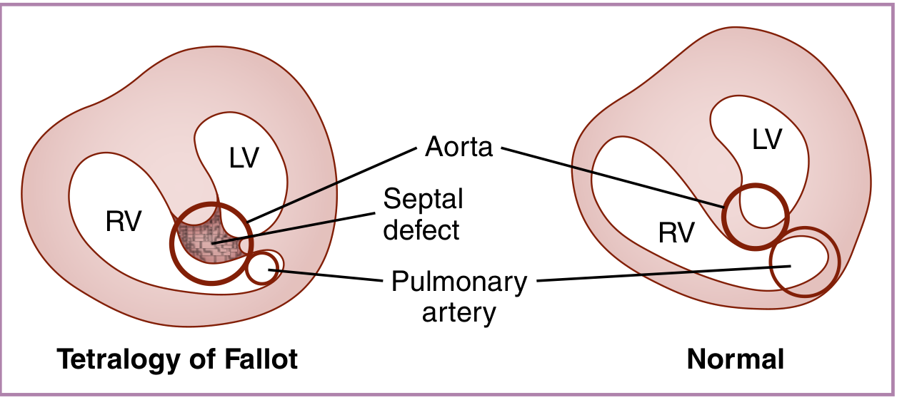
Fig: Tetralogy of Fallot (left) vs Normal (right) - note the overriding aorta, septal defect, and small pulmonary artery. (Creasy & Resnik's Maternal-Fetal Medicine)
Pathophysiology
The combination of RVOT obstruction and a large VSD results in decreased pulmonary blood flow and right-to-left shunting of deoxygenated blood across the VSD into the systemic circulation.
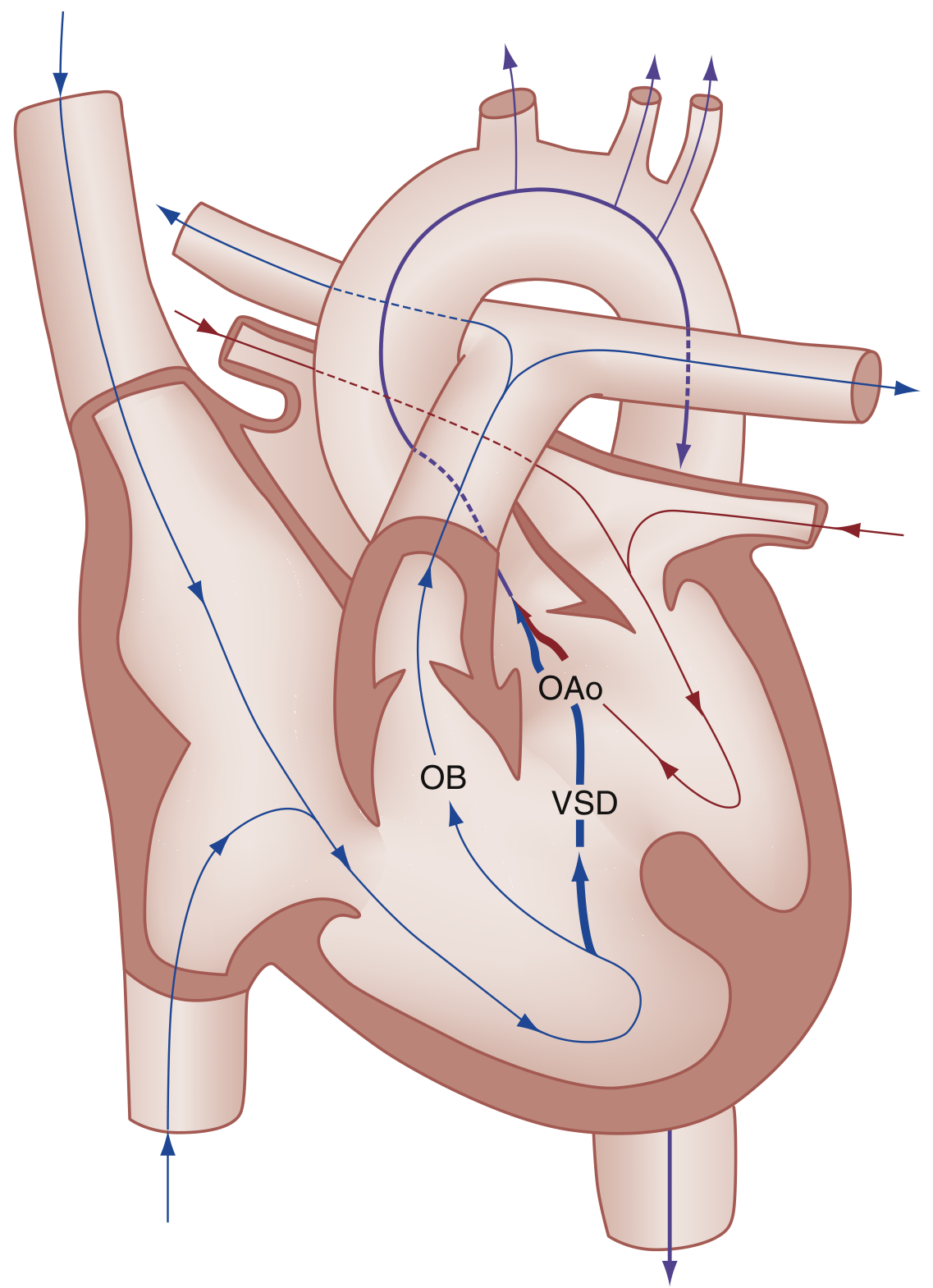
Fig: Right-to-left shunting in ToF. Deoxygenated blood from the RV shunts across the VSD into the overriding aorta (OAo). The amount depends on the degree of RVOT obstruction (OB), VSD size, and systemic vascular resistance (SVR). (Rosen's Emergency Medicine)
Clinical Features
Cyanosis
- Degree of cyanosis is directly proportional to the degree of RVOT obstruction
- "Pink Tet": mild RVOT obstruction → little or no cyanosis at birth
- Severe RVOT obstruction: profound cyanosis in the first days of life; may require prostaglandin E1 (PGE1) infusion to maintain ductal patency and pulmonary blood flow
- Chronic hypoxemia leads to compensatory polycythemia and clubbing of fingers and toes
Physical Exam
- Varying degrees of cyanosis
- Systolic ejection murmur along the left sternal border (from RVOT obstruction - the VSD itself is usually silent because it's large and unrestrictive)
Associated Anomalies
- Right-sided aortic arch (25%)
- ASD
- Anomalous origin of the left coronary artery
- Pulmonary atresia with VSD (extreme form of ToF; all RV output exits via the aorta)
Investigations
Chest X-Ray
- Boot-shaped heart ("coeur en sabot") - due to RVH causing upward elevation of the cardiac apex and a concave main pulmonary artery segment on the left heart border
- Decreased pulmonary vascular markings
- Right-sided aortic arch in ~25%
ECG
- Right ventricular hypertrophy
- Right axis deviation
Echocardiography
- Primary diagnostic modality in children
- Demonstrates all four anatomical components
- Shows large VSD and overriding aorta on parasternal long-axis view
Cardiac MRI
- Gold standard for assessing the right ventricle in repaired ToF
- Quantifies pulmonary regurgitation, RVOT anatomy, RV volumes and function
Tet Spells (Hypercyanotic/Hypoxic Spells)
A potentially life-threatening complication, with peak incidence at 2-4 months of age.
Trigger: Any acute drop in SVR (crying, defecation, feeding), hypovolemia, or tachycardia → massive right-to-left shunt → hypoxia → hyperpnea → increased venous return → more shunting (vicious cycle).
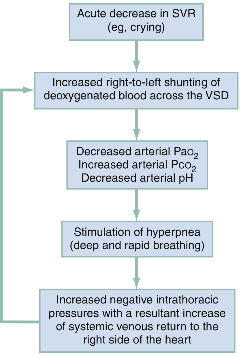
Fig: Pathophysiologic mechanisms of a hypoxic (tet) spell - a self-perpetuating cycle. (Rosen's Emergency Medicine)
Management of Tet Spells
- Knee-to-chest position (squatting equivalent) - increases SVR, reduces right-to-left shunt
- Supplemental oxygen (limited benefit alone)
- Morphine 0.1-0.2 mg/kg IV/IM (reduces hyperpnea and sympathetic drive)
- Fentanyl 1 mcg/kg IV/IM as alternative; or 1.5-2 mcg/kg intranasally
- Midazolam 0.2-0.3 mg/kg intranasally
- Sodium bicarbonate 1 mEq/kg IV if acidosis confirmed
- Ketamine 1-2 mg/kg IV or 3-5 mg/kg IM (increases SVR)
- Propranolol 0.1-0.2 mg/kg (reduces heart rate and infundibular spasm)
- Phenylephrine 0.01-0.02 mg/kg IV (direct vasoconstrictor, increases SVR)
(Rosen's Emergency Medicine, Box 165.8)
Surgical Management
Definitive Repair (Complete/Primary Repair)
- VSD closure (patch closure)
- Relief of RVOT obstruction - typically by placement of a transannular patch (across the pulmonary valve annulus to enlarge the RVOT)
- Preferred as early single-stage surgery in most centers
- Surgical mortality in the modern era is very low
Recent meta-analyses (PMID 39444206, PMID 40659877) comparing primary vs. staged repair in symptomatic neonates show primary repair is generally favored, though staged repair remains indicated in select cases.
Staged Repair (Palliation First)
Indicated when:
- Very narrow RVOT or significant central pulmonary artery hypoplasia
- Severe cyanosis in a very small/sick neonate
Modified Blalock-Taussig (BT) shunt: Anastomosis between the innominate artery and the right pulmonary artery - provides interim pulmonary blood flow until definitive repair is feasible. The shunt is taken down during subsequent definitive repair.
Post-Repair Residua and Long-Term Follow-Up
Repair is not curative - residual abnormalities are almost universal:
| Residual Problem | Notes |
|---|---|
| Pulmonary regurgitation (PR) | Most common; from transannular patch; leads to progressive RV dilation |
| RVOT aneurysm | Can develop at patch site |
| Residual RVOT obstruction | May require re-intervention |
| Arrhythmias and conduction disease | Common; risk of sudden death |
| Residual ASD or VSD | |
| Tricuspid regurgitation | |
| Aortic root dilatation |
Pulmonary valve replacement (PVR) is often required later in life due to progressive RV dilation from pulmonary regurgitation. A 2025 systematic review (PMID 40122137) compared surgical vs. transcatheter pulmonary valve replacement in ToF patients. CMR imaging is the gold standard for monitoring RV function/volumes and timing PVR.
Pregnancy in ToF
Per ACC/AHA guidelines, women with repaired ToF should undergo evaluation by an adult CHD specialist before pregnancy. In the absence of severe pulmonary hypertension, severe cardiomyopathy, or severe restrictive lung disease, pregnancy is generally well tolerated. Arrhythmia and pulmonary regurgitation are the main concerns.
Sources: Robbins & Kumar Basic Pathology | Rosen's Emergency Medicine | Creasy & Resnik's Maternal-Fetal Medicine | The Developing Human (Moore & Persaud) | Grainger & Allison's Diagnostic Radiology
This is a shared conversation. Sign in to Orris to start your own chat.