
हिस्टोलॉजी of thyroid gland and what is papilla in that?
thyroid gland histology follicles colloid parafollicular cells microscopy
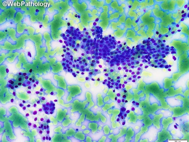
This is a light microscopy histology image of thyroid gland tissue prepared with a Romanowsky-type stain (e.g., Diff-Quik/Wright-Giemsa). The specimen shows normal-appearing follicular epithelium arranged in intact macrofollicles with abundant colloid occupying the luminal spaces. Follicular cells are evenly spaced with uniform, round to oval nuclei and scant to moderate cytoplasm, consistent with well-differentiated thyroid epithelium. The colloid background is pale blue and prominent, reflecting thyroglobulin-containing material released from follicles. The architectural pattern emphasizes large follicle units with a single layer of cuboidal to low-columnar epithelium surrounding expansive colloid pools. No prominent nuclear features of papillary carcinoma are evident, and there is no marked cellular atypia or mitotic activity in the visible fields. This image illustrates classic histology of benign thyroid tissue or colloid-rich goiter, useful for teaching follicular morphologies, thyroid folliculogenesis, and colloid dynamics. Clinically, such samples are evaluated in the context of thyroid nodules and goiter workups, with correlation to serum thyroid function tests and, when applicable, ultrasound or FNA findings. Potential educational uses include cytology-histology correlation, differential diagnosis of thyroid disease, and artifact recognition in Romanowsky-stained preparations. Images are suitable for medical education, pathology review, and database indexing. Correlative data improve student comprehension and diagnostic recall greatly.
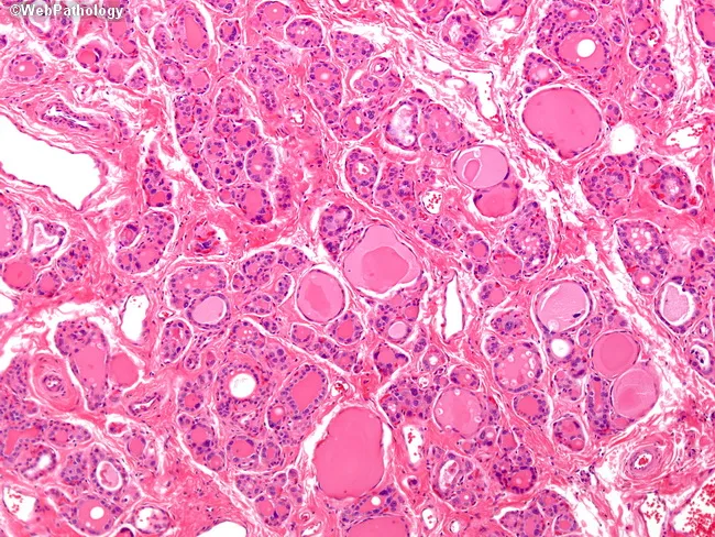
Imaging modality: Light microscopy of thyroid tissue section stained with Hematoxylin and Eosin. Specimen type: thyroid gland biopsy evaluated histologically. Imaging perspective: high-power microscopic view. Anatomical context: thyroid in the neck. Core histology demonstrates diffuse follicular hyperplasia with marked hypercellularity and reduced colloid throughout the gland. Follicles are small, compact, and tightly spaced, with scant colloid and thickened follicular epithelium. The epithelial cells show pronounced cytologic atypia, including bizarre hyperchromatic nuclei, a finding that can simulate neoplasia but is characteristic in dyshormonogenetic goiter due to inborn errors of thyroid hormone synthesis. This pattern is typically diffuse rather than nodular. Clinical significance: these histologic features correlate with congenital or neonatal hypothyroidism in severe cases and with goiter and mild hypothyroidism in milder forms; many patients present by approximately 25 years of age. Diagnostic relevance: supports dyshormonogenesis as an underlying cause of diffuse thyroid enlargement with impaired hormone production. Differential considerations include autoimmune thyroiditis and other causes of diffuse goiter; correlation with thyroid function tests and clinical history is essential. Potential clinical uses: diagnosis of dyshormonetic goiter, gene-level counseling, and guidance of hormone replacement therapy. This histology aids educational cases and research on thyroid hormone synthesis disorders, and informs patient management strategies.
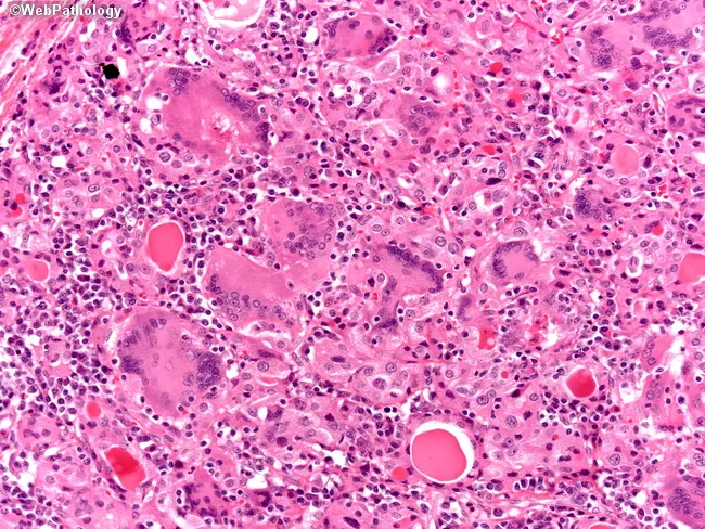
This case utilizes light microscopy of thyroid tissue stained with Hematoxylin and Eosin. The high-magnification image reveals granulomatous thyroiditis characterized by numerous foreign body giant cells and a dense chronic inflammatory infiltrate composed of lymphocytes and plasma cells. Interspersed is residual thyroid parenchyma containing colloid-filled follicles, some diminished or disrupted by inflammation. The tissue architecture shows granulomas centered around disrupted colloid with multinucleated giant cells phagocytosing colloid material, a classic appearance of subacute granulomatous (de Quervain) thyroiditis. There is relative preservation of overall gland structure with focal areas of follicular destruction. These features distinguish subacute thyroiditis from autoimmune thyroiditis (Hashimoto) and bacterial abscesses. Clinically, this pattern correlates with a transient thyrotoxic phase followed by hypothyroidism in many patients; HLA-B35 haplotype association has been reported; etiology often linked to viral trigger; most cases resolve with supportive care. The image supports diagnosis of granulomatous thyroiditis in the appropriate clinical context, guiding management toward observation and symptomatic treatment. This histology is relevant for education in endocrine pathology, differential diagnosis of thyroiditis, and research into post-viral inflammatory thyroid injury. The depiction is representative for pathology teaching slides and radiology correlations in endocrinology. Educational value for residents, fellows, and medical students with accurate histology correlations.
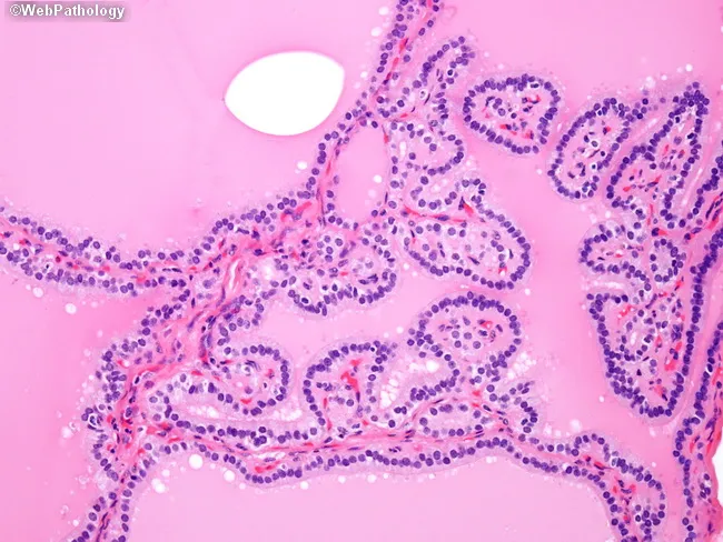
This is a brightfield, hematoxylin and eosin stained histology slide of thyroid tissue examined by light microscopy at high magnification. The specimen is thyroid gland parenchyma showing diffuse follicular hyperplasia characteristic of Graves disease. Follicles appear crowded with epithelial cells lining irregular, papillary-like folds protruding into the lumen, creating pseudo-papillary architectures that could mimic papillary thyroid carcinoma if evaluated in isolation. The nuclei within the thyrocytes are hyperplastic and dense but lack the distinctive nuclear features of papillary carcinoma, such as crowding with nuclear grooves, inclusions, Orphan Annie eye clearing, and powdery chromatin. Colloid is variably present or scant; stroma is vascular, and the overall architecture is polyfollicular with ingrowths into luminal spaces. This pattern reflects autoimmune-driven stimulation by TSH receptor autoantibodies (e.g., thyroid-stimulating immunoglobulin), resulting in diffuse goiter with hyperthyroid state. Clinically, Graves disease presents with weight loss, tachycardia, heat intolerance, and sometimes exophthalmos; histology aids diagnosis when correlated with serology and uptake studies. Key diagnostic significance: avoid mislabeling as papillary thyroid carcinoma; rely on clinical context and absence of malignant nuclear features. Potential educational uses include differential diagnosis training for thyroid pathology, immune-mediated thyroid hyperplasia vs neoplasia, and radiology-pathology correlation. This image supports teaching clinics and pathology review conferences.
thyroid gland papillary projections follicular epithelium histology diagram
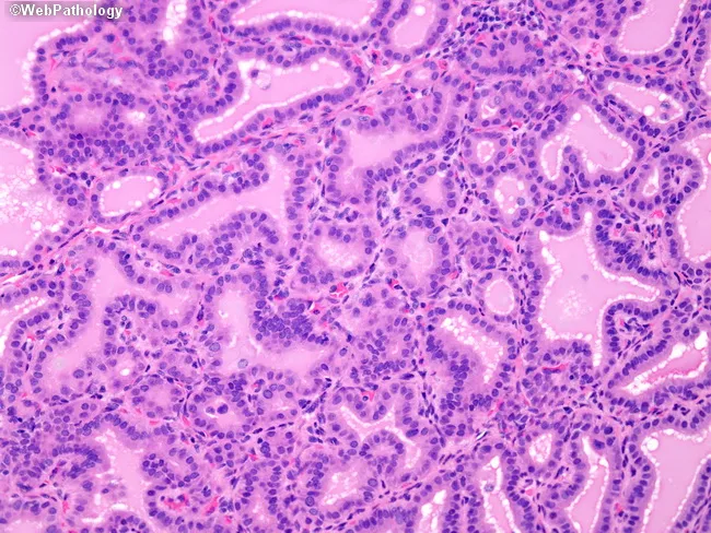
Imaging modality: light microscopy of hematoxylin and eosin–stained thyroid tissue sections. Primary subject: thyroid follicular epithelium with colloid-filled follicles. Anatomical context: thyroid gland parenchyma in the cervical region, showing follicles lined by tall columnar epithelium. Morphology: hypercellular epithelium with marked nuclear crowding creates papillary infoldings that protrude into the glandular lumens. Pale colloid is present within the follicles and margins appear scalloped. In architectural detail, papillary projections resemble papillary thyroid carcinoma but lack true fibrovascular cores, a key differentiator on histology. The follicular lumens vary in size, and colloid shows scalloping margins, consistent with benign activity rather than malignant invasion. Cytologic features include elongated nuclei with ovoid contours and scant cytoplasm; mitotic activity is not conspicuous. Immunophenotype is not assessed in this image; histology alone raises diagnostic considerations of benign hyperplastic changes including nodular hyperplasia or benign follicular adenoma with papillary-like infoldings. Diagnostic significance: recognition of papillary-like architecture without fibrovascular cores reduces the likelihood of classic papillary thyroid carcinoma, and highlights the need for correlating with nuclear features and ancillary studies. This image is relevant for surgical pathology reference, education, and differential diagnosis training in endocrine pathology. These considerations are essential for resident education, diagnostic conundrums, and interdisciplinary tumor boards.
This is a light microscopy histology image of thyroid gland tissue prepared with a Romanowsky-type stain (e.g., Diff-Quik/Wright-Giemsa). The specimen shows normal-appearing follicular epithelium arranged in intact macrofollicles with abundant colloid occupying the luminal spaces. Follicular cells are evenly spaced with uniform, round to oval nuclei and scant to moderate cytoplasm, consistent with well-differentiated thyroid epithelium. The colloid background is pale blue and prominent, reflecting thyroglobulin-containing material released from follicles. The architectural pattern emphasizes large follicle units with a single layer of cuboidal to low-columnar epithelium surrounding expansive colloid pools. No prominent nuclear features of papillary carcinoma are evident, and there is no marked cellular atypia or mitotic activity in the visible fields. This image illustrates classic histology of benign thyroid tissue or colloid-rich goiter, useful for teaching follicular morphologies, thyroid folliculogenesis, and colloid dynamics. Clinically, such samples are evaluated in the context of thyroid nodules and goiter workups, with correlation to serum thyroid function tests and, when applicable, ultrasound or FNA findings. Potential educational uses include cytology-histology correlation, differential diagnosis of thyroid disease, and artifact recognition in Romanowsky-stained preparations. Images are suitable for medical education, pathology review, and database indexing. Correlative data improve student comprehension and diagnostic recall greatly.
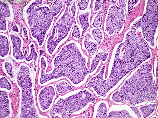
This is a light micrograph of thyroid gland tissue prepared for histopathology and stained with Hematoxylin and Eosin (H&E). The sample reveals multiple spherical to irregular follicles of varying size separated by wispy fibrous septa. The colloid-filled lumens are pink to pale pink, while the surrounding follicular epithelium is a uniform single layer of cuboidal to low-columnar cells with round to oval nuclei and inconspicuous nucleoli. No nuclear grooves, inclusions, or pseudoinclusions characteristic of papillary carcinoma are evident. The stromal background is relatively scant with no dense lymphocytic infiltrate or Hurthle cell metaplasia apparent. The architecture is reminiscent of normal thyroid parenchyma or benign nodular colloid goiter pattern, where follicle size varies and colloid predominates. There are no mitotic figures or cytologic atypia observed. This image demonstrates classic histology of endocrine gland tissue with preserved colloid and regular follicle lining, suitable as reference for normal thyroid architecture in educational, diagnostic, and research contexts. Clinically, such histology supports benign thyroid pathology when correlated with imaging and labs. In malignant differentials, absence of papillary nuclear features or tall columnar cells argues against papillary thyroid carcinoma; however, definitive diagnosis should consider capsule invasion and margin status in surgical specimens.
Thyroid Gland की Histology (ऊतक-विज्ञान)
संरचना का Overview
Thyroid Follicle - Structural & Functional Unit
Follicle की बनावट:
- Shape: Roughly spherical (गोलाकार), cyst-like compartment
- Size: 0.2 से 1.0 mm diameter
- Wall: Simple cuboidal या low columnar epithelium की single layer
- Lumen: Center में colloid - एक gel-like mass जिसमें thyroglobulin (iodinated precursor protein) भरा होता है
- Basal surface: Typical basal lamina पर टिकी होती है
- Apical surface: Colloid के साथ contact में - यहाँ microvilli होते हैं
- Histology: A Text and Atlas, p. 2011
Follicular Epithelium में दो प्रकार की Cells
1. Follicular Cells (Principal Cells)
- Function: T4 (thyroxine) और T3 (triiodothyronine) का synthesis
- Shape: Gland की functional state के अनुसार बदलती है:
- Inactive state: Flat squamous जैसी cells, colloid अधिक
- Normal state: Cuboidal cells
- Active/Stimulated state: Tall columnar cells, colloid कम
- H&E staining में: Slightly basophilic basal cytoplasm, spherical nuclei, prominent nucleoli
- Golgi apparatus: Supranuclear position में
- EM पर: rER, Golgi, lysosomes, colloid resorption droplets (CRDs) दिखते हैं
2. Parafollicular Cells (C Cells)
- Location: Follicular epithelium की periphery में, follicle की basal lamina के अंदर - लेकिन follicle lumen से कोई contact नहीं
- Function: Calcitonin का secretion (calcium metabolism regulate करता है)
- H&E में: Pale staining, solitary या small clusters में
- EM पर: Numerous small secretory vesicles (60-550 nm), prominent Golgi apparatus
- LM पर identify करना difficult होता है - immunostaining जरूरी
- Histology: A Text and Atlas, p. 2012-2014
यहाँ Textbook की Original Microscopy Image है:
Hormone Synthesis के Steps (Follicular Cell में)
- Thyroglobulin synthesis - rER में
- Thyroglobulin secretion - apical surface से colloid में
- Iodide uptake - NIS (Na+/I- symporter) द्वारा blood से
- Oxidation of iodide - Thyroid peroxidase (TPO) enzyme द्वारा
- Iodination - Thyroglobulin के tyrosine residues का → MIT, DIT बनते हैं → coupling से T3, T4
- Resorption of colloid - TSH stimulus पर receptor-mediated endocytosis
- Release - Lysosomal proteases thyroglobulin को break करती हैं → free T4 व T3 blood में
Vascular Supply
"Papilla" Thyroid में क्या होता है?
1. Papillary Projections in Graves Disease (Pseudo-papillae)
- Follicular epithelium inward fold बनाती है → lumen में protrude करती है
- ये pseudo-papillae कहलाती हैं
- Fibrovascular core नहीं होता (यह important differentiator है)
- Colloid scant होता है या scalloped margins दिखाता है
- Nuclei hyperplastic लेकिन malignant features (nuclear grooves, Orphan Annie nuclei, inclusions) absent
⚠️ Important: ये papillae benign हैं - Papillary Thyroid Carcinoma (PTC) से अलग करना जरूरी है।
2. Papillary Thyroid Carcinoma (PTC) में True Papillae
- Fibrovascular core होता है (connective tissue + blood vessel से बना stalk)
- इस core के चारों तरफ neoplastic epithelial cells की layer होती है
- Characteristic nuclear features:
- "Orphan Annie eye" nuclei (optically clear/ground glass nuclei)
- Nuclear grooves
- Nuclear pseudo-inclusions
- Nuclear crowding और overlap
- Psammoma bodies हो सकते हैं - concentrically laminated calcified structures (mineralized tombstones to mummified papillae)
Summary Table
| Feature | Normal Follicle | Graves/Hyperplasia (Pseudo-papillae) | PTC (True Papillae) |
|---|---|---|---|
| Epithelium | Simple cuboidal | Hyperplastic columnar, folded | Neoplastic, multilayered |
| Papillary projection | नहीं | हाँ (pseudo) | हाँ (true) |
| Fibrovascular core | नहीं | नहीं | हाँ |
| Nuclear features | Normal | Hyperplastic, benign | Orphan Annie, grooves, inclusions |
| Colloid | Abundant | Scant/scalloped | Minimal/absent |
| Psammoma bodies | नहीं | नहीं | हो सकते हैं |
| Nature | Normal | Benign | Malignant |
Quick Recall Points
- Structural unit = Thyroid follicle
- Follicular cells → T3, T4 (TSH से stimulated)
- Parafollicular/C cells → Calcitonin (lumen से no contact)
- Colloid = Thyroglobulin storage form
- Active thyroid → Tall columnar cells + less colloid + scalloped margins
- Inactive thyroid → Flat cells + abundant colloid
- Papilla in Graves → Pseudo-papillae, no fibrovascular core, benign
- Papilla in PTC → True papillae with fibrovascular core + malignant nuclear features
Sources: Histology: A Text and Atlas with Correlated Cell and Molecular Biology, pp. 2010-2020; Robbins & Kumar Basic Pathology, p. 729