
What do you mean by cogs of the heart? Cogs of the heart.
cogs of the heart cardiac anatomy meaning
"cogs of the heart" cardiac valves fibrous skeleton anatomy
fibrous skeleton of the heart cardiac valves rings diagram
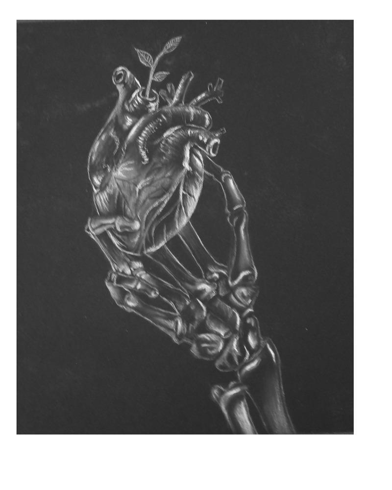
This medical illustration, titled 'Skeleton Heart,' is an anatomical diagram rendered in charcoal and white conté on a dark background, primarily serving an educational role in the medical humanities. It depicts a detailed skeletal hand and wrist—showing phalanges, metacarpals, and carpals—cradling a human heart. The heart is presented with clear anatomical landmarks, including the superior vena cava, aorta, and pulmonary trunk. A small, leafy plant sprout emerges from the superior aspect of the heart, specifically from one of the great vessels, symbolizing hope or biological regeneration. The illustration is designed to teach concepts related to cardiovascular health awareness, ischemic pain recognition, and the emotional impact of cardiac disease. It uses chiaroscuro-style shading to emphasize bone texture and cardiac valves, providing a visual metaphor for the 'warning signs' of heart failure or cardiac events. This content is suitable for medical education modules focusing on patient perspectives, prodromal symptoms of heart disease, and the psychosocial aspects of chronic illness.
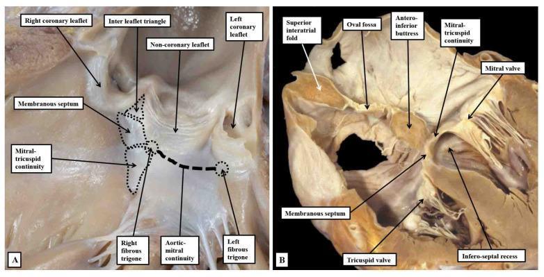
This figure consists of two clinical photographs of human heart specimens, providing a detailed anatomical study of the cardiac skeleton and outflow tracts. Panel A illustrates an infant heart with the left ventricular outflow tract (LVOT) opened. It demonstrates the structural components of the central fibrous body, including the membranous septum, right fibrous trigone, and left fibrous trigone. Key landmarks highlighted include the aortic-mitral fibrous continuity and its relationship to the interleaflet triangle between the right and non-coronary aortic sinuses. Panel B presents a 'four-chamber' section of an adult heart, emphasizing the structural support of the atrial septum. Visible features include the oval fossa, the antero-inferior buttress, and the infero-septal recess. The image depicts how the mitral-tricuspid continuity forms the roof of the infero-septal recess, providing a basal anchor for the interatrial septum. These images serve as educational materials for understanding cardiac surgical anatomy, specifically the relationships between the atrioventricular valves and the aortic root.
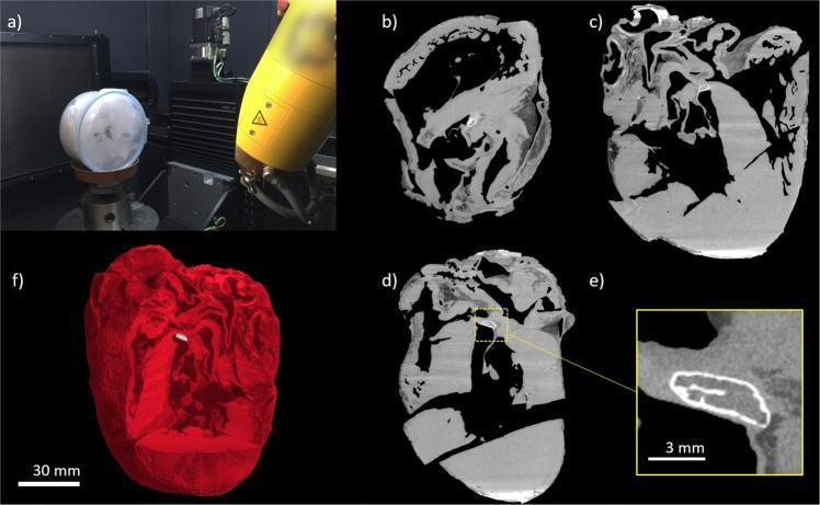
This composite image presents a high-resolution microCT analysis of a formalin-fixed heart, focusing on the identification of hyperdense structures within the cardiac skeleton. Panel (a) shows the physical setup of the microCT scanning equipment. Panels (b), (c), and (d) provide cross-sectional microCT slices of the cardiac anatomy. In these images, the cardiac chambers appear as hypodense (black) voids, while the myocardium, heart valves, and vascular walls are depicted in medium-density gray. Notably, a bright, hyperdense focal structure is visible in the valvular plane, located between the mitral and tricuspid valve regions. Panel (e) offers a magnified view of this hyperdense area, revealing a well-defined, mineralized structure with a thin, bright cortex and a less dense central region, characteristic of ectopic bone formation (os cordis) or mineralized cartilage. Panel (f) displays a 3D volumetric rendering of the heart in red, with the hyperdense bone-like structure highlighted in white within the internal anatomy. This visual material illustrates pathological mineralization and ectopic ossification within the fibrous cardiac skeleton, a significant finding in cardiovascular pathology and comparative anatomy.
"Cogs of the Heart" - The Fibrous Cardiac Skeleton
What is the Fibrous Skeleton of the Heart?
| Component | Description |
|---|---|
| 4 Fibrous Rings (Annuli Fibrosi) | Surround the orifices of all 4 heart valves |
| Right Fibrous Trigone (Central Fibrous Body) | Thickest area, where the aortic, tricuspid, and bicuspid valves meet - the "keystone" |
| Left Fibrous Trigone | Where the aortic and bicuspid (mitral) valves meet |
| Membranous Septa | Membranous portions of the interatrial, interventricular, and AV septa |
Why "Cogs"? - The 5 Key Functions
-
Structural support - Anchors the bases (leaflets and cusps) of all 4 cardiac valves and prevents them from overdilating under pressure.
-
Myocardial anchor - Atrial muscle fibers originate from the upper border of the rings; ventricular muscle fibers from the lower border. The whole pump is literally bolted to this framework.
-
Electrical insulator - The skeleton completely separates atrial from ventricular myocardium electrically. The AV node and bundle of His are the only electrical pathway through it. This allows atria to contract first, then ventricles - the coordinated "cog-turning" sequence of the cardiac cycle.
-
Maintains valve patency - Keeps the valve orifices open and patent so blood can flow through.
-
Mechanical load distribution - Distributes the mechanical forces of each heartbeat evenly across all chambers.
Visual Reference