Now I have all the material needed. Let me compose the comprehensive answers.
Complete Cell Biology Study Guide
SHORT QUESTIONS (3 MARKS EACH)
Q1. Three Organelles Bounded by a Single Membrane and Three by a Double Membrane
Single-membrane organelles
These organelles are enclosed by just one lipid bilayer:
| Organelle | Function |
|---|
| Lysosome | Contains hydrolytic enzymes; digests cellular debris and pathogens |
| Endoplasmic reticulum (ER) | Rough ER synthesizes proteins; smooth ER synthesizes lipids and detoxifies |
| Golgi apparatus | Modifies, sorts, and packages proteins for secretion or intracellular use |
Other examples: peroxisomes, vacuoles, secretory vesicles
Double-membrane organelles
These organelles have two concentric lipid bilayers (outer and inner membrane):
| Organelle | Function |
|---|
| Mitochondria | ATP synthesis via oxidative phosphorylation; site of cellular respiration |
| Nucleus | Houses the genome; controls gene expression |
| Chloroplast | Site of photosynthesis (plant cells only) |
The double membrane in mitochondria and chloroplasts reflects their endosymbiotic origin from ancient prokaryotes.
Q2. Cell Signalling - Definition and Protein Signal Pathway
Definition
Cell signalling is the process by which cells detect and respond to chemical signals from their environment. Cells use roughly two dozen families of receptors to detect extracellular molecules and physical stimuli, with each cell type bearing a distinctive set of receptor proteins that allow it to respond in a specific, programmed way.
- Junqueira's Basic Histology, p. 87
Pathway of a Protein Signal from Outside to Inside the Cell
Protein signals (e.g. hormones, growth factors, cytokines) are too large and hydrophilic to cross the plasma membrane directly. They follow this sequence:
-
Ligand synthesis and secretion - The signalling cell secretes a protein signal molecule (ligand) by exocytosis into the extracellular environment.
-
Receptor binding - The ligand binds with high affinity to a specific receptor protein on the surface of the target cell. Receptors for protein hormones are integral membrane proteins.
-
Receptor activation - Binding causes a conformational change in the receptor. Most protein signal receptors are either:
- Receptor tyrosine kinases (RTKs) - dimerize and autophosphorylate on tyrosine residues
- G-protein-coupled receptors (GPCRs) - activate associated heterotrimeric G proteins
-
Second messenger generation - Activated receptors generate intracellular second messengers (e.g. cAMP, IP3, DAG, Ca2+) that amplify the signal.
-
Protein kinase cascade - Second messengers activate protein kinases (e.g. PKA, PKC, MAPK) which phosphorylate target proteins. Second messenger-independent protein kinases such as MAPK, cyclin-dependent kinase (Cdk), and protein tyrosine kinase also play roles.
-
Cellular response - Phosphorylation events alter the activity of enzymes, structural proteins, and transcription factors, producing the final cellular response (e.g. gene expression, secretion, cell division).
- Histology: A Text and Atlas, p. 143
Q3. Prokaryotic vs Eukaryotic Cells
| Feature | Prokaryotic Cell | Eukaryotic Cell |
|---|
| Nucleus | Absent; DNA in nucleoid region | Present; DNA enclosed by nuclear envelope |
| Size | Small (1-10 µm) | Larger (10-100 µm) |
| Membrane-bound organelles | Absent | Present (mitochondria, ER, Golgi, etc.) |
| DNA | Single, circular chromosome; no histones | Multiple linear chromosomes; associated with histones |
| Ribosomes | 70S (50S + 30S subunits) | 80S (60S + 40S subunits) |
| Cell wall | Present (peptidoglycan in bacteria) | Present in plants (cellulose); absent in animals |
| Plasma membrane | Present | Present |
| Cell division | Binary fission | Mitosis/meiosis |
| Examples | Bacteria, archaea | Animal, plant, fungal, protist cells |
Diagrams
Prokaryotic cell:
        _______________
       /  Capsule       \
      | _______________  |
      ||  Nucleoid      | |
      || (circular DNA) | |
      || Ribosome (70S) | |
      ||________________| |
      |  Cell wall         |
      |  Plasma membrane   |
      |  Flagellum ------> |
       \___________________/
         Pilus
Eukaryotic (Animal) cell:
         ___________________________
        /   Smooth ER    Rough ER   \
       |   ___________  ________    |
       |  |           ||Ribosomes|  |
       |  | Nucleus   ||________|  |
       |  | (DNA +    |            |
       |  |  histones)|  Golgi     |
       |  |___________|  apparatus |
       |                           |
       |  Mitochondria  Lysosome   |
       |  Cytoskeleton             |
        \__________________________/
           Plasma membrane (single)
Q4. Stem Cells - Definition and Advantages
Definition
Stem cells are undifferentiated or partially differentiated cells capable of:
- Self-renewal - dividing to produce more stem cells
- Differentiation - developing into specialized cell types
They serve as a repair system for the body throughout life.
Advantages of Using Stem Cells
| Advantage | Explanation |
|---|
| Regenerative medicine | Can replace damaged or diseased tissues (e.g. cardiac cells after heart attack, neurons in Parkinson's disease) |
| Drug testing and development | Provide human cell models to test drug safety and efficacy without animal testing |
| Disease modelling | Patient-derived stem cells recreate diseases in vitro for mechanistic study |
| Unlimited proliferation | Can be expanded in culture to yield large quantities of cells for therapy |
| Reduced immune rejection | Autologous stem cells (from the patient) avoid immune rejection |
| Treatment of haematopoietic disorders | Haematopoietic stem cell transplantation treats leukaemia, lymphoma, aplastic anaemia |
| Understanding development | Studying stem cells reveals mechanisms of embryogenesis and organogenesis |
Q5. Categories of Stem Cells
Stem cells are classified by their potency (differentiation capacity) and origin:
By Potency
TOTIPOTENT
  │  Can form entire organism including extraembryonic tissues
  │  Example: fertilized egg (zygote), first few blastomeres
  ▼
PLURIPOTENT
  │  Can form any cell of all three germ layers
  │  Cannot form placenta/extraembryonic tissues
  │  Examples: Embryonic Stem Cells (ESCs), iPSCs
  ▼
MULTIPOTENT
  │  Can form multiple cell types within one lineage
  │  Examples: Haematopoietic stem cells (HSCs), mesenchymal stem cells
  ▼
OLIGOPOTENT
  │  Can differentiate into a few cell types
  │  Example: Lymphoid progenitor cells
  ▼
UNIPOTENT
     Can only produce one cell type, but retain self-renewal
     Example: Muscle satellite cells, spermatogonial stem cells
By Origin/Source
| Type | Source | Notes |
|---|
| Embryonic Stem Cells (ESCs) | Inner cell mass of blastocyst | Pluripotent; ethical controversies regarding embryo destruction |
| Adult (Somatic) Stem Cells | Bone marrow, adipose, gut, skin, brain | Multipotent; less ethically controversial |
| Foetal Stem Cells | Foetal tissue (umbilical cord, amniotic fluid) | More potent than adult; less controversial than ESCs |
| Induced Pluripotent Stem Cells (iPSCs) | Any adult somatic cell reprogrammed to pluripotency | Avoids embryo destruction; patient-specific |
| Cancer Stem Cells | Tumour tissue | Responsible for tumour growth and recurrence |
Q6. Advantages and Disadvantages of Induced Pluripotent Stem Cells (iPSCs)
iPSCs are adult somatic cells (e.g. skin fibroblasts) reprogrammed to a pluripotent state by introducing transcription factors (originally Oct4, Sox2, Klf4, c-Myc - the Yamanaka factors, 2006 Nobel Prize).
Advantages
| Advantage | Detail |
|---|
| No embryo destruction | Avoids the ethical controversy of destroying human embryos to obtain ESCs |
| Patient-specific | Generated from the patient's own cells - autologous transplantation reduces immune rejection |
| Unlimited supply | Can be generated from any adult cell and expanded indefinitely in culture |
| Disease modelling | Patient-derived iPSCs carry the patient's mutations, enabling personalised disease models |
| Drug screening | Patient-specific iPSC-derived cells allow personalised pharmacology testing |
| Genetic correction | Can be gene-edited (CRISPR) before differentiation to correct disease mutations |
| No HLA-matching problems | Autologous cells do not require HLA matching for transplantation |
Disadvantages
| Disadvantage | Detail |
|---|
| Tumourigenicity | c-Myc is an oncogene; integration of reprogramming vectors can cause insertional mutagenesis and tumour formation |
| Epigenetic memory | iPSCs may retain epigenetic marks from the original somatic cell, reducing full reprogramming |
| Genomic instability | Reprogramming can introduce copy number variations and point mutations |
| Inefficient reprogramming | Reprogramming efficiency is low (0.01-0.1%); cost and time are high |
| Incomplete reprogramming | Not all iPSC lines achieve true pluripotency equivalent to ESCs |
| Differentiation variability | iPSC lines vary in their differentiation potential between batches and patients |
| Safety concerns | Rigorous characterisation required before any clinical use |
Q7. Four Membrane Transport Mechanisms
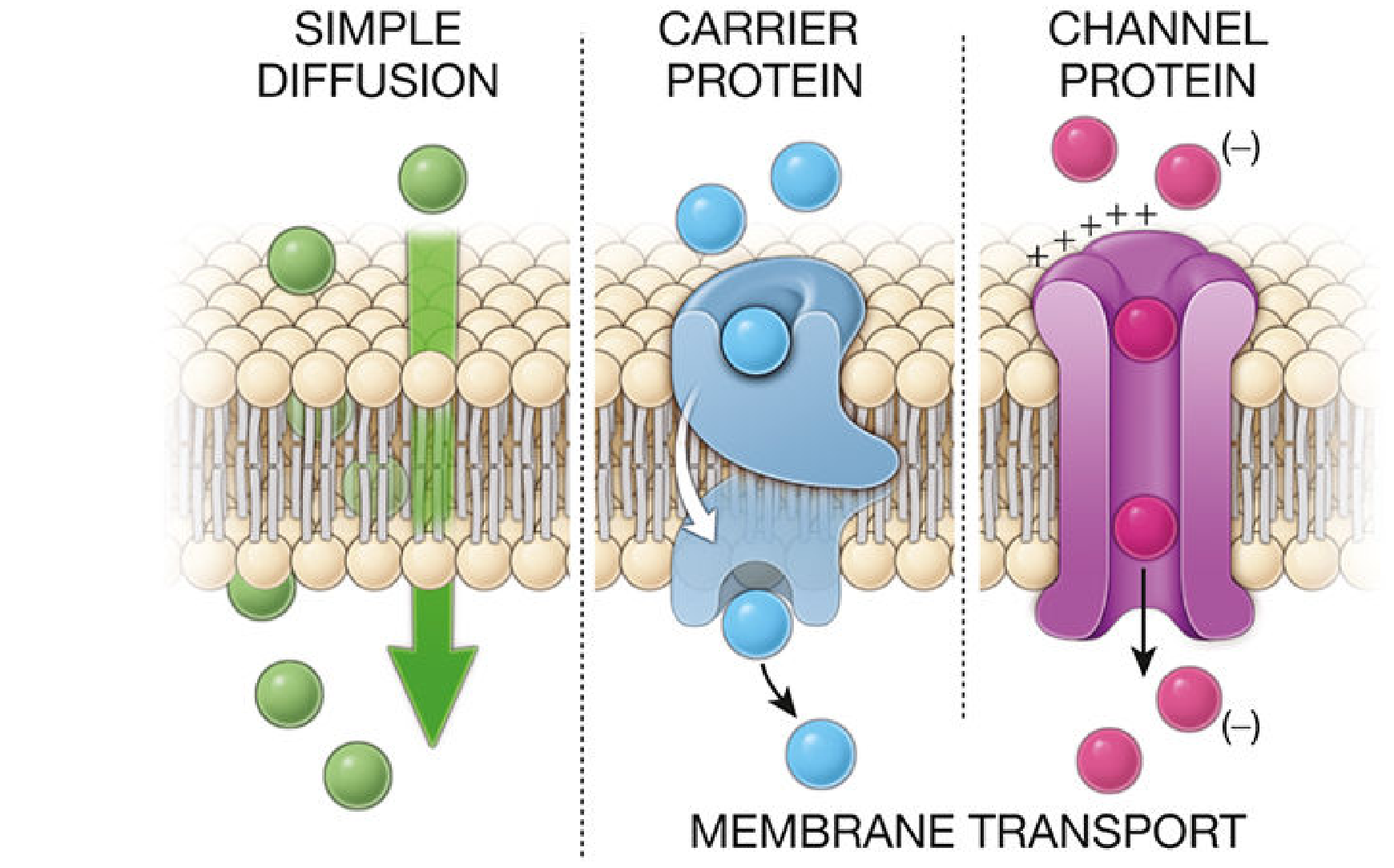
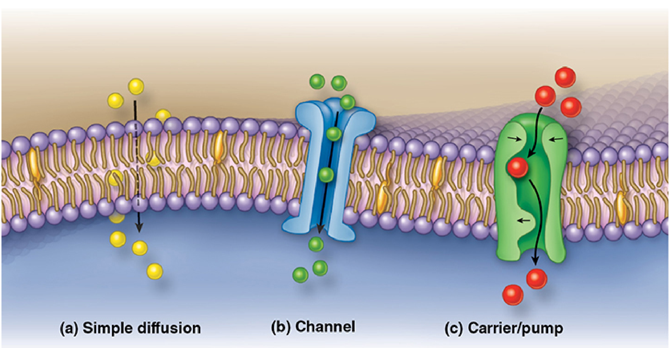
Diagram 1 - Overview of passive mechanisms
Histology: A Text and Atlas - Fig 2.9
Junqueira's Basic Histology - Fig 2-5
1. Simple Diffusion
- Mechanism: Movement of small, nonpolar, lipophilic (fat-soluble) molecules and gases directly through the lipid bilayer, down their concentration gradient.
- Energy required: None (passive)
- Direction: High concentration → Low concentration
- Examples: O₂, CO₂, N₂, ethanol, steroid hormones
- Key feature: No transport protein needed; continues until equilibrium
2. Facilitated Diffusion
- Mechanism: Movement of ions and polar/charged molecules down their concentration gradient, assisted by membrane transport proteins (channel proteins or carrier proteins).
- Energy required: None (passive); uses kinetic energy of the molecule
- Direction: High concentration → Low concentration
- Two subtypes:
- Channel-mediated: Ions (Na⁺, K⁺, Ca²⁺, Cl⁻) pass through selective ion channel proteins. Channels may be voltage-gated, ligand-gated, or mechanically gated.
- Carrier-mediated: Molecules (e.g. glucose) bind to carrier proteins which undergo conformational changes to shuttle them across (e.g. GLUT transporters)
- Examples: Glucose transport into cells, Na⁺ through ion channels
3. Osmosis
- Mechanism: Net movement of water molecules through a selectively permeable membrane from a region of high water concentration (low solute concentration / hypotonic) to a region of low water concentration (high solute concentration / hypertonic).
- Energy required: None (passive)
- Direction: Follows water potential gradient
- Driven by: Osmotic pressure difference across the membrane
- Transport protein: Water channels called aquaporins facilitate rapid water movement across cell membranes
- Key terms:
- Hypotonic solution → cell swells (gains water)
- Hypertonic solution → cell shrinks/crenates (loses water)
- Isotonic solution → no net water movement
4. Active Transport
- Mechanism: Movement of ions and solutes against their concentration or electrochemical gradient, requiring energy (ATP hydrolysis). Carried out by membrane pump proteins (ATPases).
- Energy required: ATP hydrolysis
- Direction: Low concentration → High concentration (against gradient)
- Types:
- Primary active transport: Pump directly uses ATP (e.g. Na⁺/K⁺-ATPase pump - exports 3 Na⁺ out and imports 2 K⁺ in per ATP cycle, maintaining resting membrane potential)
- Secondary active transport: Uses the electrochemical gradient established by primary pumps to co-transport another solute (symport or antiport); no direct ATP use
- Examples: Na⁺/K⁺ pump, H⁺ pump, Ca²⁺ pump
- Junqueira's Basic Histology, p. 79-80
Q8. Endocytosis and Exocytosis
Diagrams
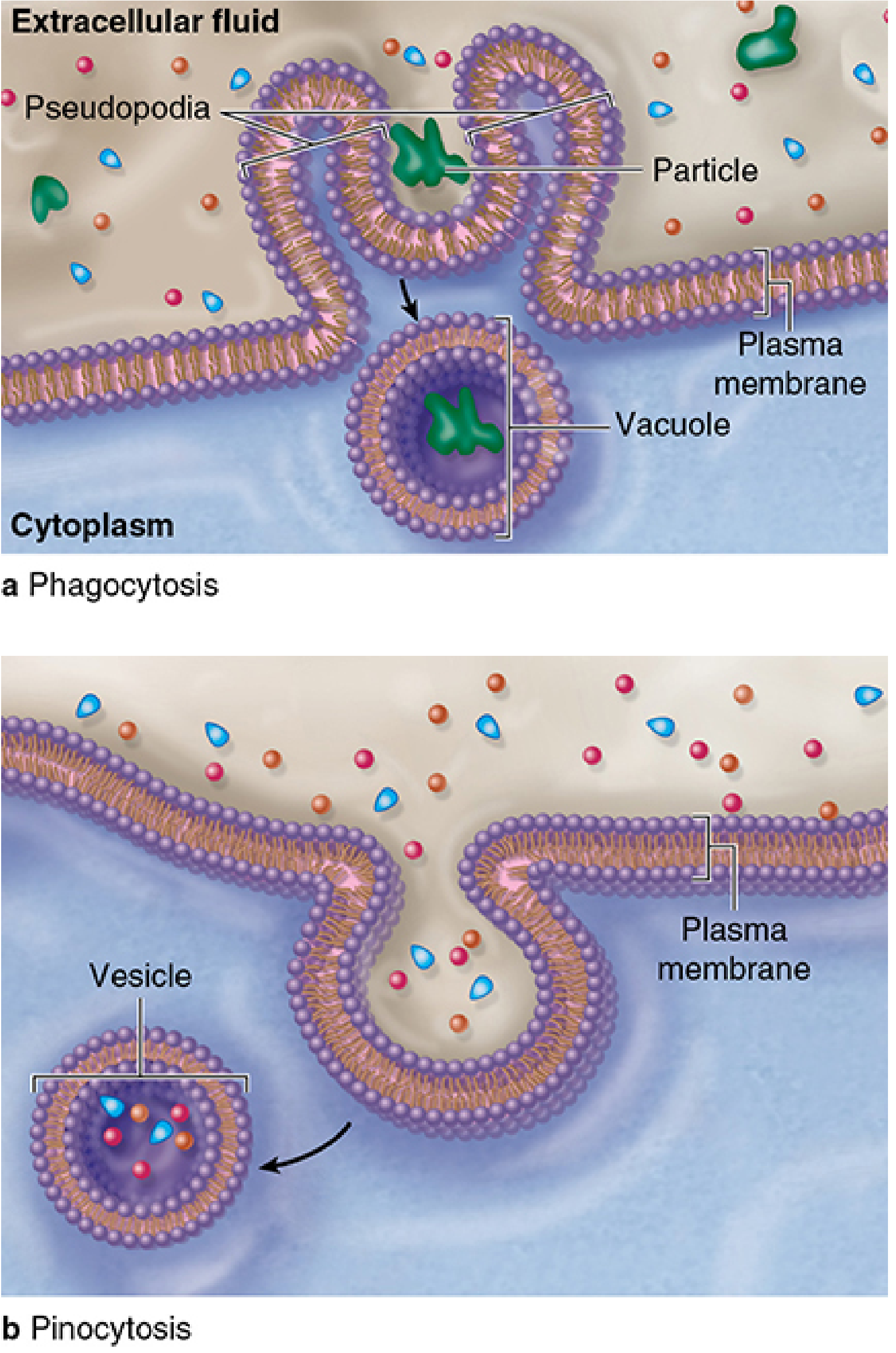
Phagocytosis and Pinocytosis:
Junqueira's Basic Histology - Fig 2-6
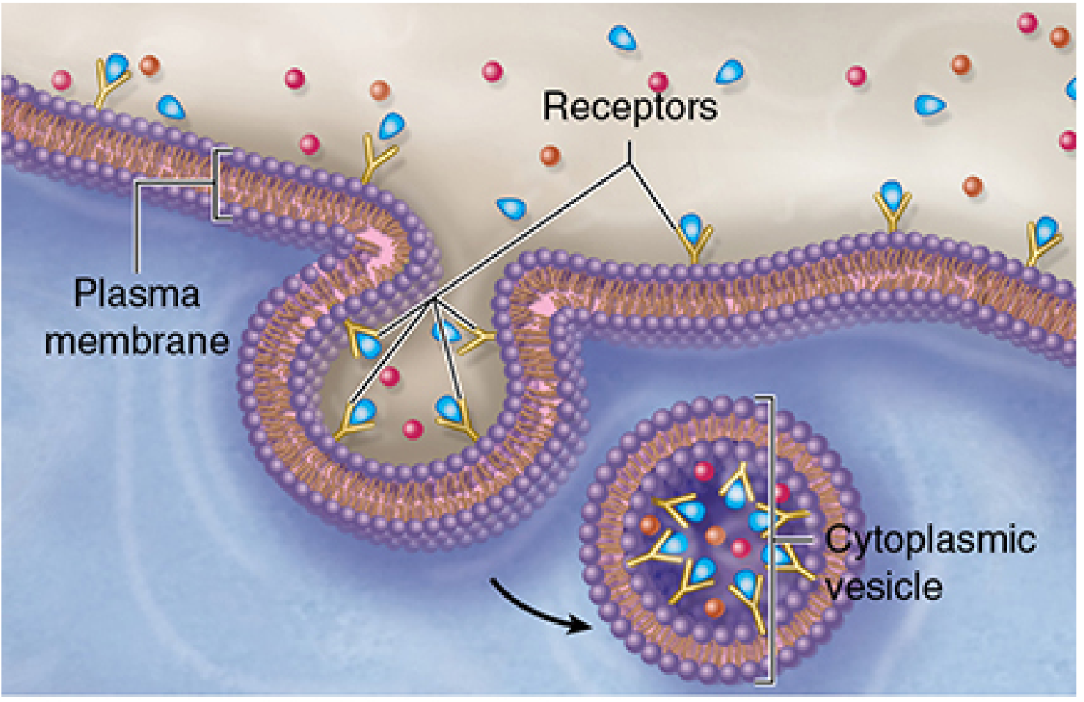
Receptor-mediated endocytosis:
Junqueira's Basic Histology - Fig 2-6c
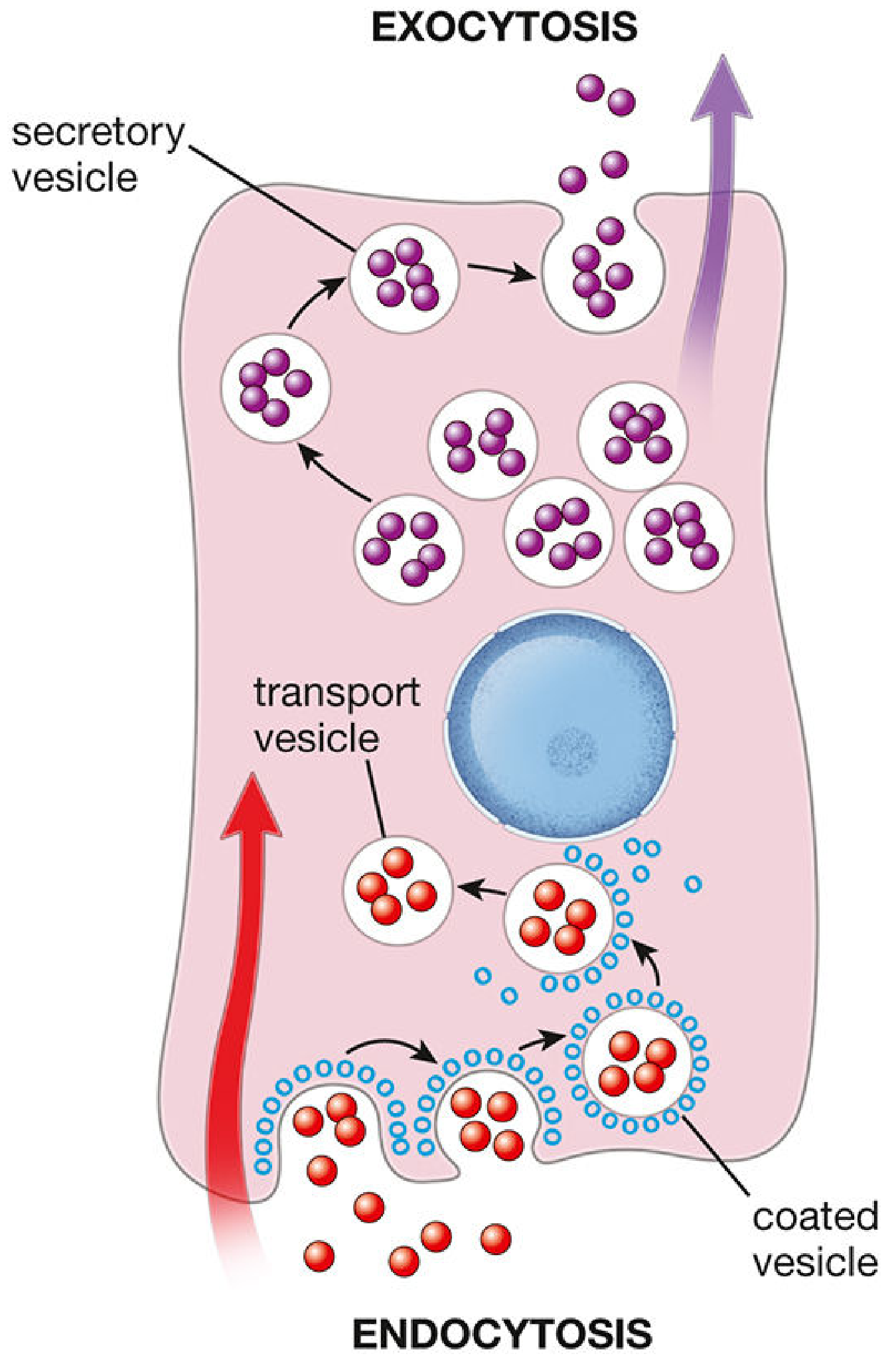
Endocytosis and Exocytosis overview:
Histology: A Text and Atlas - Fig 2.10
Endocytosis
Definition: The cellular process by which material from the extracellular environment is taken into the cell through invagination and pinching off of the plasma membrane to form intracellular vesicles.
Three major types:
a) Phagocytosis ("cell eating")
- Cell surface extends pseudopodia to surround large particles (bacteria, dead cells)
- The folds fuse to form a phagosome (large vacuole)
- Phagosome merges with a lysosome → phagolysosome → enzymatic digestion
- Performed by macrophages and neutrophils
b) Pinocytosis ("cell drinking")
- Smaller invaginations of the plasma membrane trap extracellular fluid and dissolved contents
- Pinches off as pinocytotic vesicles (~80 nm diameter)
- Vesicles either fuse with lysosomes for digestion, or travel to the opposite membrane surface for transcytosis
c) Receptor-mediated endocytosis
- Specific ligands (e.g. LDL, protein hormones, antibodies) bind to cell-surface receptor proteins
- Occupied receptors aggregate in coated pit regions lined with clathrin protein
- The pit invaginates and pinches off as a clathrin-coated vesicle (endosome)
- Clathrin is recycled; the receptor-ligand complex is sorted in the endosomal compartment
- Receptors are often returned to the cell surface (recycling endosomes) while ligands are sent to lysosomes
Exocytosis
Definition: The process by which intracellular vesicles containing secretory cargo fuse with the plasma membrane, releasing their contents into the extracellular space.
- Delivers newly synthesised proteins, lipids, and other macromolecules to outside the cell
- Triggered in many cells by a transient rise in cytosolic Ca²⁺
- Mediated by SNARE proteins which drive membrane fusion
- Maintains plasma membrane integrity by returning endocytosed membrane material
Two pathways of exocytosis:
- Constitutive secretion - products released continuously as soon as synthesised (e.g. collagen to ECM)
- Regulated secretion - products stored in vesicles and released upon signal (e.g. digestive enzymes from pancreatic acinar cells; neurotransmitters at synapses)
Note: Exocytosis and endocytosis are coupled - blocking one blocks the other (shown with tetanus/botulinum toxin experiments).
- Junqueira's Basic Histology, p. 82-86
Q9. Chemical Structure of a Single Phospholipid
A phospholipid is an amphipathic molecule consisting of:
            Choline (+ charged)
               |
          Phosphate group (- charged)
               |   ← HYDROPHILIC HEAD
          Glycerol backbone
               |
      ________/ \_________
     |                   |
 Fatty acid 1        Fatty acid 2
 (saturated -        (unsaturated -
  no double bonds)    has C=C kink)
     |                   |
     CH₂                CH₂
     |                   |
     CH₂                CH₂=CH
     |                   |
     ...                 ...     ← HYDROPHOBIC TAILS
     |                   |
     CH₃                CH₃
Full chemical structure (phosphatidylcholine example):
        CH₃
        |
H₃C—N⁺—CH₃           ← Choline (quaternary ammonium)
        |
        CH₂
        |
        CH₂
        |
    O   |
    ‖   |
HO—P—O—CH₂             ← Phosphate group
    |
    O
    |
    CH₂—O—CO—(CH₂)n—CH₃    ← sn-1 fatty acid (saturated)
    |
    CH—O—CO—(CH₂)m=...—CH₃  ← sn-2 fatty acid (unsaturated)
    |
    CH₂—OH
Key components:
- Polar head: Phosphate group + alcohol (choline, ethanolamine, serine, or inositol) - hydrophilic, water-loving
- Glycerol backbone: 3-carbon bridge linking head to tails
- Two fatty acid tails: Long hydrocarbon chains - hydrophobic, water-repelling
- Saturated tail: straight chain (palmitic acid, stearic acid)
- Unsaturated tail: kinked due to C=C double bond (oleic acid, linolenic acid)
The amphipathic nature causes phospholipids to spontaneously form bilayers in water - hydrophilic heads face outward, hydrophobic tails face inward.
Q10. Ultrastructure of Animal and Plant Cells
Animal Cell Ultrastructure
| Organelle | Membrane | Key Features |
|---|
| Plasma membrane | Single (bilayer) | Fluid mosaic; contains channels, receptors, glycoproteins |
| Nucleus | Double (nuclear envelope + pores) | Nucleolus (rRNA synthesis); chromatin (DNA + histones) |
| Mitochondria | Double | Cristae (inner membrane folds); matrix; 70S ribosomes; circular DNA |
| Rough ER | Single (continuous with nuclear envelope) | Studded with ribosomes; protein synthesis and modification |
| Smooth ER | Single | Lipid synthesis, detoxification, Ca²⁺ storage |
| Golgi apparatus | Single (stacked cisternae) | Cis face (receives) → trans face (dispatches); post-translational modification |
| Lysosomes | Single | Acid hydrolases; pH ~5; digest cellular debris |
| Ribosomes | None | 80S (60S + 40S); free in cytoplasm or attached to RER |
| Centrosome/Centrioles | None | 9+0 microtubule triplet arrangement; cell division |
| Cytoskeleton | None | Microfilaments (actin), intermediate filaments, microtubules |
| Peroxisomes | Single | Catalase; fatty acid oxidation, H₂O₂ breakdown |
Plant Cell Ultrastructure
Plant cells have all the above EXCEPT centrioles and lysosomes (vacuole takes over lysosome function), PLUS:
| Additional Structure | Membrane | Key Features |
|---|
| Cell wall | None (extracellular) | Cellulose microfibrils; provides rigidity and support |
| Chloroplasts | Double | Thylakoids (contain chlorophyll, stacked into grana); stroma; own DNA |
| Central vacuole | Single (tonoplast) | Large; provides turgor pressure; storage; pH regulation |
| Plasmodesmata | Extension of plasma membrane | Channels through cell walls connecting adjacent cells |
LONG QUESTIONS (6 MARKS EACH)
L1. Fluid Mosaic Model of the Plasma Membrane
Definition and History
The Fluid Mosaic Model was proposed by Singer and Nicolson in 1972. It describes the plasma membrane as a dynamic, two-dimensional fluid structure in which proteins are embedded within or associated with a phospholipid bilayer, free to move laterally - like a mosaic of tiles that can slide around.
Composition
1. Phospholipid Bilayer (the "fluid" component)
- The backbone of the membrane consists of two layers of phospholipids arranged tail-to-tail
- Hydrophilic heads face outward (toward water - extracellular fluid and cytoplasm)
- Hydrophobic tails face inward (away from water), forming the hydrophobic core
- The bilayer is fluid at body temperature (~37°C), allowing lateral movement of lipids and proteins
- Cholesterol molecules are interspersed among the phospholipids:
- Reduces membrane fluidity at high temperatures
- Prevents solidification at low temperatures
- Maintains optimal membrane flexibility
2. Proteins (the "mosaic" component)
Membrane proteins perform most of the functional tasks of the membrane:
| Type | Location | Examples |
|---|
| Integral (intrinsic) proteins | Penetrate the hydrophobic core; many span the full bilayer (transmembrane proteins) | Ion channels, carrier proteins, receptors, ATPases |
| Peripheral (extrinsic) proteins | Loosely attached to the inner or outer surface; easily removed | Cytoskeletal anchors, signalling enzymes |
| Lipid-anchored proteins | Covalently bound to lipid molecules inserted in the bilayer | G proteins, some cell-surface enzymes |
3. Carbohydrates (glycocalyx)
- Short oligosaccharide chains covalently attached to membrane lipids (glycolipids) and proteins (glycoproteins)
- Located exclusively on the extracellular surface
- Functions: cell recognition, cell adhesion, immune identity (ABO blood groups), receptor binding, protection from mechanical/chemical damage
Labelled Diagram
EXTRACELLULAR SPACE
      │
      │  Carbohydrate chains (Glycocalyx)
      │  ⌇⌇⌇⌇⌇⌇⌇⌇⌇
══════╪══╪══╪═══╪══╪════════════════════════════
● ● ● │  │  │   │  │ ● ● ● ● ● ● ● ● ● ● ● ●    ← Phospholipid heads
║ ║ ║ ┼──┼──┼───┼──┼ ║ ║ ║ ║ ║ ║ ║ ║ ║ ║ ║ ║    ← Fatty acid tails
║ ║ ║ │  │  │   │  │ ║ ║ ║ ║ ║ ║ ║ ║ ║ ║ ║ ║
║ ║ ║ │  │ [G]  │  │ ║ ║ ║ ║ ║ ║ ║ ║ ║ ║ ║ ║
║ ║ ║ [A]  [B]  [C] ║ ║ ║ ║ [Chol]║ ║ ║ ║ ║ ║   ← Integral proteins
║ ║ ║ │           │ ║ ║ ║ ║ ║ ║ ║ ║ ║ ║ ║ ║ ║    & cholesterol
● ● ● │           │ ● ● ● ● ● ● ● ● ● ● ● ●    ← Phospholipid heads
══════╪═══════════╪════════════════════════════
      │           │
   [Peripheral] [Peripheral]
     protein      protein
      │
CYTOPLASM
Key:
- [A] = Transmembrane integral protein (e.g. channel/transporter)
- [B] = Transmembrane receptor protein
- [C] = Peripheral protein (inner leaflet, e.g. G protein)
- [G] = Glycoprotein (extracellular carbohydrate chain)
- [Chol] = Cholesterol molecule
Properties of the Fluid Mosaic Model
| Property | Detail |
|---|
| Fluidity | Lipids and many proteins can move laterally within their leaflet; limited flip-flop between leaflets |
| Selective permeability | Membrane controls what enters and exits the cell |
| Asymmetry | Inner and outer leaflets differ in lipid and protein composition; carbohydrates on outer only |
| Lipid rafts | Specialised microdomains rich in cholesterol and saturated fatty acids; less fluid; concentrate signalling proteins |
L2. Structure and Functions of Subcellular Organelles
1. Mitochondria
Structure:
- Bounded by two membranes
- Outer membrane: smooth, contains porins (VDAC channels) that make it permeable to small molecules
- Inner membrane: highly folded into shelf-like projections called cristae - greatly increases surface area for ATP synthesis
- Contains the electron transport chain proteins (Complexes I-IV) and ATP synthase (Complex V)
- Intermembrane space: between the two membranes; protons are pumped here during oxidative phosphorylation
- Matrix: enclosed by the inner membrane; contains:
- TCA (Krebs) cycle enzymes
- Circular mitochondrial DNA (~16,569 base pairs in humans)
- 70S ribosomes (like bacteria - evidence of endosymbiotic origin)
- Mitochondria-specific tRNAs and rRNAs
Functions:
- Primary function: ATP synthesis via oxidative phosphorylation - "powerhouse of the cell"
- Pyruvate oxidation and acetyl-CoA generation
- TCA cycle (citric acid cycle)
- Beta-oxidation of fatty acids
- Calcium homeostasis
- Regulation of apoptosis (release of cytochrome c)
- Heat generation (thermogenesis via uncoupling proteins)
2. Nucleus
Structure:
- Largest organelle; bounded by a double membrane (nuclear envelope)
- Outer nuclear membrane: continuous with the rough ER; studded with ribosomes
- Inner nuclear membrane: lined by the nuclear lamina (intermediate filaments that provide structural support)
- Nuclear pores (~3,000-4,000 per nucleus): large protein complexes (~120 nm) that perforate the nuclear envelope and regulate bidirectional traffic of molecules (mRNA out; transcription factors, histones in)
- Nucleoplasm: gel-like interior containing:
- Chromatin: DNA wrapped around histone octamers (nucleosomes) - heterochromatin (condensed, transcriptionally inactive) and euchromatin (dispersed, transcriptionally active)
- Nucleolus: dense, spherical sub-region; site of ribosomal RNA (rRNA) synthesis and ribosome assembly; disappears during mitosis
Functions:
- Stores and protects the genetic information (DNA)
- Transcription (DNA → mRNA)
- Pre-mRNA processing (splicing, capping, poly-A tail addition)
- Ribosome biogenesis (in nucleolus)
- Controls all cell activities - gene expression, cell cycle, differentiation
3. Chloroplast
Structure (plant cells only):
- Bounded by two membranes
- Outer membrane: smooth and freely permeable
- Inner membrane: contains protein transporters; selectively permeable
- Intermembrane space: between the two membranes
- Stroma: gel-like interior containing:
- Circular chloroplast DNA
- 70S ribosomes
- Starch granules
- Calvin cycle enzymes (carbon fixation)
- Thylakoids: flattened, membrane-bound sacs within the stroma, arranged in stacks called grana (singular: granum); connected by lamellae (intergranal thylakoids)
- Thylakoid membranes contain chlorophyll and other pigments, embedded in Photosystems I and II, and the electron transport chain
Functions:
- Photosynthesis:
- Light-dependent reactions (on thylakoid membranes): Capture light energy, split water (O₂ released), generate ATP and NADPH
- Light-independent reactions / Calvin cycle (in stroma): Use ATP and NADPH to fix CO₂ into glucose (G3P)
- Synthesis of fatty acids, amino acids
- Own DNA and ribosomes allow semi-autonomous function
4. Golgi Apparatus
Structure:
- Stack of 4-8 flattened membrane-bound cisternae (like a stack of hollow pancakes)
- Cis face (entry/receiving face): oriented toward the RER; receives vesicles budding from the RER
- Trans face (exit/dispatch face): oriented toward the plasma membrane; buds off secretory vesicles
- Sorted into: cis Golgi network → medial Golgi → trans Golgi network (TGN)
Functions:
- Post-translational modification of proteins: glycosylation (addition of sugar chains), phosphorylation, sulfation, proteolytic cleavage
- Sorting and packaging of proteins and lipids into vesicles for correct destinations (lysosomes, plasma membrane, secretion)
- Synthesis of complex polysaccharides and glycolipids
- Quality control - proteins that fail quality checks are sent to lysosomes for degradation
5. Rough Endoplasmic Reticulum (Rough ER)
Structure:
- Network of membrane-bound flattened sacs (cisternae) and tubules
- Continuous with the outer nuclear membrane
- Cytoplasmic surface is studded with 80S ribosomes (giving the "rough" appearance)
Functions:
- Protein synthesis: Ribosomes on the RER translate mRNAs encoding secretory, membrane, and lysosomal proteins; the nascent polypeptide is co-translationally inserted into the ER lumen
- Protein folding and modification: Glycosylation (N-linked); disulfide bond formation; chaperone assistance
- Protein quality control: Misfolded proteins are retrotranslocated and degraded (ERAD pathway)
- Vesicle budding: Correctly folded proteins are packaged into COPII-coated vesicles for transport to the Golgi
6. Smooth Endoplasmic Reticulum (Smooth ER)
Structure:
- Network of branching tubules; no ribosomes on its surface (hence "smooth")
- Continuous with the rough ER
- Relative amount varies by cell type (abundant in liver, adrenal cortex, gonads, muscle)
Functions:
- Lipid synthesis: Phospholipids, cholesterol, and triglycerides
- Steroid hormone synthesis: In adrenal cortex, gonads (testosterone, oestrogen, cortisol)
- Drug detoxification: Liver SER - cytochrome P450 enzymes oxidise and conjugate drugs and toxins for excretion
- Calcium storage and release: In skeletal muscle (called sarcoplasmic reticulum) - Ca²⁺ release triggers muscle contraction
- Glycogen metabolism: Glucose-6-phosphatase activity
7. Lysosomes
Structure:
- Small (~0.1-1.2 µm), membrane-bound vesicles
- Bounded by a single membrane (the lysosomal membrane)
- Lysosomal membrane contains:
- ATP-driven H⁺ pump (V-ATPase) maintaining interior at pH ~4.5-5.0
- Lysosomal membrane proteins (LAMP-1, LAMP-2) that protect it from its own enzymes
- Interior contains ~60 hydrolytic enzymes (acid hydrolases):
- Proteases (cathepsins), lipases, nucleases, glycosidases, phosphatases
- All optimally active at acidic pH; safe at cytoplasmic pH ~7.2
Functions:
- Intracellular digestion: Digest engulfed bacteria (phagolysosomes), worn-out organelles (autophagy), and macromolecules
- Autophagy: Recycling of damaged organelles - they are enclosed in autophagosomes which fuse with lysosomes
- Receptor-mediated endocytosis: Processing of endocytosed ligands
- Antigen processing: In immune cells (present peptides on MHC molecules)
- Bone resorption: Osteoclasts secrete lysosomal enzymes extracellularly to degrade bone matrix
Lysosomal storage diseases (e.g. Gaucher's disease, Tay-Sachs) result from deficiency of specific lysosomal enzymes, causing accumulation of undegraded substrates.
8. Ribosomes
Structure:
- Not membrane-bound - no surrounding membrane
- Dense ribonucleoprotein particles made of rRNA + protein
- Two subunits that associate during translation:
| Cell Type | Ribosome | Large Subunit | Small Subunit |
|---|
| Eukaryotic (cytoplasmic) | 80S | 60S (28S, 5.8S, 5S rRNA + ~49 proteins) | 40S (18S rRNA + ~33 proteins) |
| Prokaryotic / Mitochondrial / Chloroplast | 70S | 50S (23S, 5S rRNA) | 30S (16S rRNA) |
- The ribosome has three tRNA binding sites: A site (aminoacyl), P site (peptidyl), E site (exit)
Functions:
- Protein synthesis (translation): Decode mRNA codons and catalyse peptide bond formation between amino acids
- Free ribosomes (cytoplasm): synthesise proteins destined for the cytoplasm, nucleus, mitochondria, peroxisomes
- Membrane-bound ribosomes (on RER): synthesise secretory proteins, membrane proteins, lysosomal proteins
The ribosome's peptidyl transferase activity resides in the 23S/28S rRNA (a ribozyme) - meaning the RNA catalyses the reaction, not the protein component.
*Sources: Histology: A Text and Atlas with Correlated Cell and Molecular Biology, 8th ed (Pawlina); Junqueira's Basic Histology - Text and Atlas, 17th ed; Histology: A Text and Atlas (Gartner & Hiatt)*All 12 questions are fully answered. Here is a quick summary of what's covered:
All questions have been answered above. Here's a quick index:
| Q | Topic | Marks |
|---|
| Q1 | Single vs double membrane organelles | 3 |
| Q2 | Cell signalling + protein signal pathway | 3 |
| Q3 | Prokaryotic vs eukaryotic cells with diagrams | 3 |
| Q4 | Stem cell definition + advantages | 3 |
| Q5 | Categories of stem cells (by potency and origin) | 3 |
| Q6 | iPSC advantages and disadvantages | 3 |
| Q7 | 4 membrane transport mechanisms with diagrams | 3 |
| Q8 | Endocytosis and exocytosis with diagrams | 3 |
| Q9 | Phospholipid chemical structure | 3 |
| Q10 | Animal and plant cell ultrastructure | 3 |
| L1 | Fluid mosaic model - structure, composition, labelled diagram | 6 |
| L2 | Organelle ultrastructure and functions (all 7 organelles) | 6 |
Key points to remember for exams:
- Single membrane: Lysosome, ER, Golgi, vacuole, peroxisome
- Double membrane: Nucleus, mitochondria, chloroplast
- Active transport is the only process that uses ATP and moves substances against their gradient
- Osmosis specifically refers to water movement; aquaporins are the water channels
- iPSCs were discovered by Yamanaka (2006); the main risk is tumourigenicity from c-Myc oncogene
- The fluid mosaic model (Singer & Nicolson, 1972) features lateral fluidity, membrane asymmetry, and lipid rafts
- Ribosomes are the only organelle with no membrane whatsoever