
Help me study with arterial line and IABP with pictures wherever relevant including recent advancements with reference as standard anaesthesia text books
Reading File
Now let me run parallel searches across the medical library and PubMed:
Finding Sources
Finding Sources
Searching PubMed
"arterial line" OR "arterial catheter" anesthesia complications
Searching PubMed
"intra-aortic balloon pump" outcomes perioperative
Now let me search for more specific sections and read the key textbook content in parallel:
Finding Sources
Finding Sources
Finding Sources
Searching PubMed
"intra-aortic balloon pump" versus "mechanical circulatory support" cardiogenic shock
Excellent - now let me read the key textbook sections in parallel:
Reading File
Reading File
Reading File
Reading File
I found an image. Let me fetch it and continue reading more textbook content:
Loading Image
Reading File
Reading File
Reading File
Let me get more content from Barash and Miller on arterial lines, and look for more images:
Reading File
Reading File
Finding Sources
Reading Article
PMID: 41178577
Excellent! I have a great image URL for the damping waveform. Let me fetch it and get remaining key content:
Loading Image
Reading File
Reading File
Reading File
Let me fetch those waveform images from Miller's and get remaining IABP content:
Loading Image
Loading Image
Reading File
Reading File
Let me get the fast-flush test image and a couple more key sections:
Loading Image
Reading File
Reading File
Let me fetch a couple more critical images and get the normal waveform data:
Loading Image
Reading File
Searching PubMed
"arterial line" ultrasound guided placement first-pass success
I now have comprehensive content. Let me update the todos and compile the study guide:---
Arterial Line and IABP - Comprehensive Study Guide
Sources: Miller's Anesthesia 10e, Barash Clinical Anesthesia 9e, Braunwald's Heart Disease, Fischer's Mastery of Surgery 8e, Tintinalli's Emergency Medicine, Morgan & Mikhail's Clinical Anesthesiology 7e
PART 1: ARTERIAL LINE (Intra-arterial Blood Pressure Monitoring)
1. Principles of Operation
Intra-arterial blood pressure monitoring uses a fluid-filled catheter connected via stiff, low-compliance tubing to a pressure transducer. The transducer converts mechanical displacement of a silicon crystal into voltage changes, which are amplified, filtered, and displayed as the arterial pressure waveform.
"Intra-arterial blood pressure monitoring uses fluid-filled tubing to transmit the force of the pressure pulse wave to a pressure transducer that converts the displacement of a silicon crystal into voltage changes."
- Barash, Cullen & Stoelting's Clinical Anesthesia, 9e
The system can be described by a second-order differential equation governed by two critical properties:
- Natural frequency (fn) - the frequency at which the system resonates
- Damping coefficient (ζ) - the speed of decay of oscillations
The arterial pressure trace contains frequencies from 1-30 Hz; optimal dynamic response requires the system's fn and ζ to be appropriately matched to this range.
2. Indications
From Barash 9e and Miller 10e:
- Need for continuous, beat-to-beat BP monitoring - haemodynamically labile patients, anticipated rapid BP changes
- Major surgery - cardiac, thoracic, vascular, neurosurgery (beach-chair position)
- Anticipated pharmacologic manipulation - vasopressors, inotropes, antihypertensives
- Repeated arterial blood sampling - ABGs, electrolytes, glucose during CPB
- Failure of non-invasive BP measurement - morbid obesity, arrhythmias, non-pulsatile CPB flow
- Supplementary diagnostic information - pulse pressure variation (PPV), systolic pressure variation (SPV) for fluid responsiveness
"During nonpulsatile CPB, noninvasive blood pressure recordings are not accurate. Intra-arterial monitoring provides continuous, real-time, beat-to-beat assessment."
- Miller's Anesthesia, 10e
3. Cannulation Sites
| Site | Advantages | Key Notes |
|---|---|---|
| Radial artery | Most popular; accessible; dual blood supply from ulnar artery | Preferred site; Allen test has poor predictive value |
| Ulnar artery | Useful after failed radial attempt | Non-tapered catheters preferred |
| Brachial artery | Complications similar to radial | No collateral protection to hand; median nerve risk |
| Axillary artery | Patient comfort and mobility; useful in vasoconstrictive states | Risk of cerebral embolisation; avoid prolonged high-pressure flushing |
| Femoral artery | Largest accessible vessel; best in low-flow states; easy access | CDC guidelines: prefer axillary/brachial over femoral due to infection risk; distal to inguinal ligament to avoid retroperitoneal haematoma |
| Dorsalis pedis | Collateral = posterior tibial artery | Higher systolic estimates; popular in paediatrics |
"Radial artery thrombosis can be minimized by using small catheters, avoiding polypropylene-tapered catheters, and reducing the duration of cannulation."
- Barash, 9e
The Allen Test - historically used to assess collateral circulation. However, its prognostic value has NOT been confirmed and routine use is no longer widely recommended. Even a satisfactory Allen test does not protect against distal emboli from the catheter itself.
4. Cannulation Techniques
Three standard techniques (Barash 9e):
- Direct arterial puncture - standard needle-over-catheter approach
- Guidewire-assisted (Seldinger technique) - especially for larger vessels (femoral, axillary)
- Transfixion-withdrawal method - intentional posterior wall puncture, then gradual withdrawal
Ultrasound guidance is strongly supported by evidence, particularly:
- As a rescue method following a failed attempt
- In difficult patients (small vessels, obesity, low-flow states, patients on mechanical support without pulsatile flow)
- The benefit decreases with clinical experience for routine radial cannulation
- Miller's 10e notes: "The degree of benefit decreases with clinical years of experience placing radial catheters"
5. The Normal Arterial Waveform
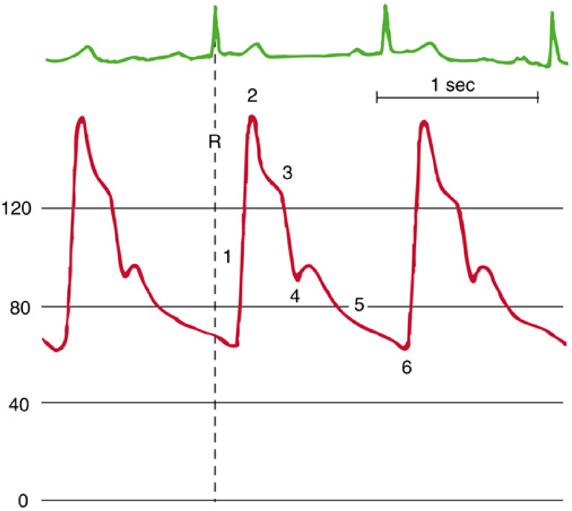
Figure: Normal arterial waveform. The systolic upstroke starts 120-180 ms after the ECG R wave (time for LV depolarisation, isovolumetric contraction, AV opening, ejection, and pressure wave propagation). Components:
- Systolic upstroke
- Systolic peak pressure
- Systolic decline
- Dicrotic notch - marks aortic valve closure
- Diastolic runoff
- End-diastolic pressure
(Miller's Anesthesia, 10e - from Mark JB. Atlas of Cardiovascular Monitoring)
6. Transducer Setup: Zeroing and Leveling
These are two distinct procedures (Miller 10e):
- Zeroing: Exposes the transducer to atmospheric pressure → establishes that as the reference (zero point)
- Leveling: Positions this reference point relative to the patient's body
Standard reference level = Mid-thoracic level (halfway between anterior sternum and bed surface in supine patient) - closest to mid-left atrial position
Neurosurgery exception: Zero at the level of the Circle of Willis (ear) when upright/beach-chair position - accounts for the hydrostatic column between heart and brain.
7. Dynamic Response - Natural Frequency and Damping
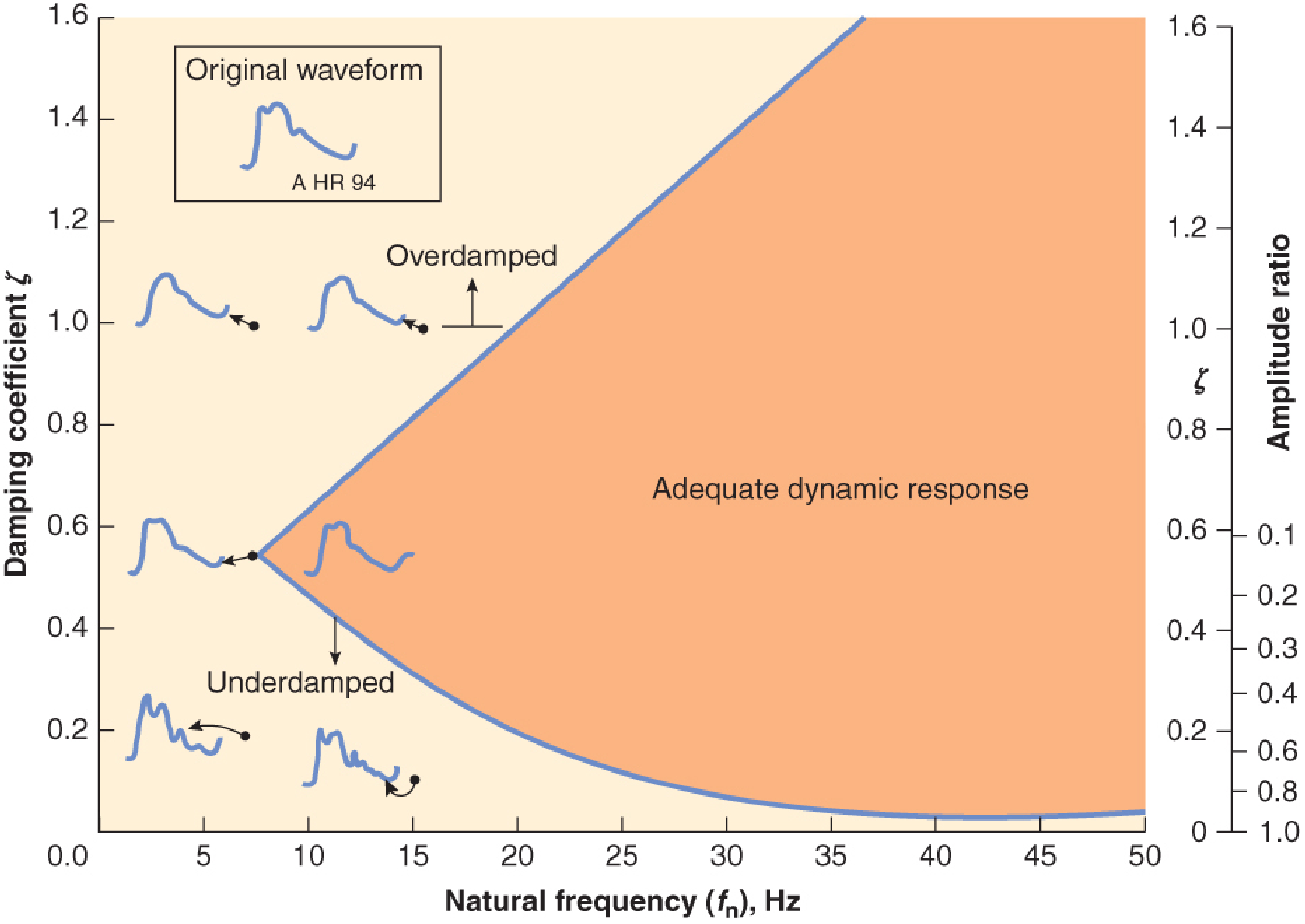
Figure: Barash 9e - The shaded (orange) zone represents adequate dynamic response. The underdamped region (low ζ, low fn) overestimates systolic pressure; the overdamped region underestimates it.
Key clinical rules:
- Underdamped systems: overestimate systolic by 15-30 mmHg, amplify artifacts - caused by excess catheter length, air bubbles, stiff tubing
- Overdamped systems: underestimate systolic, widen the waveform - caused by clots, air, kinked tubing, loose connections
- Mean arterial pressure (MAP) is preserved in both - the most reliable measurement
To improve fidelity: Use stiff short tubing, minimise stopcocks, eliminate air bubbles, use appropriate catheter size.
8. The Fast-Flush Test
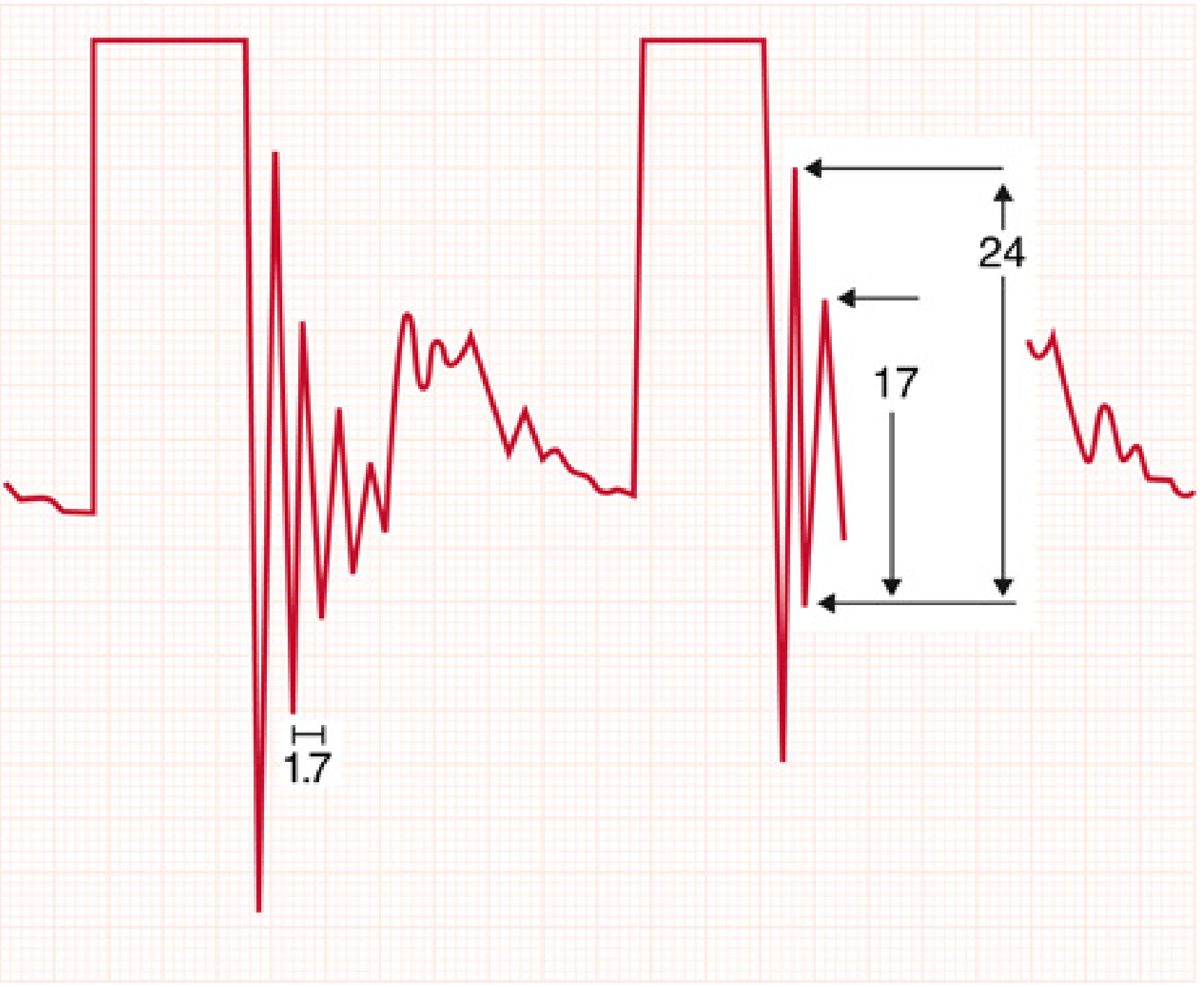
Figure: Fast-flush test (Miller 10e). Two square-wave artifacts on standard 1-mm grid paper at 25 mm/s:
- Natural frequency = 1 ÷ period of one oscillation cycle (measured in mm × paper speed)
- Amplitude ratio = height of second peak ÷ height of first peak → used to derive damping coefficient
In the example: period = 1.7 mm → fn = 14.7 Hz; amplitude ratio = 17/24 = 0.71
9. Overdamped vs Underdamped Waveforms
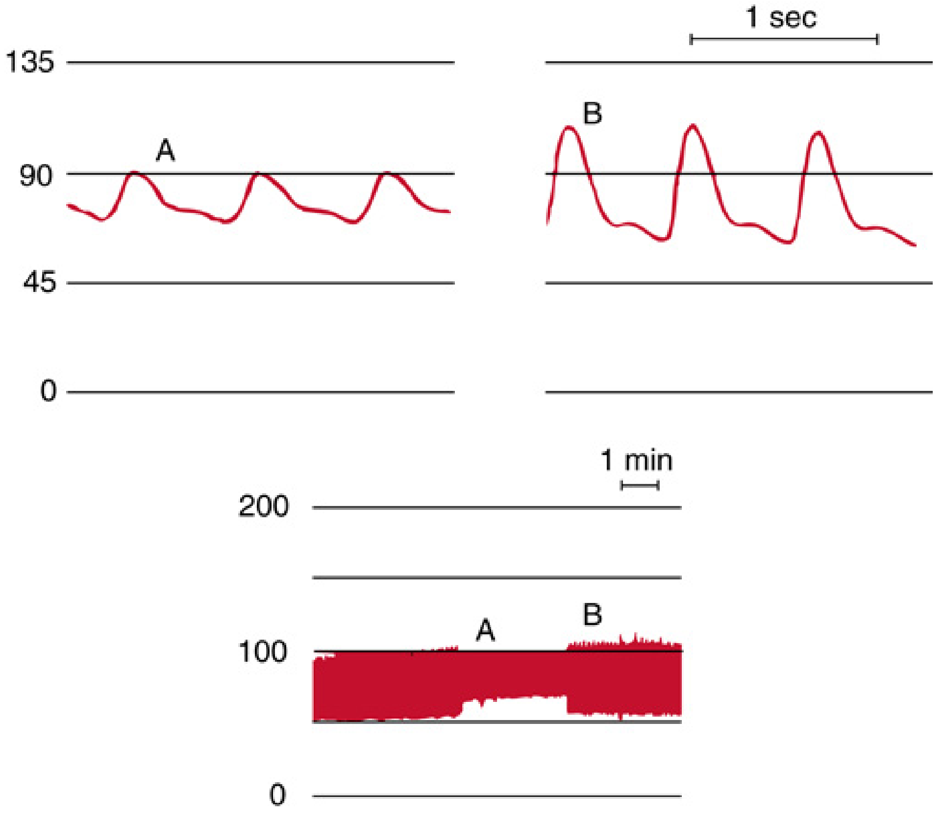
Figure: Miller 10e, Fig 32.21. Overdamped waveform (A) shows diminished pulse pressure compared to normal (B). Despite damping, MAP remains unchanged - this is a key clinical pearl.
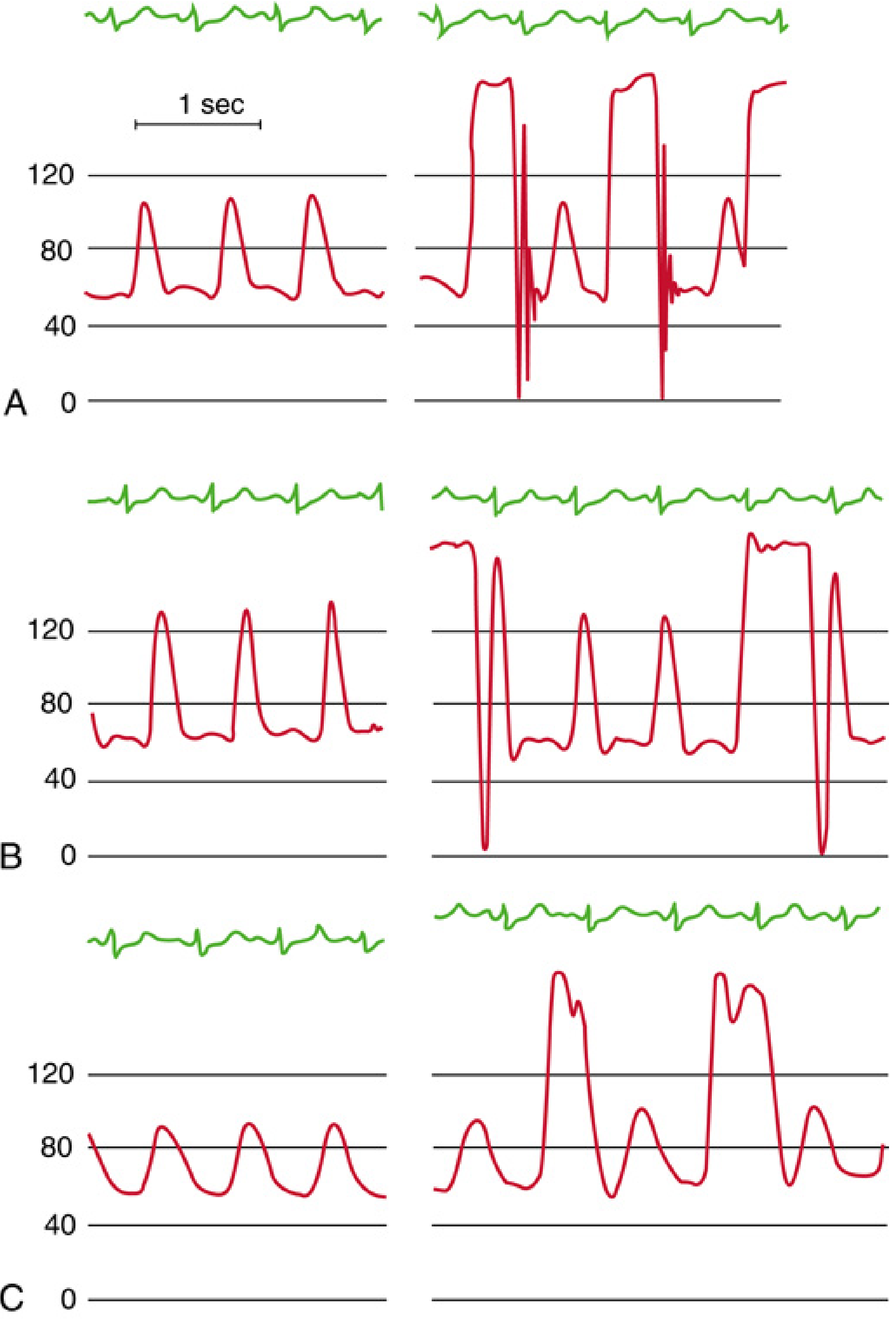
Figure: Miller 10e, Fig 32.22. Effect of air bubbles: (A) Normal system, fn = 17 Hz. (B) 0.1 mL air bubble - paradoxical BP increase due to reduced fn. (C) 0.5 mL bubble - spurious hypotension. This is why removing all air from the system is mandatory.
10. Distal Pulse Amplification
As the pressure wave travels to the periphery (Miller 10e):
- Systolic peak rises
- Diastolic pressure falls
- Pulse pressure widens
- Dicrotic notch appears later and more blunted
- MAP remains almost unchanged
This means radial artery systolic pressures are typically higher than central aortic systolic pressures. Inter-individual variation is large - up to 14% of patients show >15 mmHg difference between brachial and radial readings.
Post-CPB radial-femoral gradient: On initiation of bypass, radial artery mean pressure falls and may be >20 mmHg lower than femoral artery pressure - resolves in most patients within 1 hour of rewarming.
11. Pulse Pressure Variation (PPV) and Fluid Responsiveness
From Barash 9e (thoracic anaesthesia context):
- PPV > 13% predicts fluid responsiveness
- PPV < 9% predicts non-responsiveness
- PPV 9-13% = grey zone
- Requires controlled ventilation, regular cardiac rhythm, closed chest
12. Arterial Line - Site Selection in Special Cases
- Mediastinoscopy: Right arm catheter monitors for innominate artery compression (loss of signal = compression → CNS risk). Alternatively, left radial artery + right-hand pulse oximeter.
- Cardiac surgery with radial arraft harvest: Place line on the dominant side (graft usually from non-dominant arm).
- Lateral decubitus: Transducer remains at heart level regardless of arm position - invasive pressure unaffected; NIBP cuff will differ between arms by hydrostatic gradient.
13. Complications
From Miller 10e (Box 32.4) and Barash 9e:
| Complication | Notes |
|---|---|
| Distal ischaemia / thrombosis | Contributing factors: atherosclerosis, diabetes, low CO, vasoconstrictors, prolonged cannulation |
| Pseudoaneurysm / AV fistula | Rare; more with difficult placement |
| Haemorrhage | From disconnection; meticulous tightening of all connections |
| Air embolism (cerebral) | Retrograde flow from high-pressure flushing of central sites (axillary) |
| Infection / bacteraemia | ~1.3% catheter-associated bloodstream infection rate (ICU data) |
| Peripheral neuropathy | Median or radial nerve injury (radial site); median nerve at brachial site |
| Misinterpretation of data | Equipment misuse, poor calibration |
Recent Evidence (2026): A systematic review and meta-analysis of 39 studies (19,018 arterial catheters, ICU setting) found: all-cause failure = 13% (95%CI 7.6-19.5%); non-infectious failure = 19.8%; catheter-associated bloodstream infection = 1.3%; local infection = 1.2%. Up to 1 in 5 ICU arterial catheters fail, requiring system-wide interventions.
PART 2: INTRA-AORTIC BALLOON PUMP (IABP)
1. History and Device Description
First described by Harken in 1958, the IABP is the most commonly used mechanical circulatory support (MCS) device worldwide.
Device anatomy (Braunwald's Heart Disease):
- Double-lumen, 7.5-8.0 French catheter
- Polyethylene balloon at distal end
- One lumen connects to pump console for gas delivery
- Helium is used as inflation gas - low viscosity for rapid transfer; rapidly absorbed into blood if balloon ruptures (safety advantage over air)
Balloon position: Descending thoracic aorta, just distal to the left subclavian artery, extending to just above the renal arteries.
2. Mechanism of Action
The IABP works on the principle of counterpulsation - timed precisely opposite to the cardiac cycle:
| Phase | Timing | ECG Trigger | Haemodynamic Effect |
|---|---|---|---|
| Inflation | Onset of diastole | Middle of T wave (repolarisation); just after dicrotic notch on aortic trace | ↑ diastolic aortic pressure → ↑ coronary perfusion pressure |
| Deflation | Onset of systole | Peak of R wave | ↓ afterload → ↓ LV wall stress → ↓ myocardial O₂ consumption |
Net haemodynamic effects:
- ↑ Diastolic blood pressure (diastolic augmentation)
- ↓ Systolic blood pressure (afterload reduction)
- ↓ Aortic end-diastolic pressure (predeflation pressure falls)
- Modest ↑ cardiac output (~0.5-1.0 L/min)
- ↑ Coronary artery blood flow
- ↓ Myocardial oxygen consumption
"Patients must have some level of LV function and electrical stability for an IABP to be effective because any increase in cardiac output depends on the work of the heart itself."
- Braunwald's Heart Disease
3. The IABP Waveform
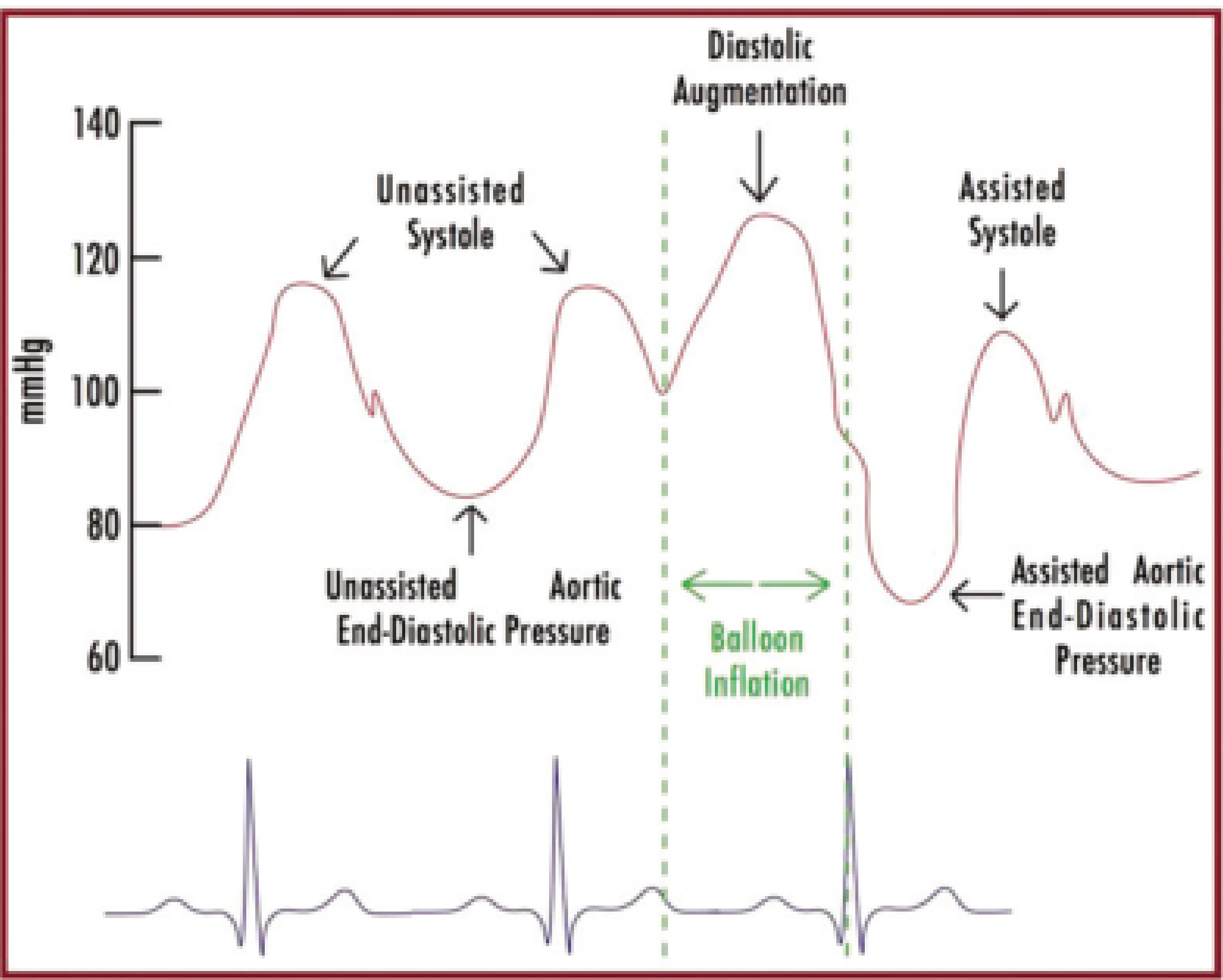
Figure: Fischer's Mastery of Surgery 8e, Fig 6.4. Classic 1:2 counterpulsation tracing showing:
- Unassisted systole: baseline LV ejection pressure (~115 mmHg)
- Diastolic augmentation: balloon inflation creates a second, higher peak (~125 mmHg) - the hallmark of effective IABP
- Assisted aortic end-diastolic pressure: falls below unassisted end-diastolic pressure - reflects afterload reduction
- Assisted systole: lower peak than unassisted systole - due to reduced afterload
The ECG is shown below for timing correlation.
4. IABP Timing: Correct vs Incorrect
| Timing Error | Consequence |
|---|---|
| Early inflation (before aortic valve closure) | Increases LV afterload, ↑ O₂ consumption - dangerous |
| Late inflation (after dicrotic notch) | Suboptimal diastolic augmentation |
| Early deflation (before systole) | Incomplete afterload reduction; potential for reflux |
| Late deflation (into systole) | Increases afterload - may ↑ O₂ consumption |
Correct timing: inflation at the dicrotic notch; deflation just before the next systolic upstroke.
5. Optimal Factors for IABP Effectiveness
From Braunwald's Heart Disease:
- Balloon position in the aorta
- Blood displacement volume
- Balloon diameter relative to aortic diameter
- Timing of inflation (diastole) and deflation (systole)
- Patient's own heart rate, blood pressure, and vascular resistance
- Residual LV contractile function
6. Indications
Established indications:
- Acute MI complicated by cardiogenic shock (historically; see IABP-SHOCK II below)
- Acute decompensated heart failure with hypotension
- Prophylaxis for high-risk PCI
- Ventricular septal defect or acute mitral regurgitation (papillary muscle rupture) complicating MI
- Bridge to definitive treatment (transplant, LVAD) during complete heart failure
- Post-CABG low-output syndrome
- Refractory unstable angina / recurrent ischaemia
"For the patient with refractory myocardial ischaemia or haemodynamic instability despite optimal medical therapy, intra-aortic balloon counterpulsation can reduce the incidence of ischaemic episodes until revascularisation can be performed."
- Goldman-Cecil Medicine
7. Contraindications
Absolute:
- Severe aortic regurgitation (diastolic inflation worsens regurgitation, ↑ LV volume overload)
- Aortic dissection (balloon inflation may propagate dissection)
Relative:
- Aortic aneurysm
- Significant coagulopathy
- Severe peripheral arterial disease at insertion site
- Uncontrolled sepsis
8. Insertion
Standard approach:
- Percutaneous, via left femoral artery (most common), under fluoroscopic or ultrasound guidance
- Seldinger technique with a sheath
- Position confirmed with fluoroscopy or chest X-ray (tip at 2nd-3rd intercostal space / 2 cm distal to left subclavian origin)
Alternative route - Axillary (Subclavian) IABP:
This is an increasingly important modern approach (Braunwald's 2022):
- Used for ambulatory bridging to transplant or durable LVAD
- Driven by the October 2018 UNOS Heart Allocation Policy revision - patients on temporary MCS receive higher priority for donor heart allocation than those on durable MCS
- In the largest published series (195 patients): 68% successfully bridged to transplant (120) or LVAD (13)
- Unique complications: balloon folding within the aorta; catheter tip entering an aortic branch vessel - serial surveillance X-rays are essential
Operative placement: Via ascending aorta or axillary artery - used when femoral access is impossible.
9. Comparison with Other MCS Devices
| Device | Mechanism | Flow | Access | Support Level |
|---|---|---|---|---|
| IABP | Counterpulsation (pneumatic) | ↑ ~0.5 L/min | Percutaneous (femoral/axillary) | Partial |
| Impella 2.5/CP | Microaxial pump across AV | 2.5-3.5 L/min | Percutaneous (femoral) | Partial-Full |
| Impella 5.0 | Microaxial pump | 5 L/min | Operative (axillary/aorta) | Full |
| TandemHeart | Centrifugal (LA→femoral) | 2-4 L/min | Percutaneous (transseptal) | Partial |
| VA-ECMO | Centrifugal + oxygenator | 4-6 L/min | Percutaneous or operative | Full |
(Braunwald's Heart Disease, Table 59.2)
IABP + VA-ECMO: When VA-ECMO is used alone, the increase in systemic afterload may cause LV distension. Concomitant IABP, LV vent, atrial septostomy, or a percutaneous LV-to-aorta VAD may be needed for LV unloading.
10. IABP-SHOCK II Trial - Key Evidence
The landmark IABP-SHOCK II Trial (RCT, multicenter) evaluated IABP in AMI + cardiogenic shock with planned early revascularization (PCI or CABG):
"At 30 days, 119 patients in the IABP group (39.7%) and 123 patients in the control group (41.3%) had died (RR with IABP 0.96; 95% CI 0.79-1.17; P = 0.69). No significant differences were found in secondary endpoints."
- Braunwald's Heart Disease
Key conclusion: IABP counterpulsation does NOT reduce 30-day mortality in AMI complicated by cardiogenic shock when early revascularisation is planned. No benefit on time to haemodynamic stabilisation, ICU stay, serum lactate, catecholamine dose, or renal function.
This has shifted the current approach away from routine IABP use in this setting, with Impella devices showing more promise (systematic review: Frain & Rees, Perfusion 2024, PMID 34479465).
11. Complications of IABP
From Fischer's Mastery of Surgery 8e and Braunwald's Heart Disease:
The Benchmark Registry (IABP in AMI) reported:
- Major complications: 2.6%
- All complications: 8.1%
| Complication | Details |
|---|---|
| Limb ischaemia | Most common major complication; distal limb ischaemia from thrombosis or reduced flow around catheter |
| Bleeding | At insertion site; worsened by anticoagulation |
| Infection / bacteraemia | ~9.2% with axillary approach in bridge-to-transplant series |
| Stroke | Embolism; ~2.6% with axillary IABP |
| Thrombocytopenia | Mechanical destruction of platelets by balloon |
| Balloon rupture / gas embolism | Rare; helium minimises risk |
| Aortic injury | Perforation, dissection |
| Mesenteric ischaemia | ~3% with axillary IABP |
| Left arm ischaemia | ~3.5% with axillary IABP |
| Death | Rare; related to underlying disease severity |
12. Weaning IABP
- Reduce counterpulsation ratio progressively: 1:1 → 1:2 → 1:3
- Monitor for haemodynamic deterioration at each step
- Remove when MAP maintained >70 mmHg, CI >2.2 L/min/m², low-dose or no vasopressors
13. Recent Advances and Future Directions
IABP
- Axillary IABP for ambulatory bridging - allows patients to walk, exercise, and recover conditioning while awaiting transplant (driven by UNOS 2018 policy change)
- Fully ambulatory counterpulsation device - under investigation; wearable external counterpulsation for outpatient use in less advanced HF (Braunwald's Heart Disease, Fig 59.11)
- IABP + VA-ECMO combination - recognised as a strategy to prevent LV distension during ECMO support
Arterial Lines
- Ultrasound-guided cannulation - now the standard of care as rescue; evidence for routine use in difficult patients
- Pulse contour analysis CO monitoring - continuous cardiac output from the arterial waveform (LiDCO, PiCCO, FloTrac/Vigileo systems) - uses area under the arterial curve related to stroke volume and arterial compliance
- Continuous radial artery BP (tonometry) - ClearSight/Finapres devices use servo-controlled finger photoplethysmography as a non-invasive arterial line alternative
- Arterial catheter failure (2026 data) - Up to 1 in 5 ICU arterial catheters fail; recent systematic review highlights the need for standardised care bundles (Levido et al., Anaesthesia 2026)
Key Memory Mnemonics
IABP Indications - "CASH PUMP":
C - Cardiogenic shock (AMI)
A - Acute decompensated HF
S - Septal defect (post-MI VSR)
H - High-risk PCI prophylaxis
P - Post-CABG low output
U - Unstable angina refractory
M - Mitral regurgitation (acute)
P - Bridge to transplant/LVAD
Arterial Line Complications - "ITHHEFM":
Ischaemia, Thrombosis, Haemorrhage, Haematoma, Embolism, Fistula, Misinterpretation
Quick Reference Summary Table
| Arterial Line | IABP | |
|---|---|---|
| Purpose | Continuous BP monitoring + sampling | Haemodynamic support |
| Primary site | Radial artery | Femoral artery (descending aorta) |
| Catheter size | 20G (radial) | 7.5-8.0 French |
| Gas used | N/A | Helium |
| Key contraindication | Poor collateral flow (relative) | AR, aortic dissection |
| Key complication | Thrombosis, infection | Limb ischaemia, bleeding |
| Critical trial | N/A | IABP-SHOCK II (no mortality benefit in AMI+CS) |
| Recent advance | US-guided insertion; pulse contour CO | Axillary approach for ambulatory bridging |
Sources:
- Miller's Anesthesia, 2-Volume Set, 10e (Chapter 32 and 50)
- Barash, Cullen & Stoelting's Clinical Anesthesia, 9e (Chapters 26 and 38)
- Braunwald's Heart Disease, 15e (Chapter 59 - Mechanical Circulatory Support)
- Fischer's Mastery of Surgery, 8e (Chapters on Haemodynamic Support)
- Tintinalli's Emergency Medicine, Comprehensive Study Guide (Chapter on Cardiogenic Shock)
- Morgan & Mikhail's Clinical Anesthesiology, 7e
- Levido A et al. "Incidence of peripheral arterial catheter failure and complications in intensive care." Anaesthesia 2026. PMID 41178577 - Systematic Review/Meta-analysis
- Frain K, Rees P. "IABP versus percutaneous Impella in emergency revascularisation for MI and cardiogenic shock." Perfusion 2024. PMID 34479465 - Systematic Review
This is a shared conversation. Sign in to Orris to start your own chat.