
Emphysema with pictures patho
Reading File
Now let me search the medical library for emphysema pathology content and images in parallel.
Finding Sources
Finding Sources
Good results. Now let me read the main pathology content from Robbins (the gold standard for pathology) and search for images simultaneously.
Reading File
Reading File
Finding Sources
Excellent content and image URLs found. Let me continue reading the Robbins pathology text for pathogenesis and fetch the key images in parallel.
Reading File
Loading Image
Loading Image
Loading Image
Excellent images. Now let me fetch the pathogenesis flowchart and gross pathology images, and read the remaining pathogenesis text.
Loading Image
Reading File
Reading File
Excellent content gathered. Now let me fetch the bullous emphysema gross image and the Robbins Basic pathology morphology images, plus do a quick web search for the latest evidence.
Loading Image
Finding Sources
Searching PubMed
emphysema pathogenesis protease antiprotease
I have comprehensive content and images. Let me now compile the full answer.
Emphysema - Pathology with Pictures
Definition
Emphysema is defined as irreversible enlargement of the airspaces distal to the terminal bronchiole, accompanied by destruction of alveolar walls, without significant fibrosis. It is classified by its anatomic distribution within the lung acinus.
- Robbins, Cotran & Kumar Pathologic Basis of Disease, p. 635
- Robbins & Kumar Basic Pathology, p. 447
Types of Emphysema
Classification is based on which part of the acinus (the unit distal to the terminal bronchiole) is destroyed. Only the first two types cause clinically significant COPD.
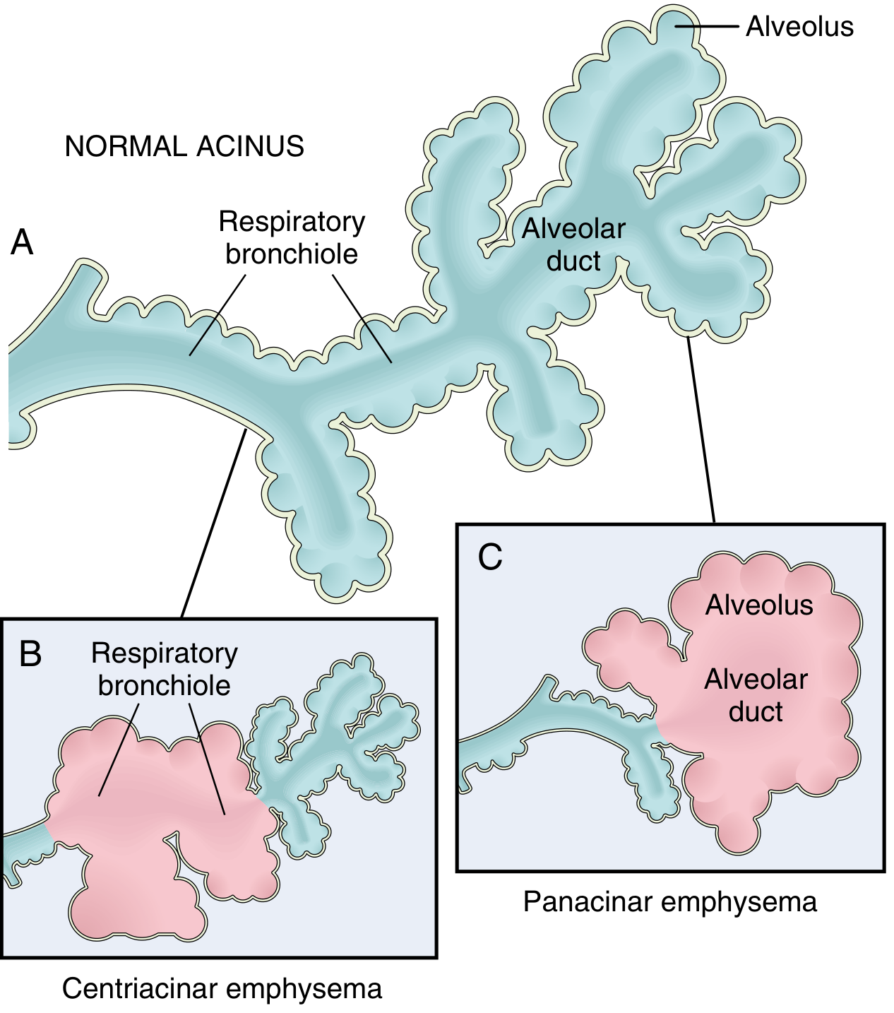
Fig. 15.6 - Patterns of emphysema. (A) Normal acinus structure. (B) Centriacinar emphysema - dilation begins at the respiratory bronchioles. (C) Panacinar emphysema - distension begins at the alveolus and alveolar duct. (Robbins, Cotran & Kumar)
1. Centriacinar (Centrilobular) Emphysema
- Most common type - constitutes >95% of clinically significant cases
- The central/proximal parts of the acinus (respiratory bronchioles) are destroyed; distal alveoli are spared
- Both emphysematous and normal airspaces exist within the same acinus
- Predominantly affects the upper lobes, especially apical segments
- Strongly linked to heavy cigarette smoking
2. Panacinar (Panlobular) Emphysema
- The entire acinus is uniformly enlarged - from respiratory bronchioles to terminal alveoli
- Preferentially affects the lower lobes and anterior lung margins
- Classically associated with α1-antitrypsin (AAT) deficiency (compounded by smoking)
-
80% of homozygous Pi-ZZ patients develop symptomatic panacinar emphysema
3. Distal Acinar (Paraseptal) Emphysema
- The proximal acinus is normal; distal part (adjacent to pleura and lobular septa) is involved
- Characteristic: multiple enlarged airspaces 0.5 mm to >2.0 cm, forming cyst-like structures and bullae
- Clinically important as the cause of spontaneous pneumothorax in young adults
4. Irregular (Paracicatricial) Emphysema
- Irregular acinar involvement; almost always associated with scarring
- Usually clinically insignificant
Pathogenesis
The central mechanism is protease-mediated destruction of the extracellular matrix of alveolar walls, driven by an imbalance between tissue-damaging forces and protective mechanisms.
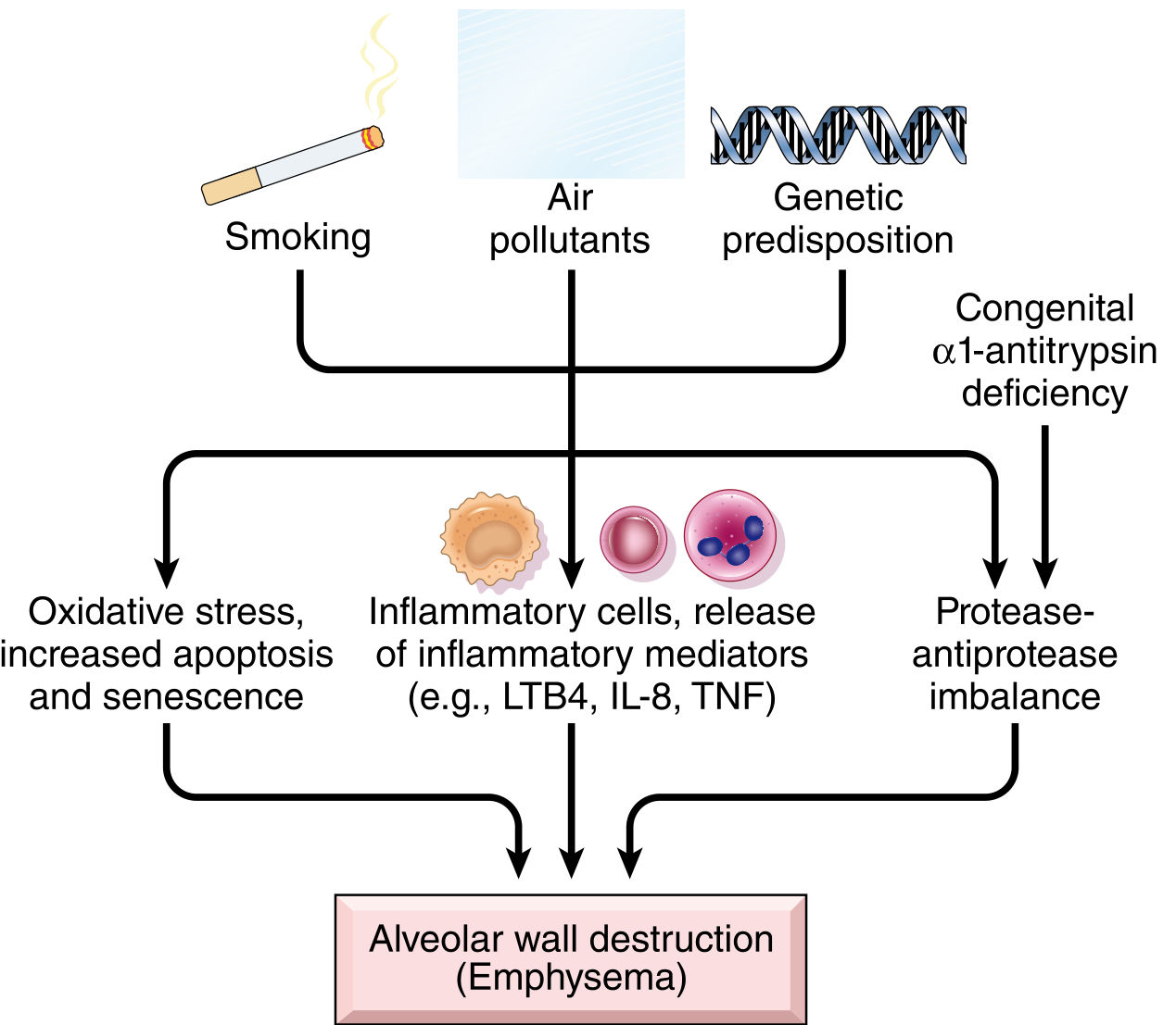
Fig. 15.8 - Pathogenesis of emphysema. (Robbins, Cotran & Kumar)
Key Mechanisms:
1. Toxic Injury and Inflammation
- Inhaled smoke and noxious particles damage respiratory epithelium and recruit inflammatory cells
- Inflammatory mediators released: LTB4, IL-8, TNF, and others
- Chronic accumulation of neutrophils, macrophages, and CD4+/CD8+ T cells in affected lung
2. Protease-Antiprotease Imbalance
- Inflammatory cells release proteases (especially elastase from neutrophils) that break down elastin and other matrix components
- Normally counteracted by α1-antitrypsin (AAT), the primary elastase inhibitor
- In AAT deficiency (Pi-ZZ genotype, chromosome 14), this protection fails - resulting in unopposed elastin degradation
- Matrix metalloproteinases (MMPs) from macrophages also play a role
3. Oxidative Stress
- Cigarette smoke and inflammatory cells generate reactive oxygen species (ROS)
- Oxidants cause tissue damage, endothelial dysfunction, and amplify inflammation
- NRF2 (encoded by NFE2L2) is the key cellular sensor/protector against oxidant damage - genetic variants in NRF2 increase susceptibility
4. Airflow Obstruction Mechanism
- Destruction of elastic tissue in alveolar walls reduces radial traction on surrounding respiratory bronchioles
- Without this traction, bronchioles collapse during expiration - causing functional airflow obstruction even without mechanical blockage
Morphology (Gross and Microscopic)
Gross Pathology
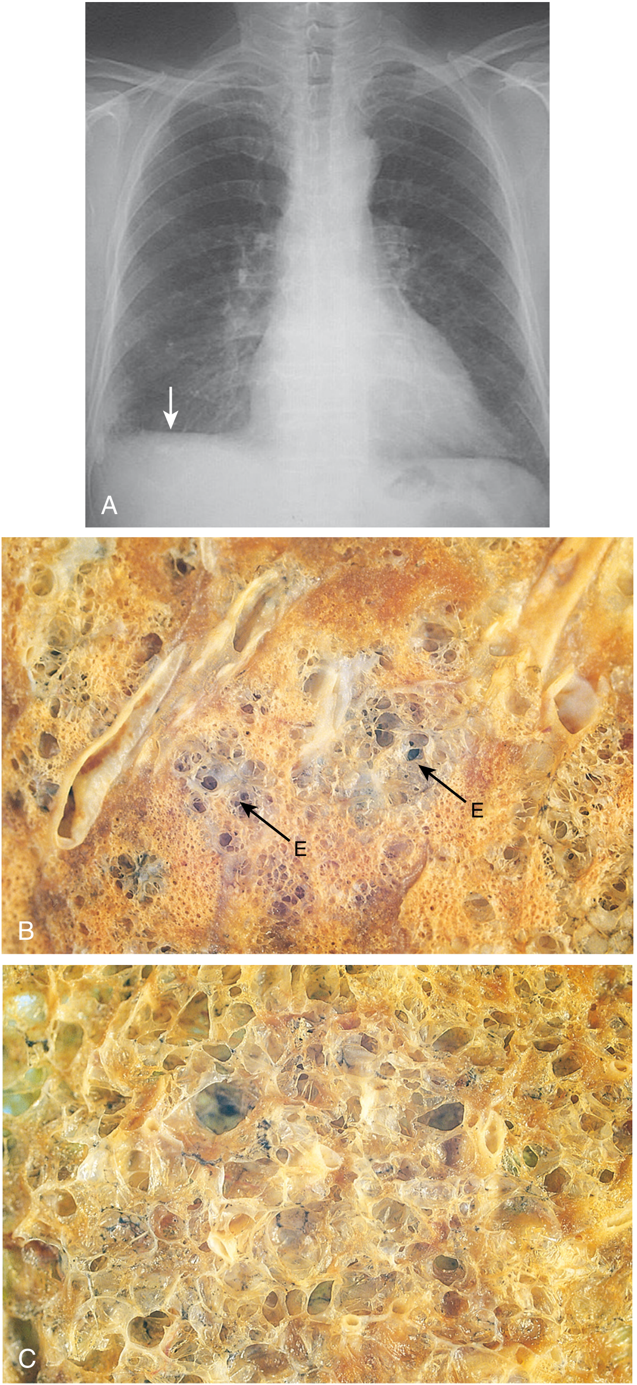
Fig. 15.7 - Emphysema. (A) CXR: hyperinflated lungs, flattened diaphragm. (B) Centriacinar emphysema - focal emphysematous spaces (E) surrounded by normal tissue. (C) Panacinar emphysema - uniformly enlarged airspaces throughout. (Robbins, Cotran & Kumar)
- Advanced emphysema produces voluminous lungs that overlap the heart anteriorly and flatten the diaphragm
- In smoking-related disease, the upper two-thirds of the lungs are most severely affected
- Large alveoli are easily visible on cut sections of fixed lung
- Panacinar emphysema: pale, voluminous lungs that often obscure the heart at autopsy
Bullous Emphysema (Gross)
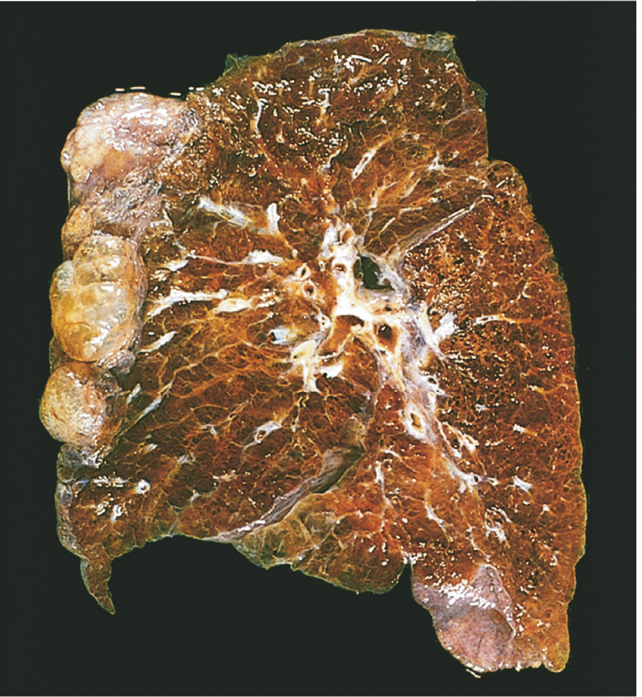
Fig. 15.9 - Bullous emphysema. Large subpleural bullae visible (upper left). (Robbins, Cotran & Kumar)
Microscopic Pathology
- Abnormally large alveoli separated by thin septa with focal centriacinar fibrosis
- Loss of alveolar attachments to the outer wall of small airways
- Pores of Kohn so enlarged that septa appear to "float" or protrude blindly (club-shaped ends)
- Decreased capillary bed area as walls are destroyed
- Advanced: blebs/bullae compressing respiratory bronchioles and vasculature
- Pulmonary hypertension changes from chronic hypoxemia and capillary loss
Summary Table
| Feature | Centriacinar | Panacinar | Paraseptal |
|---|---|---|---|
| Location in acinus | Proximal (respiratory bronchioles) | Entire acinus | Distal (near pleura/septa) |
| Lung distribution | Upper lobes | Lower lobes | Upper half, subpleural |
| Cause | Smoking | AAT deficiency ± smoking | Unknown |
| Clinical significance | COPD (most common) | COPD + AAT deficiency | Spontaneous pneumothorax |
| Bullae? | Rarely | Occasionally | Characteristic |
Sources: Robbins, Cotran & Kumar Pathologic Basis of Disease (pp. 634-638) | Robbins & Kumar Basic Pathology (pp. 447-451)
Complications
Complications of emphysema include cor pulmonale, respiratory failure, pneumothorax, pulmonary hypertension, and secondary polycythemia. Let me pull the full textbook content with images.
Searching sources...
Finding Sources
Finding Sources
The FTS search pulled unrelated results. Let me search more specifically in the COPD/emphysema sections of Robbins and respiratory medicine texts.
Finding Sources
Finding Sources
Good leads. Now let me read the Robbins COPD clinical features/complications section and the cor pulmonale section, plus look for the COPD summary box in Robbins.
Reading File
Reading File
Finding Sources
Excellent content. Now let me fetch the cor pulmonale image and search for more complications content (secondary polycythemia, lung cancer, respiratory failure).
Loading Image
Finding Sources
Finding Sources
I have all the core content needed. Let me now read the Tintinalli clinical features section for the full complications picture, and fetch the pneumothorax image.
Reading File
Finding Sources
I have comprehensive content. Let me fetch the CXR image and compile the full answer.
Loading Image
All content and images gathered. Here is the full compiled answer:
Complications of Emphysema
Emphysema produces progressive complications through two main mechanisms: airflow obstruction causing gas-exchange failure, and loss of the capillary bed causing pulmonary vascular changes. The clinical picture also varies depending on the relative mix of emphysema vs. chronic bronchitis ("pink puffer" vs. "blue bloater").
1. Respiratory Failure (Acute and Chronic)
The most directly lethal complication. As emphysema advances, particularly when FEV1 falls below 1 L:
- Hypoxemia worsens progressively - initially only on exertion, later at rest
- Hypercapnia develops due to ventilation-perfusion mismatch and loss of alveolar surface area
- Oxygenation deteriorates further during acute exacerbations, exercise, and sleep
- Acute respiratory failure is a major cause of death - often triggered by superimposed bacterial or viral infection
Tintinalli's Emergency Medicine, p. 509 | Robbins, Cotran & Kumar, p. 638
2. Pulmonary Hypertension
The pathway from emphysema to pulmonary hypertension runs through two mechanisms:
- Hypoxic vasoconstriction - chronic alveolar hypoxia causes sustained constriction of pulmonary arterioles
- Loss of capillary bed - destruction of alveolar walls eliminates pulmonary capillaries, reducing the vascular cross-sectional area and raising pulmonary vascular resistance
The result is a sustained rise in pulmonary arterial pressure, eventually exceeding the right ventricle's compensatory capacity.
3. Cor Pulmonale (Right-Sided Heart Failure)
The direct downstream consequence of sustained pulmonary hypertension.
- Right ventricular pressure overload leads first to right ventricular hypertrophy, then to RV dilation and failure
- RV wall may thicken to >1.0 cm (normal ~3 mm)
- The enlarged RV can distort and compress the left ventricle (D-sign on cross-section)
- Clinical signs: peripheral edema, ascites, raised JVP, tricuspid regurgitation
- In pure emphysema, cor pulmonale tends to be a late/end-stage finding (more common and earlier in chronic bronchitis)
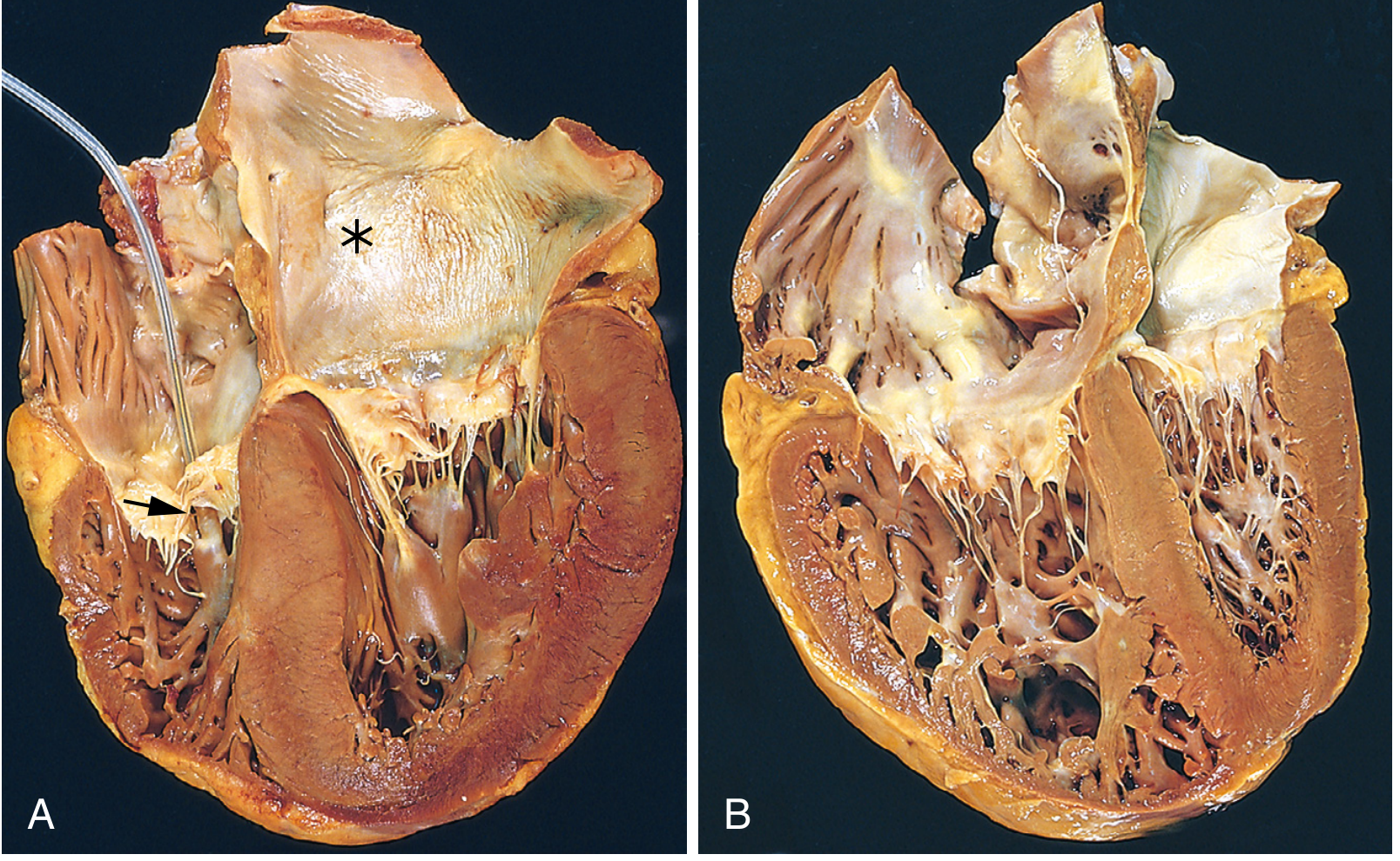
Fig. 12.19B - Cor pulmonale. The right ventricle (left side of image) is markedly dilated with a thickened free wall. The left ventricle shape is distorted. (Robbins, Cotran & Kumar, p. 522)
4. Secondary (Reactive) Polycythemia
- Chronic hypoxemia stimulates erythropoietin release from the kidneys
- This drives increased red cell production (erythrocytosis/secondary polycythemia)
- Clinically visible as facial vascular engorgement - a clinical sign of severe COPD
- Polycythemia increases blood viscosity, which in turn worsens pulmonary hypertension and raises cardiovascular risk (stroke, thrombosis)
Tintinalli's Emergency Medicine, p. 509
5. Spontaneous Pneumothorax
- Subpleural blebs and bullae (characteristic of paraseptal emphysema and advanced disease) can rupture spontaneously
- Smoking-related emphysema is the most common cause of secondary spontaneous pneumothorax
- Can be acutely life-threatening in a patient with already compromised lung function - even a small pneumothorax can precipitate respiratory failure
- Distinguished from primary spontaneous pneumothorax (young, tall, healthy adults) by its severity and the lack of reserve
Fishman's Pulmonary Diseases and Disorders | Schwartz's Principles of Surgery
The CXR below shows the hyperinflated lungs with flattened diaphragms and attenuated vascular markings - the radiological substrate from which these complications arise:
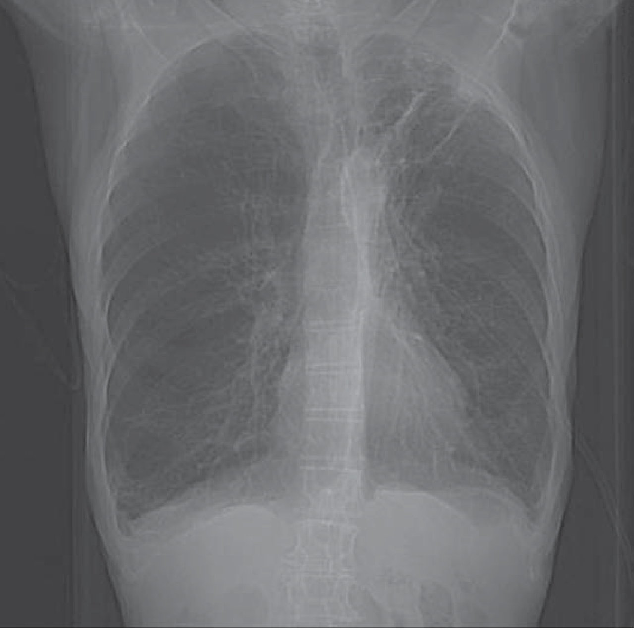
Fig. 70-1 - CXR in COPD. Note hyperaeration, flattened diaphragms, attenuated vascular shadows. (Tintinalli's Emergency Medicine)
6. Acute Exacerbations (AECOPD)
- Superimposed bacterial (e.g., H. influenzae, S. pneumoniae, M. catarrhalis) or viral infections precipitate acute worsening
- Lead to sudden-onset increased dyspnea, worsening gas exchange, and may trigger acute respiratory failure
- A major cause of hospitalization and death in COPD patients
- Mucus plug formation has been shown to cause increased all-cause mortality in COPD
7. Weight Loss and Cachexia
- The increased work of breathing in severe emphysema leads to markedly elevated caloric expenditure
- Combined with poor intake, this produces progressive weight loss that can be so severe as to mimic occult malignancy
- The "pink puffer" phenotype is classically thin and barrel-chested
8. Lung Cancer (Increased Risk)
- Independent of smoking, the chronic inflammatory milieu and structural lung destruction in emphysema is associated with increased lung cancer risk
- Minor hemoptysis in COPD should always raise suspicion for an underlying malignancy
Summary: Complications Cascade
Emphysema
├── Airflow obstruction + gas exchange failure
│ ├── Hypoxemia → secondary polycythemia
│ ├── Hypercapnia → CO₂ narcosis, somnolence
│ └── Acute respiratory failure (death)
│
├── Pulmonary vascular changes
│ ├── Hypoxic vasoconstriction
│ ├── Capillary bed destruction
│ └── Pulmonary hypertension → Cor pulmonale → RHF (death)
│
├── Structural / mechanical
│ └── Bulla/bleb rupture → Pneumothorax (can be fatal)
│
└── Systemic
├── Weight loss / cachexia
├── Secondary polycythemia
└── Increased lung cancer risk
Sources: Robbins, Cotran & Kumar Pathologic Basis of Disease (pp. 636-638, 522) | Tintinalli's Emergency Medicine (p. 509) | Fishman's Pulmonary Diseases and Disorders | Schwartz's Principles of Surgery
Lung abscess
Your credits are exhausted. You can purchase a top-up or subscribe to a plan to continue using Orris. If you're already subscribed, your daily credits will reset tomorrow.
This is a shared conversation. Sign in to Orris to start your own chat.