
Radiological of kidneys and urinary tract (sonography,contrast radiological methods,radionuclide sctinigraphy,cv,mri) indications advantages disadvantages Include anatomy and physiology in short also, pathology too mbbs
"urinary tract"[MeSH Terms] AND "diagnostic imaging"[MeSH Terms]
intravenous urogram normal kidneys ureters bladder
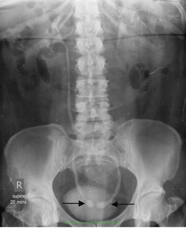
This diagnostic image is a supine anteroposterior (AP) radiograph from an intravenous urogram (IVU), taken 20 minutes post-contrast administration. The film primarily demonstrates the urinary tract, highlighting pathognomonic findings of bilateral ureteroceles. Centrally within the pelvis, the distal ureters exhibit symmetric, well-circumscribed bulbous dilations as they enter the bladder lumen, classically described as the 'cobra head' or 'spring onion' sign. These dilations are outlined by a thin, radiolucent halo, representing the ureterocele wall. Proximal to these structures, the ureters show persistent filling or 'columnization' of contrast. The kidneys show normal contrast enhancement without evidence of hydronephrosis or calyceal blunting. The lumbar spine and bony pelvis appear radiographically normal. This visual serves as a classic educational example of simple adult-type ureteroceles in a patient with recurrent urinary tract infections, illustrating the diagnostic utility of excretory urography in identifying congenital anomalies of the vesicoureteric junction.
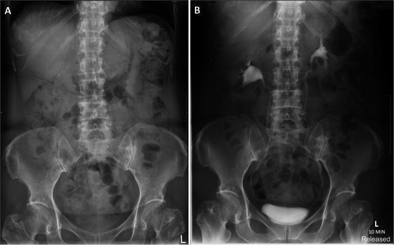
This diagnostic image pair features two anteroposterior radiographs of the abdomen and pelvis, comparing a plain film with a follow-up intravenous urogram (IVU). Image A is a non-contrast interval plain radiograph (KUB) showing the lumbar spine, pelvic girdle, and proximal femora with normal skeletal alignment. The soft tissue shadows of the kidneys and psoas muscles are faintly visible, with no evidence of residual contrast or abnormal calcifications. Image B is a follow-up IVU taken at the 10-minute excretory phase. It demonstrates normal opacification of the bilateral renal pelvicalyceal systems, showing smooth calyces and renal pelvis morphology. Contrast material is seen transitioning into the ureters, and the urinary bladder is partially opacified and well-contoured. Crucially, image B confirms the absence of contrast extravasation or pooling, indicating resolution of any prior urinary tract injury. This comparison is used in urological follow-up to ensure the structural integrity of the collecting system and the normal transit of contrast to the bladder.
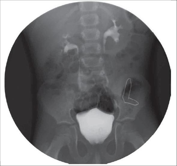
This abdominal X-ray is an Intravenous Urogram (IVU) of a pediatric patient, demonstrating the anatomy and drainage of the upper and lower urinary tract. The diagnostic image shows opacified bilateral renal collecting systems with contrast material filling the renal pelves and calyces. The right kidney is positioned slightly higher than the left, aligned with the upper lumbar vertebrae. Both kidneys exhibit a normal configuration with resolution of previous hydronephrosis. The ureters are visualized as thin, radio-opaque lines descending toward the pelvis. The urinary bladder is seen centrally within the pelvic cavity, appearing densely and uniformly opacified with smooth contours, indicating normal filling. An artificial white outline is present over the left iliac wing for anatomical highlighting. This image serves as post-operative follow-up following surgical correction of obstructive uropathy (posterior urethral valves and orthotopic ureterocele), illustrating restored drainage and normal functional anatomy of the renal units.
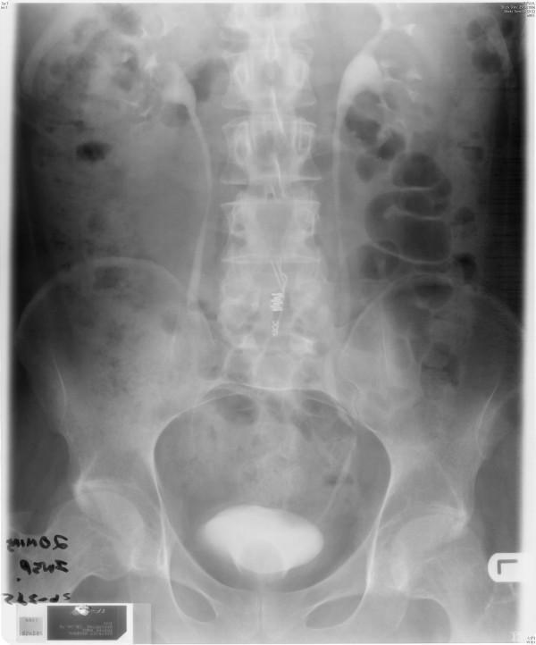
This diagnostic image is an abdominal X-ray radiography showing a 20-minute film from an Intravenous Urogram (IVU). The image captures the abdomen and pelvis, specifically focusing on the renal system. Contrast excretion is visible bilaterally, highlighting the renal pelvis and calyces of both kidneys, which appear to have normal morphology and position. The ureters are visualized as thin, radiopaque lines descending toward the pelvis. The urinary bladder is partially opacified with contrast in the pelvic region, showing a smooth contour without obvious filling defects. The lumbar spine and pelvic bones are clearly visible and appear intact. Notably, there is an absence of previously reported medical hardware (such as Brindley stimulator cables) in the left flank area, indicating successful removal. The image serves to demonstrate normal renal excretory function and the patency of the collecting system following a surgical intervention for infection or device removal.
Radiological Evaluation of the Kidneys and Urinary Tract (MBBS Notes)
1. Applied anatomy (short)
- Kidneys: retroperitoneal, T12 to L3. The right kidney sits slightly lower than the left because of the liver. Each is about 11 cm long, and the two are within 1.5 cm of each other in length.
- Layers: cortex (glomeruli, convoluted tubules), medulla (pyramids), and calyces. Minor calyces join into major calyces, which join into the renal pelvis, which becomes the ureter at the PUJ.
- Ureter: about 25 cm long, with three constrictions where stones lodge: the PUJ, the pelvic brim (crossing the iliac vessels), and the VUJ.
- Bladder and urethra: the bladder is a muscular reservoir (about 400 to 500 mL). The male urethra is divided into prostatic, membranous, bulbar and penile parts.
- Vascular supply: the renal artery arises from the aorta at L1 to L2. Accessory renal arteries are common.
2. Applied physiology (short)
- Blood flow and filtration: the kidneys receive about 20% of cardiac output. Normal GFR is about 120 mL/min.
- Functions: filtration, tubular reabsorption and secretion, concentration of urine, acid-base and fluid balance, and renin, erythropoietin and vitamin D activation.
- Why this matters for imaging:
- Contrast media and radiotracers are cleared by filtration (DTPA, iodinated contrast) or by tubular secretion (MAG3, hippuran).
- DMSA binds proximal tubular cells, so it shows functioning cortex.
- Impaired GFR means poor opacification on contrast studies and a higher risk of contrast nephropathy.
3. Pathology: what imaging is looking for
| Group | Examples |
|---|---|
| Congenital | Agenesis, ectopic or horseshoe kidney, duplex system, PUJ obstruction, ureterocele, posterior urethral valves, polycystic kidney |
| Obstruction | Calculi, strictures, tumours, BPH. Results in hydronephrosis, hydroureter and later cortical thinning |
| Infection | Acute pyelonephritis, renal abscess, pyonephrosis, TB (calcification, "putty kidney", calyceal destruction), reflux nephropathy with scarring |
| Neoplasm | Renal cell carcinoma, Wilms tumour, angiomyolipoma, transitional cell carcinoma (renal pelvis, ureter, bladder) |
| Vascular | Renal artery stenosis, infarct, renal vein thrombosis |
| Medical renal disease | Small echogenic kidneys in CKD, diabetic nephropathy |
| Trauma | Contusion, laceration, urine leak, pedicle injury |
4. Imaging methods
A. Plain radiograph (KUB)
- Indications: suspected radio-opaque stones (about 90% are visible), nephrocalcinosis, calcified lesions such as TB, and follow-up of stones. It is also the scout film before IVU.
- Advantages: cheap, quick and widely available.
- Disadvantages: low soft-tissue contrast. Uric acid stones are radiolucent. Bowel gas and faeces obscure the view, and it uses ionising radiation.
B. Sonography (ultrasound)
- Indications:
- Hydronephrosis and obstruction
- Renal size, cortical thickness and echogenicity (CKD)
- Cyst versus solid mass
- Stones (acoustic shadow), polycystic kidney and congenital anomalies
- Bladder wall, post-void residual and prostate size
- Renal transplant (with Doppler)
- Antenatal screening and paediatric first-line imaging
- Guidance for biopsy and nephrostomy
- Advantages:
- No radiation and no contrast, so it is safe in pregnancy, children and renal failure
- Portable, real-time, cheap and repeatable
- Doppler assesses vascularity and resistive index
- Disadvantages:
- Operator dependent and limited by obesity and bowel gas
- Poor for mid-ureteric stones and gives no functional data
- Cannot reliably exclude small urothelial tumours
- Dose and contrast: none for either.
C. Contrast radiology
- Technique: a scout KUB, then IV non-ionic contrast. Films are taken at about 5 to 10 minutes (nephrogram and calyces), 15 to 20 minutes (ureters) and post-micturition (bladder).
- Indications:
- Haematuria and renal colic (historically)
- Delineating collecting-system anatomy, such as duplex systems and PUJ obstruction
- Post-operative or post-traumatic urine leak
- Advantages: shows anatomy and some function of the whole tract on one study. It is cheap and widely available.
- Disadvantages:
- Contrast reactions and nephrotoxicity
- Radiation
- Poor in renal failure
- Poor for characterising masses
- Largely replaced by CT urography. Grainger & Allison's Diagnostic Radiology states CTU is now the primary method for evaluating the upper urinary tracts, and is better than ultrasound and excretory urography for detecting upper tract urothelial cancer.
- Contraindications: previous severe contrast reaction, renal failure (relative), pregnancy, myeloma or dehydration (relative), and metformin therapy (caution).
- Method: contrast is injected through a ureteric catheter at cystoscopy.
- Indications: non-functioning kidney on IVU, contrast allergy, ureteric strictures, and tumour delineation.
- Advantages: excellent opacification that does not depend on renal function.
- Disadvantages: invasive and needs anaesthesia. It carries a risk of infection and ureteric injury.
- Method: contrast is injected through a needle or a nephrostomy tube.
- Indications: failed retrograde study, or an existing nephrostomy.
- Disadvantages: invasive, with a risk of bleeding and sepsis.
- MCUG indications: vesicoureteric reflux (graded I to V), posterior urethral valves, and neurogenic bladder in children with recurrent UTI.
- RGU indications: urethral stricture and urethral trauma.
- Advantages: the best method for reflux and urethral detail.
- Disadvantages: catheterisation is distressing, gonadal radiation is a concern in children, and it carries an infection risk.
- Indications: renal artery stenosis, haemorrhage or embolisation, pre-operative mapping in donors, and arteriovenous malformation.
- Disadvantages: invasive, with a risk of contrast nephropathy and puncture-site complications. CT or MR angiography is now preferred for diagnosis.
D. Computed tomography (CT, CTU)
- Indications:
- Haematuria. ACR guidelines name CTU as first-line imaging for frank or persistent microscopic haematuria unless a benign cause is already established (Grainger & Allison's Diagnostic Radiology).
- Unenhanced CT KUB is the gold standard for stones.
- Staging of renal masses, trauma, complicated infection or abscess, and retroperitoneal pathology.
- Advantages: fast, high resolution, three-phase study (unenhanced, nephrographic, excretory), and sensitive for small stones and tumours.
- Disadvantages:
- High radiation dose, especially for the three-phase protocol
- Iodinated contrast risk (allergy, contrast-induced nephropathy)
- Overuse is a concern
- Gives limited functional information
E. MRI and MR urography
- Indications:
- Characterising indeterminate renal masses (cyst versus tumour, angiomyolipoma, venous extension of tumour into the IVC)
- Staging of bladder and prostate cancer
- Patients who cannot have CT, such as children, pregnancy, contrast allergy and renal impairment
- Congenital anomalies (including disorders of sex development) and MR angiography for renal artery stenosis
- Support: Campbell-Walsh Urology states MR urography is indicated when radiation or iodinated contrast is contraindicated. Grainger & Allison's also recommends MRU in these patients.
- Advantages: no ionising radiation, the best soft-tissue contrast, multiplanar imaging, and functional MR urography can be done.
- Disadvantages:
- Expensive, with long scan times and limited availability
- Poor for stones and small calcifications
- Contraindicated with pacemakers and some metal implants, and claustrophobia limits use
- Gadolinium carries a risk of nephrogenic systemic fibrosis when eGFR is below 30
- Motion artefact
F. Radionuclide scintigraphy
| Agent | Handling | Use |
|---|---|---|
| 99mTc-DTPA | Glomerular filtration | GFR, perfusion, obstruction |
| 99mTc-MAG3 | Tubular secretion | Best dynamic renogram: differential function, obstruction (with frusemide), transplant, poor renal function |
| 99mTc-DMSA | Binds proximal tubules | Static cortical imaging: scars, acute pyelonephritis, differential function, ectopic or horseshoe kidney |
- Indications:
- PUJ or ureteric obstruction (diuretic renography)
- Split (differential) function before nephrectomy
- Renal scarring after reflux or UTI (done at least 3 to 6 months after infection)
- Renal transplant complications (rejection, ATN, urine leak)
- Renovascular hypertension (captopril renography)
- Direct isotope cystography for reflux (low radiation)
- Diuretic renography details (Campbell-Walsh Urology):
- The patient must be well hydrated and have an empty bladder, because a full bladder gives false-positive results.
- The renogram curve has an early peak and then a rapid descent when normal.
- A rising curve or a plateau suggests obstruction.
- Advantages: physiological and quantitative, the most accurate measure of split function, low radiation dose, and no nephrotoxic contrast.
- Disadvantages:
- Poor anatomical detail
- Radiation (small), and it needs a nuclear medicine department
- Interpretation is affected by hydration, a full bladder and poor function
- A focal DMSA defect is not specific. Campbell-Walsh Urology says any process that replaces, injures or destroys normal cortex will give an abnormal scan.
5. Comparison table
| Modality | Best for | Radiation | Contrast | Functional data |
|---|---|---|---|---|
| KUB | Radio-opaque stones | Yes | No | No |
| Ultrasound | First-line screening, hydronephrosis, children, pregnancy | No | No | No |
| IVU | Anatomy of the collecting system | Yes | Iodinated | Some |
| RGP, antegrade | Obstruction level | Yes | Local | No |
| MCUG, RGU | Reflux, urethra | Yes | Local | No |
| CT, CTU | Stones, haematuria, masses, trauma | High | Iodinated | Limited |
| MRI, MRU | Mass characterisation, staging, patients who cannot have CT | No | Gadolinium (optional) | Possible |
| Scintigraphy | Split function, obstruction, scars | Low | None (radiotracer) | Yes |
6. Quick modality choice by problem
- Renal colic: non-contrast CT KUB, with ultrasound in pregnancy and children.
- Haematuria: CTU, with ultrasound plus cystoscopy in low-risk patients and MRU if CT is contraindicated.
- Hydronephrosis in an infant: ultrasound, then MAG3 renogram for obstruction, and MCUG if reflux is suspected.
- Recurrent UTI in a child: ultrasound, MCUG, and a DMSA scan for scars.
- Renal mass: ultrasound, then CT or MRI.
- Renal artery stenosis: Doppler ultrasound, then CT or MR angiography, with captopril renography as an option.
- Urethral injury or stricture: retrograde urethrogram.
- Renal failure: ultrasound first.