Short note on RS Cells
Reading File
Finding Sources
Finding Sources
Reading File
Reading File
Loading Image
Here is a short note on Reed-Sternberg (RS) cells, based on Robbins' Pathologic Basis of Disease:
Reed-Sternberg (RS) Cells
Definition
RS cells are the pathognomonic neoplastic cells of Hodgkin Lymphoma (HL). Their identification is essential for diagnosis, but they must be seen in an appropriate reactive cellular background to confirm HL.
Morphology
Classic (Diagnostic) RS Cell:
- Large cell (~45 µm in diameter)
- Bilobed or multinucleated nucleus
- Each lobe contains a large, prominent, inclusion-like "owl-eye" nucleolus (5-7 µm - roughly the size of a small lymphocyte)
- Abundant pale cytoplasm
- Surrounded by reactive cells: lymphocytes, eosinophils, plasma cells, macrophages
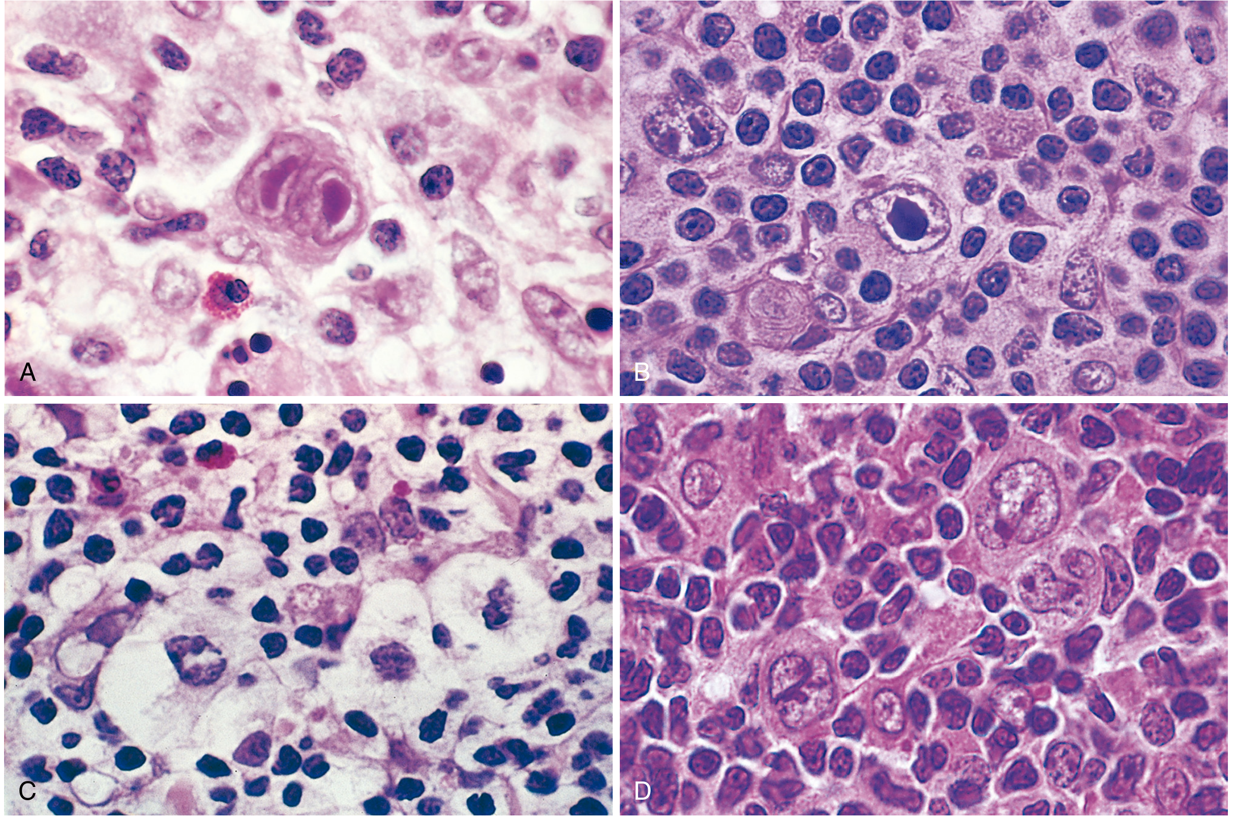
RS Cell Variants
| Variant | Features | Associated Subtype |
|---|---|---|
| Mononuclear (Hodgkin cell) | Single nucleus with large inclusion-like nucleolus | Mixed cellularity, lymphocyte-rich |
| Lacunar cell | Folded/multilobate nucleus, pale cytoplasm, sits in an empty space (artifact of formalin fixation) | Nodular sclerosis |
| Lymphohistiocytic (L&H / "Popcorn" cell) | Multiple in-folded nuclear membranes, small nucleoli, fine chromatin, abundant pale cytoplasm | Nodular lymphocyte predominant HL |
Cell of Origin
RS cells originate from germinal center or post-germinal center B cells, established by clonal IGH gene rearrangements and somatic hypermutation patterns in single isolated RS cells. Despite this B-cell origin, classic RS cells paradoxically fail to express most B-cell genes including immunoglobulin.
Pathogenesis
- NF-κB activation is the central oncogenic event; it rescues "crippled" germinal center B cells from apoptosis
- EBV (Epstein-Barr Virus): Present in ~30-70% of cases depending on subtype. EBV-encoded LMP-1 directly activates NF-κB
- In EBV-negative cases, loss-of-function mutations in IκB or TNFAIP3 activate NF-κB
- RS cells are aneuploid with frequent gains at chromosome 2p (REL oncogene) and chromosome 9p (PD-L1/PD-L2), the latter helping immune evasion
Immunophenotype
| Feature | Classic HL RS cells | Nodular LP (Popcorn cells) |
|---|---|---|
| CD15 | + | - |
| CD30 | + | - |
| CD20 | - | + |
| CD45 | - | + |
| EBV | Variable (subtype-dependent) | - |
| PAX5 | Weak + | Strong + |
Reactive Microenvironment
RS cells secrete cytokines (IL-5, IL-10, M-CSF) and chemokines (eotaxin) that attract a polymorphic reactive infiltrate. The recruited cells (eosinophils, T cells) in turn feed back to activate CD30 and CD40 on RS cells, further upregulating NF-κB. RS cells also express PD-L1 and PD-L2, which suppress cytotoxic T-cell responses and enable immune escape.
Conditions with RS-Like Cells (Must Exclude)
Cells resembling RS cells can be seen in:
- Infectious mononucleosis
- Solid tissue cancers (metastatic)
- Certain non-Hodgkin lymphomas
RS cells are therefore diagnostic of HL only when identified in the correct morphological and immunophenotypic context.
Source: Robbins, Cotran & Kumar - Pathologic Basis of Disease (Robbins Pathology), Chapter 13 - Lymphoid Neoplasms
This is a shared conversation. Sign in to Orris to start your own chat.