
Ankylosing spondylitis
Reading File
Now let me search the medical library and PubMed in parallel for comprehensive coverage.
Finding Sources
Searching PubMed
"ankylosing spondylitis"[MeSH Terms] AND treatment
Searching the Web
ankylosing spondylitis 2025 2026 guidelines treatment update
Good. Now let me read the key textbook sections in parallel - Goldman-Cecil (best for comprehensive rheumatology), the radiology textbook, and search for more specific content.
Reading File
Reading File
Searching PubMed
"ankylosing spondylitis"[MeSH Terms] AND "biologics"[tiab]
Excellent content retrieved. Let me now read the main Goldman-Cecil block for the full clinical details and fetch the key radiology images.
Reading File
Reading File
Loading Image
Loading Image
Now let me get the Goldman-Cecil main rheumatology block for the core clinical content on AS.
Finding Sources
Loading Image
Now let me get the main Goldman-Cecil rheumatology section on AS (block11 around line 3935).
Reading File
Now let me get the main rheumatology chapter content - I need to find the dedicated chapter (Chapter 244 referenced in Goldman-Cecil).
Finding Sources
Finding Sources
Firestein & Kelley's has a dedicated chapter on AS. Let me read it.
Reading File
Let me now read the Firestein dedicated AS chapter (block15 at line 7807).
Reading File
Now let me read the main dedicated Firestein AS chapter (block6 at line 3120) for the core epidemiology, pathophysiology, and clinical content.
Reading File
I now have rich content from multiple authoritative textbooks. Let me compile the comprehensive answer.
Ankylosing Spondylitis (AS)
Overview
Ankylosing spondylitis is a chronic, progressive inflammatory spondyloarthropathy that primarily targets the axial skeleton - the sacroiliac joints, spine, and large peripheral joints. It is the prototypic form of axial spondyloarthritis (AxSpA). The term itself derives from Greek: ankylos (bent/fused) and spondylos (vertebra).
Epidemiology
-
Prevalence: ~0.1-0.5% of the general population; varies by ethnic group and HLA-B27 frequency
-
Age of onset: Most commonly 15-25 years; rarely begins after age 40
-
Sex: Affects men 2-3x more than women, though women are increasingly recognized (often milder or delayed diagnosis)
-
Genetics: Heritability exceeds 95%. Sibling recurrence risk is 9.2% vs. 0.1-0.4% in the general population
-
Firestein & Kelley's Textbook of Rheumatology, p. 3122
Pathogenesis
HLA-B27 and Genetics
- HLA-B27 is the strongest genetic susceptibility factor: >90% of White AS patients carry it (vs. ~8% of the general population), giving a relative risk of 50-100
- Only ~2% of HLA-B27-positive individuals develop AS, so additional genetic and environmental factors are required
- Over 80 risk loci outside the HLA region have been identified, including:
- ERAP1/ERAP2 (aminopeptidase genes) - involved in peptide trimming for HLA presentation; ERAP1 association is only found in HLA-B27-positive individuals (genetic epistasis)
- IL-23R, IL-12B, IL-27 - genes in the IL-23/IL-17 pathway (key therapeutic target)
Immune Mechanisms
-
The IL-23/IL-17 axis plays a central role: IL-23 drives Th17 differentiation, which produces IL-17A/F; this promotes entheseal inflammation
-
Notably, anti-IL-17 drugs are effective in AS but anti-IL-23 drugs are not - suggesting IL-23-independent sources of IL-17 at entheses (likely innate immune cells)
-
TNF-alpha also mediates spinal and sacroiliac inflammation
-
Pathological process: enthesitis (inflammation at ligament/tendon insertions into bone) → new bone formation → syndesmophytes → fusion
-
Firestein & Kelley's Textbook of Rheumatology, p. 3154-3166
Clinical Features
Axial (Spine and Sacroiliac)
- Inflammatory back pain is the hallmark - insidious onset, age <40, duration >3 months, worse with rest, improved with exercise, associated with morning stiffness >1 hour
- Sacroiliitis - bilateral and symmetrical; buttock pain may radiate down the thighs
- Reduced spinal mobility in all planes (Schober's test < 5 cm)
- Progressive thoracic kyphosis, loss of lumbar lordosis
- Chest wall restriction: fusion of costovertebral and sternoclavicular joints limits rib cage expansion to <2.5 cm (measured at 4th intercostal space)
Peripheral Joint Involvement
- Up to 30% of patients have peripheral arthritis (hips are the most important - the second most commonly affected joint after the SIJ)
- Hip involvement leads to diffuse joint space loss and characteristic entheseal new bone around the femoral head (can mimic femoroacetabular impingement)
Extra-articular Manifestations
| Feature | Frequency | Notes |
|---|---|---|
| Anterior uveitis | ~25-40% | Acute, unilateral, recurrent; HLA-B27-associated |
| Inflammatory bowel disease | ~5-10% | Crohn's > UC |
| Psoriasis | ~10% | |
| Pulmonary (fibrobullous upper lobe disease) | 1-4% | Bilateral upper lobe fibrosis and cyst formation; no effective therapy |
| Aortic regurgitation | rare | Ascending aortitis |
| Cardiac conduction defects | rare | |
| Cauda equina syndrome | rare | Late complication |
Late Complications
-
Osteoporosis - from chronic inflammation and abnormal biomechanical loading
-
Spinal fractures - the rigid, fused spine is paradoxically osteopenic and highly vulnerable; transverse fractures through fused segments are highly unstable and potentially catastrophic
-
Spondylodiscitis (Andersson lesion) - inflammatory destructive lesion at unfused disc levels in an otherwise fused spine; can mimic infection
-
Restrictive lung disease - from rib cage fixation; diaphragm compensates but eventually may cause respiratory failure
-
Goldman-Cecil Medicine, p. 3943-3945; Grainger & Allison's Diagnostic Radiology, p. 3659-3661
Diagnosis
Modified New York Criteria (for AS)
Clinical criteria:
- Low back pain and stiffness for >3 months, improved with exercise and not relieved by rest
- Limited lumbar spine motion in sagittal and frontal planes
- Limited chest expansion (relative to normal values for age and sex; <2.5 cm at 4th ICS)
Radiological criterion:
- Bilateral sacroiliitis grade ≥2, OR unilateral sacroiliitis grade 3-4
Definite AS = radiological criterion + ≥1 clinical criterion
ASAS Classification Criteria (axial SpA, broader)
Allows diagnosis before radiographic sacroiliitis (non-radiographic axial SpA) using MRI evidence of active inflammation or HLA-B27 + ≥2 other SpA features.
Laboratory
- HLA-B27: positive in >90% of White patients (lower in other ethnic groups, e.g., ~70% in African Americans)
- Elevated CRP/ESR: in active disease (~50-60% of patients; normal does not exclude active disease)
- RF and ANA: negative (hence "seronegative" spondyloarthropathy)
- Henry's Clinical Diagnosis and Management by Laboratory Methods: HLA-B27 testing is confirmatory but not sufficient alone
Imaging
Plain Radiography
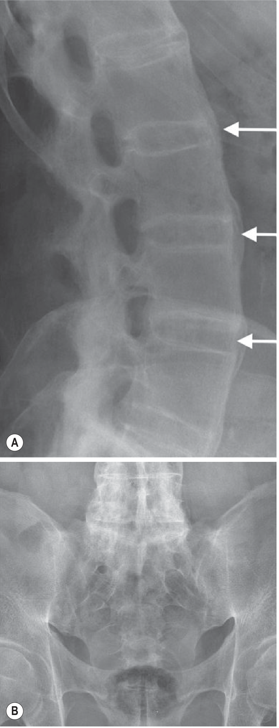
- Sacroiliac joints: erosion, subchondral sclerosis, eventually ankylosis (bilateral, symmetrical)
- Spine:
- Romanus lesions - sclerotic "shiny corners" at vertebral body corners (earliest X-ray finding; due to enthesitis at Sharpey fiber insertions)
- Squared vertebrae - due to erosion and anterior longitudinal ligament ossification
- Syndesmophytes - thin, vertical bone outgrowths (distinguishable from the coarser, horizontal osteophytes of OA or the "chunky" syndesmophytes of psoriatic/reactive arthritis)
- Bamboo spine - complete fusion of vertebral bodies and posterior elements (end-stage)
Fig. 44.17 - MRI of sacroiliac joints in AS: T1-weighted image shows erosion and joint space loss; T2 fat-suppressed shows extensive subchondral oedema (active disease):
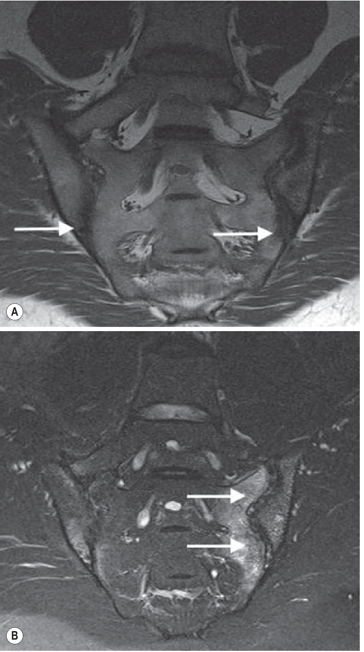
Fig. 44.18 - Plain radiograph showing Romanus lesions (sclerotic vertebral corners) and bridging syndesmophytes in the thoracic spine:
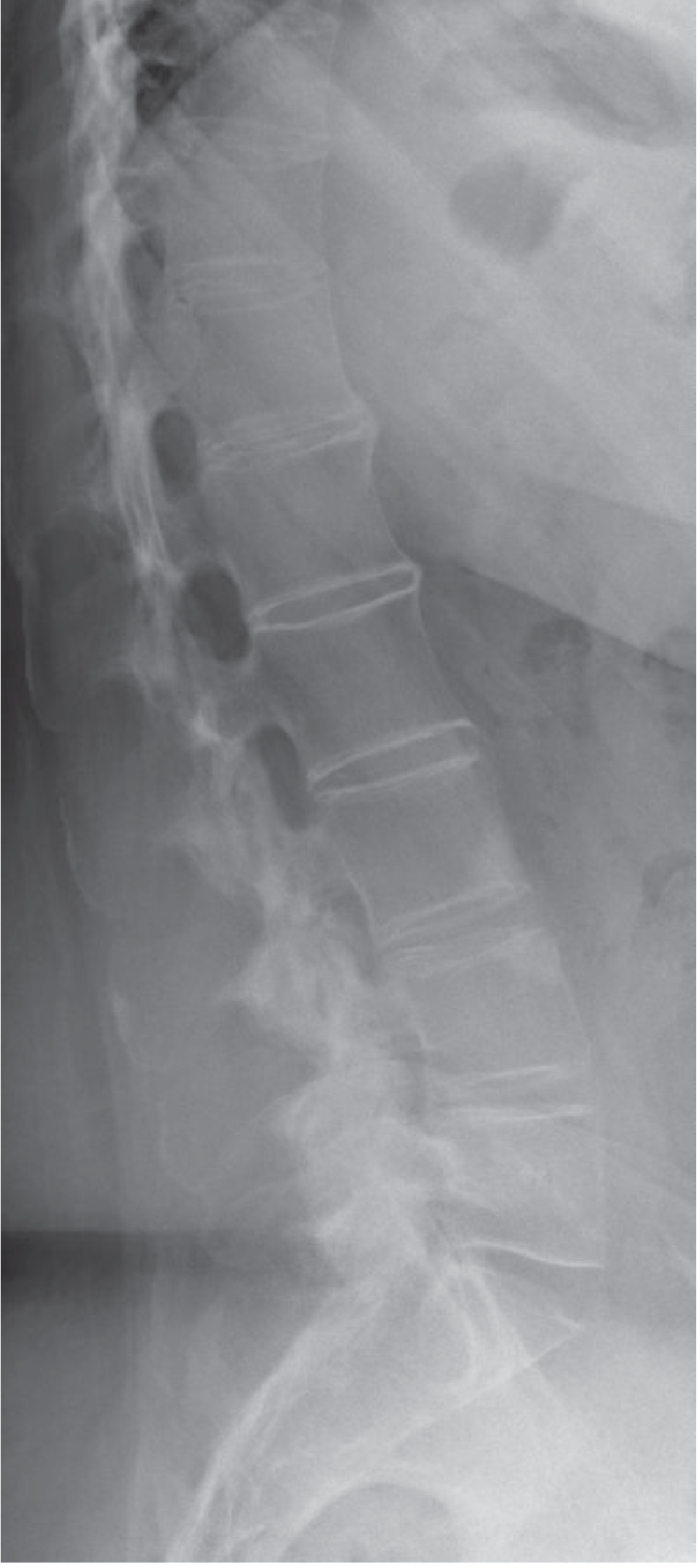
Fig. 44.20 - Advanced bamboo spine with vertical syndesmophytes (A) and complete bilateral sacroiliac joint fusion (B):
MRI (preferred for early disease)
- Most sensitive and specific for early sacroiliitis and spinal inflammation
- Detects subchondral bone marrow oedema before plain radiograph changes appear
- Key distinguishing feature: AS-related oedema is greater in craniocaudal dimension than transverse, and is not associated with disc dehydration or height loss (vs. degenerative disease)
- STIR sequences best for detecting active inflammation
CT
-
Best for detecting structural damage, erosions, and degree of ankylosis
-
Not preferred for early disease (radiation, poor sensitivity for marrow oedema)
-
Grainger & Allison's Diagnostic Radiology, p. 3621-3662
Treatment
Non-Pharmacological
- Exercise and physiotherapy are strongly recommended and improve cardiorespiratory fitness, spinal mobility, and pain - must be started concurrently with drug therapy
- Posture training, hydrotherapy, supervised group exercise
- Smoking cessation (smokers have worse outcomes and more rapid radiographic progression)
NSAIDs (First-Line)
- NSAIDs at maximum tolerated doses are the cornerstone of treatment for active disease
- Continuous treatment is preferred over on-demand use for persistent symptoms
- No particular NSAID is superior to another; patient-specific factors (GI risk, cardiovascular risk) guide selection
- Treatment failure is defined as inadequate response to at least two different NSAIDs
- ACR guidelines: strong evidence for NSAIDs; strong caution against systemic glucocorticoids for active AS
Biologic DMARDs (Second-Line)
TNF Inhibitors (TNFi) - first biologic choice:
Five TNFi are approved and show remarkable efficacy similarity:
- Adalimumab, etanercept, infliximab, certolizumab pegol, golimumab
- Improve symptoms, quality of life, CRP, anemia, sleep, and MRI inflammation scores
- Early and continuous treatment inhibits radiographic progression (syndesmophyte formation)
- Special considerations: For patients with comorbid inflammatory bowel disease or recurrent uveitis, prefer infliximab or adalimumab over etanercept (TNFi monoclonal antibodies > etanercept for these conditions)
- TNFi are also effective in non-radiographic axial SpA
IL-17 Inhibitors - alternative biologic:
- Secukinumab, ixekizumab (IL-17A inhibitors) - effective in AS
- Bimekizumab (IL-17A and IL-17F dual inhibitor) - also effective
- Preferred in patients who fail or cannot tolerate TNFi, or with predominant skin involvement
- Note: anti-IL-23 agents (guselkumab, risankizumab) do NOT work in AS despite working in psoriatic arthritis
JAK Inhibitors (Third-Line/Later):
- Tofacitinib (JAK inhibitor) - FDA-approved December 2021 for AS in patients with inadequate response to or intolerance of TNFi
- ASAS20 achieved in 56.4% vs. 12.5% placebo at 16 weeks; response seen by 2-4 weeks
- Safety: hepatic events (2.3%), herpes zoster (2.3%), serious infections (0.8%)
- Upadacitinib - also approved for AS
Certolizumab for Non-Radiographic AxSpA
- First anti-TNF agent FDA-approved for non-radiographic axial SpA specifically
Surgery
-
Total hip arthroplasty for severe hip involvement
-
Spinal osteotomy for severe fixed kyphotic deformity
-
Special anesthetic consideration: endotracheal intubation must be performed with great caution due to rigid cervical spine; hyperextension can cause fracture; cricoarytenoid joint involvement may limit mouth opening
-
Firestein & Kelley's Textbook of Rheumatology, p. 7809-7823; Goldman-Cecil Medicine, p. 3948
Disease Monitoring
- BASDAI (Bath Ankylosing Spondylitis Disease Activity Index) - validated patient-reported measure
- ASDAS (Ankylosing Spondylitis Disease Activity Score) - incorporates CRP; preferred by guidelines
- Serial MRI for assessing response to biologics
- Serial plain radiographs every 2 years to monitor structural progression
Prognosis
-
Clinical course is highly variable
-
Strongest predictor of new syndesmophyte formation is presence of syndesmophytes at baseline
-
Significant impact on work disability and quality of life
-
Osteoporosis occurs in proportion to disease duration and activity
-
With early biologic treatment, radiographic progression can be significantly retarded
-
Late complications (bamboo spine, fractures, cauda equina syndrome, upper lobe fibrosis) are largely preventable with effective therapy
-
Goldman-Cecil Medicine, p. 1004-1008
Sources: Firestein & Kelley's Textbook of Rheumatology (2-Volume Set); Goldman-Cecil Medicine International Edition; Grainger & Allison's Diagnostic Radiology; ACR/SAA/SPARTAN Guidelines (2019 update)
This is a shared conversation. Sign in to Orris to start your own chat.